

Dr.SUBASHINI S,Dr.RUKSANA AYUB K
ABSTRACT
Aim:
To evaluate epidemiology, microbiological features as well as antibiotic susceptibility patterns of Nocardia from cases keratitis seen over a period of 6 years in a tertiary eye care hospital.
Methods:
Microbiology records of 49 cases of culture proven Nocardia keratitis diagnosed from 2012 to 2017 were reviewed retrospectively. The outcome data included isolation rate, predisposing factors, demography (age and sex), utility of conventional diagnostic methods, microbiological profile and antibiotic sensitivity patterns.
Results:
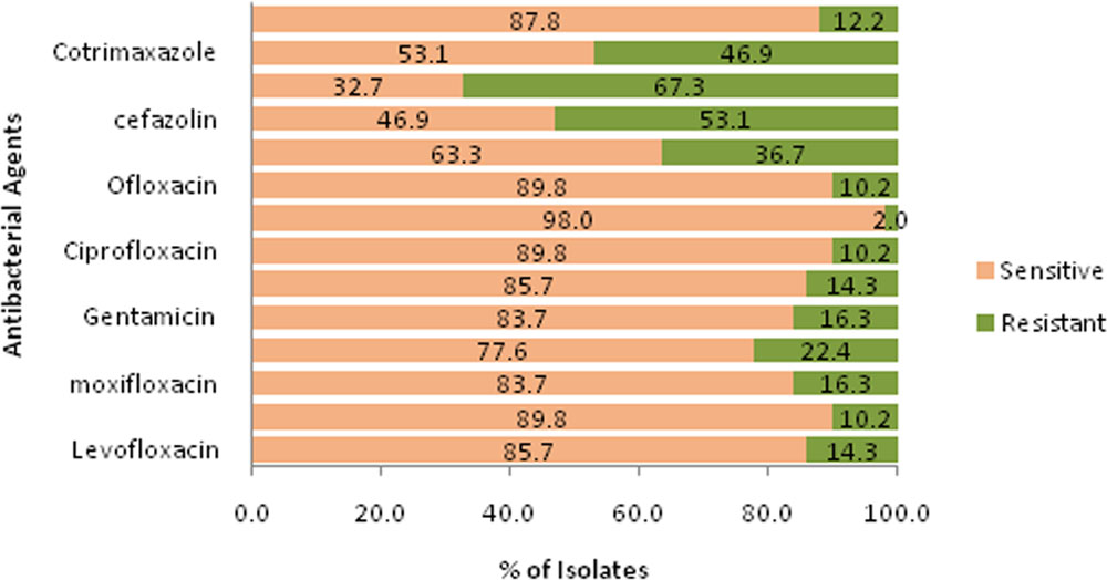
A total of 49 (2.1%) Nocardia keratitis were isolated from 2370 culture proven ocular specimens. Most of the cases were male (n=34, 69.4%) between the mean age group 56.51 and it ranges from 27 to 85 years. All 49 cases were identified by culture and confirmed by 1% acid fast staining. Out of them, 80.8% and 76.6%were positive for KOH mount and Gram staining respectively. Upon in vitro susceptibility testing, 98% showed sensitivity for amikacin and 89.8% showed sensitive for ciprofloxacin.
Conclusion:
Ocular nocardiosis is a rare entity among all other infection. Early accurate diagnosis by culture and confirming it with 1% acid fast staining yielding better clarity to clinicians to start appropriate treatment.
INTRODUCTION
Nocardia is a thin, filamentous, aerobic, gram positive bacteria that found in fresh and marine water, soil, dust and decaying vegetation.Nocardia keratitis may be misdiagnosed clinically, as it is uncommon and the picture may resemble mycotic keratitis or keratitis caused by atypical mycobacteria.2In addition, Antibiotic susceptibility testing of Nocardiais technically difficult and time-consuming. To start appropriate therapy, apart from clinical examination, precise laboratory diagnosis and the results of in vitro anti-bacterial susceptibility testing are important.2In this study, we describe 49 patients with culture-proven Nocardia infections from corneal scrapings. Besides epidemiological details such as demography, and seasonal variation, the results of in vitro anti-bacterial susceptibility and efficacy of conventional diagnostic techniques in diagnosing nocardiosis are also described.
MATERIALS AND METHODS
Cases with cultureproven ocular nocardiosis were identified from microbiology laboratory database among all ocular specimens collected from patients with keratitis. During 6 years of study period (January2012- December2017) 2370 corneal scrapings samples were collected and processed for microbiological investigations. Culture methods involved direct inoculation of specimens on to 5% sheep blood agar, chocolate agar, non-nutrient agar, potato dextrose agar, thioglycollate, and brain–heart infusionbroth.
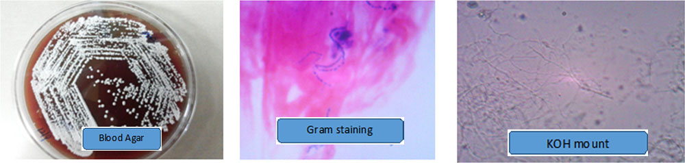
The inoculated potato dextrose agar was incubated at 25oC to isolate fungi and examined daily up to 3 weeks. To isolate bacteria, inoculated sheep blood agar plates were incubated under aerobic and anaerobic conditions; chocolate agar was incubated with 5% carbon dioxide, and thioglycollate and brain–heart infusion broth aerobically at 37oC. The material obtained by scraping was also spread onto labelled slides in a thin, even manner for 10% potassium hydroxide (KOH) wet mount, Gram stainingand Giemsa staining. Acid fast stains (Ziehl-Neelsen and Kinyoun’s) were performed in suspected cases. A culture was considered positive when there was growth of the same organism on two or more media on ‘C’ streak (inoculum site), confluent growth at the site of
Inoculation on one solid medium, growth in one medium with consistent direct microscopy findings, growth of the same organism on repeated corneal scraping and growth consistent with clinical signs. The isolated Nocardia species were identified by standard bacteriological
methods. All isolates were tested for susceptibility to antibacterial agents such as, gentamicin, ciprofloxacin, vancomycin, gentamycin, ofloxacin, gatifloxacin, cefotaxime, ceftazidime and co-trimoxazole by Kirby Bauer discdiffusion method following guidelines by National Committee for Clinical Laboratory Standards
Data analysis:
Isolation rate estimates are provided with 95% confidence intervals (CIs) with normal approximation. Annual and monthly isolation figureswere presented in line diagrams for trend analysis. Statistical significance of trend was tested using chi-square test for trends. A P-value <0.05 was considered significant.
Results:
A total of 49 nocardial isolates were identified among 2370 culture-positive specimens and the isolation rate was estimated to be 2.1% (95% CI: 1.6, 2.7%) during the 6-year study period. The rest of the pathogens included bacteria (14.9%), fungi (81.2%), mixed (0.8%), and Acanthamoeba (1.0%) (table1)
| Isolates | Number of cases | Isolation rate (% (95% CI)) |
| Only bacteria | 353 | 14.9 (13.5, 16.4) |
| Only fungi | 1925 | 81.2 (79.6, 82.7) |
| Only parasite | 24 | 1.0 (0.6, 1.5) |
| Mixed | 19 | 0.8 (0.5, 1.2) |
| Nocardia species | 49 | 2.1 (1.6, 2.7) |
| Total | 2370 | 100 |
Table 1
Seasonal trend: The annual isolation ranged from 1 (2017) to 12 (2013 and 2016) (Figure 1). The annual trend was statistically significant in the isolation of Nocardia species yield (P=0.01), implying that there was significant variability in isolation rate over years. Similarly, when the monthly trend was analysed, the isolation numbers ranged from 1(March) to 12 (October) (Figure 2). There was statistically significant monthly trend (P=0.0025) in the isolation of Nocardia species, suggesting that the infection rate varies all through the year and was seasonal.
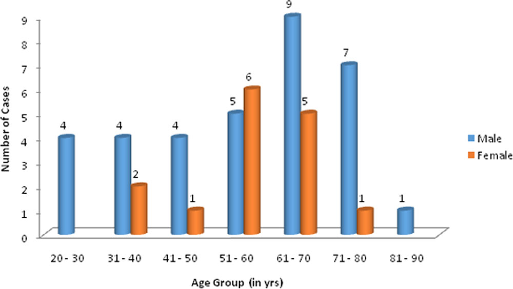
Demographics:The mean age is 56.51 years and it ranges from 27 to 85 years. (Figure 3). Men were infected more (71.4%) than women (28.4%) (Figure 3).
Discussion
In this study, the isolation rate of Nocardia was observed as 2.1%. When compared with Sridhar et al4 (1.7%), Srinivasan et al5 (1.6%), Bharathi et al2 (1.4%) it was high but low as compared with Manikandan et al3 (3.1%). In contrast to other studies, analysis of annual and monthly isolation revealed that there is significant seasonal variation over 6 years and increased in the month of October (n=12). Manikandan et al and Bharathi et al did not find any seasonal variation
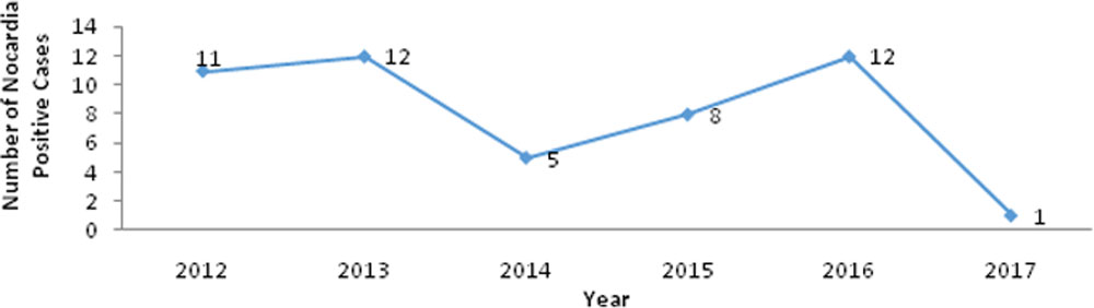
Figure 1:Yearly trend of isolation of Nocardia species
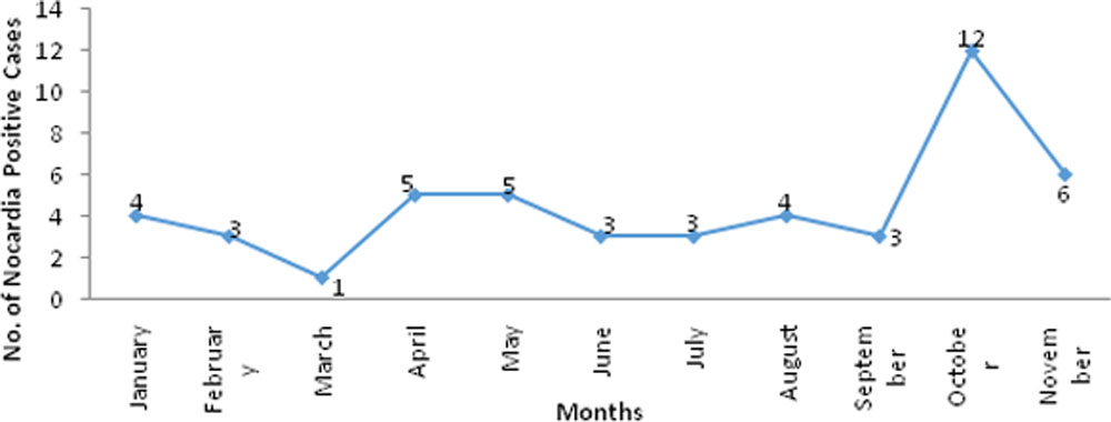
Figure 2:Monthly trend of isolation of Nocardia species
The main limitations of our study are its retrospective nature and the study being conducted at a tertiary care hospital, where a significant number of patients are referrals after initial treatment.Future investigations based on longer time series may allow testing of the cyclic patterns detected in this study. To conclude, Nocardia keratitis can be identified by Gram stain in most of the cases. 1% acid-fast stain should be performed in all suspected cases.
Amikacin can be used as monotherapy in the treatment. Similar to Manikandan et al study3, most of the patients affected were elderly adults from 51 to 70 years of age. (Mean age 56.51 years). Although they did not have any underlying immunodeficient condition, age related immunocompromisation makes them vulnerable group.
Men (71.4%) were more affected than women (28.4%) in this study. In most of the published reports, male predominance was clearly delineated due to their more outdoor activities.In this study, Nocardia positivity was shown after Gram stain examination and KOH mount 76.6% & 80.8%% respectively.
It is similar to Manikndan et al study3 (69.4%) Sridhar et al study where 10 out of 15 cases showed positivity. In this study, amikacin (98%) was found to be the most effective drug against Nocardia similar to Manikandan et al study3(98.7%),Sridhar et al4 study and Hussian et al6(100%);We observed a sensitivity of 89.8% to ciprofloxacin among our isolates. Bharathi et al2Maikandan et al3had reported that 93.5% & 97% of the strains were sensitive to ciprofloxacin.We observed that vancomycin sensitivity 87.8% was similar to Bharathi et al study2 (78.5%).
References:
- Srinivasan M, Reddy J. Ocular Nocardiosis. Journal of TNOA 1998;38:25-29.
- Bharathi MJ, Ramakrishnan R, Vasu S, Meenakshi R,Chirayath A, Palaniappan R. Nocardia asteroides keratitis in South India. Ind J Med Microbiol 2003;21: 31–36
- Manikandan P, Bhaskar M, Revathi R, Anita R, Lakshmi LA, Narendran V. Isolation and antimicrobial susceptibility pattern of Nocardia among people with culture-proven ocular infections attending a tertiary care eye hospital in Tamilnadu, South India. Eye. 2007;21(8):1102.
- Sridhar MS, Sharma S, Reddy MK, Mruthyunjay P, Rao GN. Clinicomicrobiological review of Nocardia keratitis. Cornea. 1998;17(1):17-22.
- Matulionyte R, Rohner P, Uckay I, Lew D, Garbino J. Secular trends of nocardial infection over 15 years in a tertiarycare hospital. J Clin Pathol 2004;57: 807–812.
.
Leave a Comment