

Dr.Ganesh Venkataraman, Dr.Prasanna venkataraman,Dr.NABEED N P R,Dr.Premanand Chandran
Tunnel and track technique for Aurolab Aqueous Drainage Implant (AADI) placement
Abstarct
AIM: To evaluate the safety and efficacy of the scleral tunnel-track technique used in Aurolab aqueous drainage implant (AADI)-350, in preventing tube exposure through conjunctiva. DESIGN: Retrospective, hospital based case series. MATERIALS AND METHODS: A retrospective analysis of 13 patients who underwent AADI for refractory glaucoma. These patients had undergone scleral tunnel-track technique to prevent tube exposure &had atleast6 month follow-up. Outcome measures were intraocular pressure (IOP) control and rates of tube related complications. RESULTS: 13 eyes of 13 patients were included in the study. Mean follow up was 10.61±0.72 months. There was significant reduction in terms of both IOP reduction and number of AGM (anti-glaucoma medication) postoperatively. There were no tube related complications. CONCLUSION: Scleral tunnel track technique promises to be a safe alternative to donor sclera/corneal patch graft in reducing tube exposure.
Introduction
Refractory glaucomas pose a major challenge to the glaucoma surgeon. With many risk factors for failure, including previous surgery, chronic usage of antiglaucoma medications (AGM), the surgeon has to decide on the appropriate surgical plan. Glaucoma drainage devices have become a valuable tool in the management of these glaucomas. But these devices have their own set of complications, some of which can be vision threatening. We have modified our surgical approach to address these issues. We present here a modified technique of tube placement which does away with corneal or scleral patch graft.
Aurolab Aqueous Drainage Implant (AADI) 350 is a non-valved drainage implant made up of NuSil silicone elastomer and is available in surface area of 350 mm2, plate length of 32 mm and tube length of 35 mm. The device has lateral wings which are designed to be positioned under the rectus muscle. It also has fixation holes to help anchor the end plate about 8-10 mm posterior to the limbus. As the device has larger surface area as compared to other valved devices it helps to achieve a lower target pressure in the long term.
The tube has a relatively long subconjunctival course and is conventionally covered by donor material such as sclera, dura, or pericardium to prevent conjunctival erosion over the tube hence reducingthe chances of tube erosion and subsequent complications such as tube infection and endophthalmitis.
We present a retrospective analysis of 13 patients who underwent AADI-350, in whom the sclera “tunnel – track” technique was used for tube insertion into the anterior chamber. This technique of tube placement avoids scleral/corneal patch graft, reduces the profile of the implant, increases patients’ comfort, reduces operative time and there is less dependency on eye banks for tissue procurement.
MATERIALS & METHODS:
PATIENT POPULATION:
Patients for this study were recruited from the Glaucoma clinic in Aravind Eye Hospital, Coimbatore – a tertiary eye care hospital in South India. Consecutive patients who were operated upon by a single surgeon over the period from July 2014 to June 2017 using a modified surgical technique for the implantation of AADI 350 were included. Patients were selected for surgery because they had glaucoma uncontrolled on maximum tolerated medical therapy and were considered at high risk of failure for trabeculectomy or had previously failed trabeculectomy. Patients who had undergone Ahmed Glaucoma Valve implantation and placement of the tube in the vitreous cavity and implantation done in any other quadrant other than superotemporal quadrant were excluded from the study. This study was conducted in accordance with the Declaration of Helsinki after institutional review board approval.
DATA COLLECTION:
Preoperative data collection included recording of age, sex, diagnosis, number of preoperative antiglaucoma medications, preoperative visual acuity, and intraocular pressure (IOP). All patients included had completed 6 month follow-up. Postoperative data included visual acuity, IOP, and number of antiglaucoma medications used postoperatively and for every subsequent postoperative visit at day 1, day 7, 1 week, 1 month, 3 months, 6 months and every 2 months thereafter. Any intraoperative or postoperative complication was recorded. All available follow up data were used.
OUTCOME MEASURES:
The primary outcome measure was IOP control.
Success criteria:
Complete was defined as a post-operative IOP between 6 and 21 mmHg, without additional topical or systemic medications. If this defined IOP control was achieved with it use of antiglaucoma medication it was considered as a qualified success
Failure criteria:
- IOP < 6 mm Hg or > 21 mm Hg on two visits after 3 months
- Additional glaucoma surgical intervention to control IOP
- Visual acuity of no light perception (NLP)
- Persistent corneal decompensation
The secondary outcome measure was tube related complications, including tube exposure, extrusion, migration and tube-related endophthalmitis.
SURGICAL TECHNIQUE:
All the surgeries were performed under local anesthesia by a single surgeon. AADI-350 model was implanted in all 13 eyes in the superotemporal quadrant. A limbal based conjunctival flap was fashioned, adequate dissection carried underneath and relaxing cuts given for adequate exposure. After identifying and hooking the superior and lateral rectus muscles, the wings of the plate were inserted between them. It was anchored between these two muscles with 9-0 nylon suture and secured 10-12 mm away from limbus. The tube patency was tested and then completely occluded, close to the plate with 7-0 polyglactin suture.
In our modified technique, we created a scleral tunnel proximally and a distal conventional tube track of about 4 mm using a 23G bent needle into the anterior chamber. The tunnel is created between two partial thickness parallel grooves about 4 mm apart and the proximal groove about 3-4 mm away from the plate. The distal groove will be about 4 mm away from the limbus. From the scleral groove closer to the limbus, a needle (23‑gauge) track was made into the AC almost in the same plane as the scleral tunnel and anterior chamber was entered in a plane parallel to iris. After assessing the length of the tube, it was trimmed bevel facing the corneal endothelium. Intact tube was passed through this scleral tunnel-track and into anterior chamber
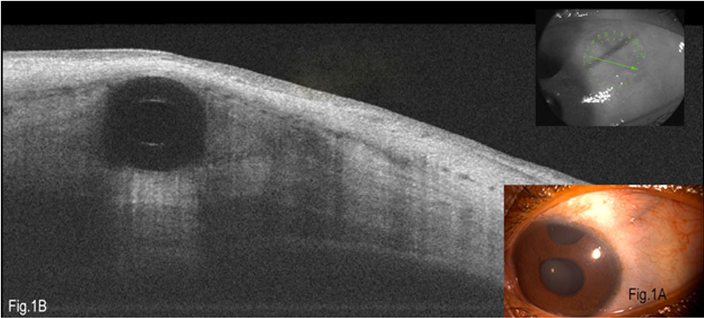
Fig 1A- shows the superotemporal tube in situ; Fig 1B- Anterior segment- OCT image showing the tunnel-track and the tube in situ. Note adequate coverage of tube all around by scleral tissue without internal/external erosion. This is an adolescent patient of congenital glaucoma with a previous trabeculectomy with trabeculotomy.
The tubewas secured with 9-0 nylon sutures close to the plate and also to the sclera at the point of entry into sclera tunnel. Scleral incision which was 4 mm behind the limbus is secured with 9-0 nylon suture. Conjunctiva was closed with 7-0 polyglactin in a running fashion. Patients received a combination of gatifloxacin 0.3% and dexamethasone 0.1% eye drops six times daily which was tapered over 6weeks. Homatropine 2% eye drop was used for 2 weeks and stopped after 2 weeks. Anti-glaucoma medications were continued after the surgery and titrated according to IOP measurements in the follow-up visits.
RESULTS
During the study period 17 eyes of 17 patients were operated with this technique. 4 eyes were excluded as they did not comply with 6 month follow-up criteria. The remaining 13 eyes were analyzed. All the surgeries were performed by an experienced single surgeon.
Of the 13 patients, 10 were males and 3 were females. The mean age was 47.69 years (SD 23.7, with range between 8-78 years). The diagnoses were, congenital glaucoma(2), Glaucoma in pseudophakia (2), Primary angle closure glaucoma (2), Post keratoplasty glaucoma (2), Pseudoexfoliation glaucoma (1), Traumatic glaucoma (1), Uveitis glaucoma (1), Primary open angle glaucoma (1), Juvenile open angle glaucoma(1).
We analyzed the number of prior ocular surgeries each patient underwent, before the AADI implantation. Nine eyes had 1 (69.23%), Two eyes had 2 (15.38%) and one eye had 4 (7.69%) surgeries previuosly. In our study, 3 patients had prior combined cataract and glaucoma surgery (23.08%) and 2 patients each had prior penetrating keratoplasty with intraocular lens implantation, congential glaucoma surgery (Trabeculectomy with Trabeculotomy) and small incision cataract surgery.
In the analysis of the lens status, 7 eyes were pseudophakic (53.85%), 5 phakic (38.46%) and 1 aphakic. None of our phakic patients developed any corneal decompensation.
All 13 patients had a minimum of 6 month follow up. At 6 months, 7 patients achieved complete success, 5 achieved qualified success and 1 patient failed to achieve IOP control.
Table: 1 . Demographics
| No | Age | Diagnosis | Previous number of surgeries | Previous surgeries | Preop
IOP |
Preop
No. Of AGM |
Post op IOP at 6 months | Postop no. Of AGM at 6 months | ||
| 1 | 53 | Uveitic glaucoma | 0 | Nil | 30 | 4 | 15 | 0 | ||
| 2 | 61 | PKP glaucoma | 4 | IOL, explantation, PKP+SFIOL, CPC Diode | 35 | 5 | 7 | 0 | ||
| 3 | 78 | PACG | 1 | Combined surgery | 16 | 2 | 16 | 0 | ||
| 4 | 20 | Glaucoma in pseudophakia | 1 | IOL | 36 | 5 | 16 | 0 | ||
| 5 | 70 | PXG | 1 | Combined surgery | 18 | 1 | 6 | 0 | ||
| 6 | 44 | Traumatic glaucoma | 2 | Sclerocorneal tear repair, IOL | 19 | 3 | 16 | 0 | ||
| 7 | 71 | POAG | 1 | Combined surgery | 27 | 3 | 12 | 0 | ||
| 8 | 18 | Congenital glaucoma | 1 | Trabeculectomy +
Trabeculotomy |
36 | 4 | 14 | 4 | ||
| 9 | 57 | Glaucoma in pseudophakia | 1 | IOL | 38 | 4 | 16 | 1 | ||
| 10 | 74 | PKP glaucoma | 2 | IOL, PKP | 52 | 4 | 15 | 1 | ||
| 11 | 41 | PACG | 1 | Pterygium surgery | 50 | 6 | 17 | 4 | ||
| 12 | 25 | JOAG | 1 | Trabeculectomy | 34 | 5 | 14 | 4 | ||
| 13 | 8 | Congenital glaucoma | 1 | Trabeculectomy+
Trabeculotomy |
26 | 3 | 25 | 3 | ||
Key: PXG – Pseudoexfoliation glaucoma; POAG- Primary open angle glaucoma; PACG- Primary angle closure glaucoma; JOAG- Juvenile open angle glaucoma; PKP- Penetrating keratoplasty; IOL – Intraocular lens implantation; Combined surgery- Combined cataract+glaucomasurgery ; SFIOL- Scleral fixated IOL; CPC Diode- Diode laser cyclophotocoagulation.
1-7: Complete success; 8-12: Qualified success ; 13- Failure.
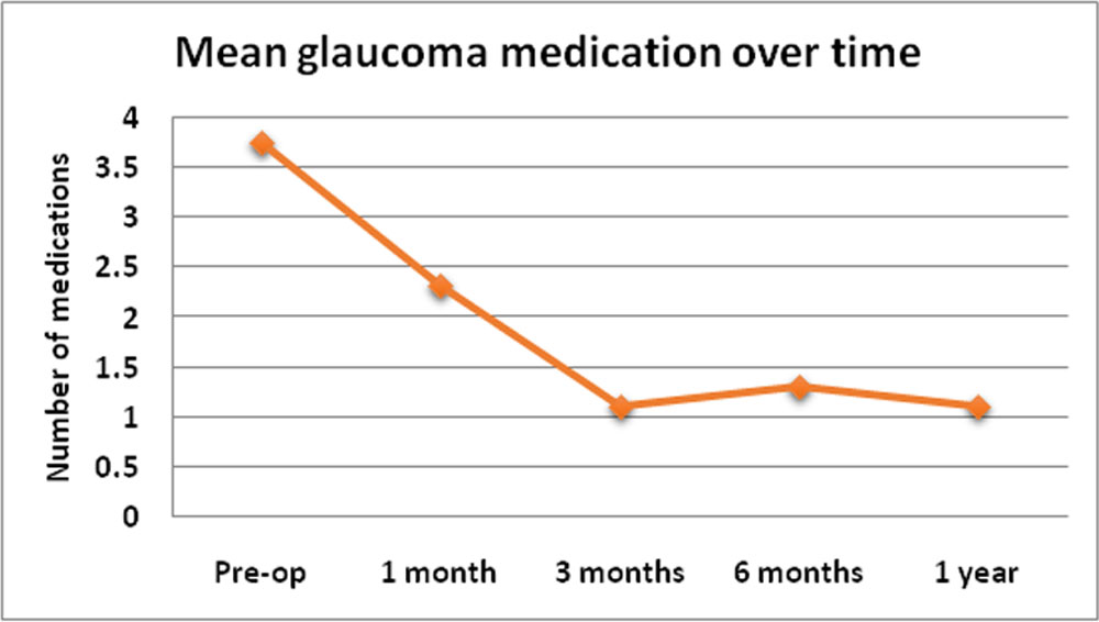
Fig 2. Shows the number of anti-glaucoma medications (AGM) over time. The graph shows a sharp decline following surgery and plateau is reached by about 3 to 6 months. Mean number of AGM preoperatively was 3.76 (±1.36). It declined to 2.3(±1.49) at 1 month, 1.1(±1.66) at 3 months, 1.31(±1.75) at 6 months, 1.1(±1.37) at 1 year with a statistically significant difference.
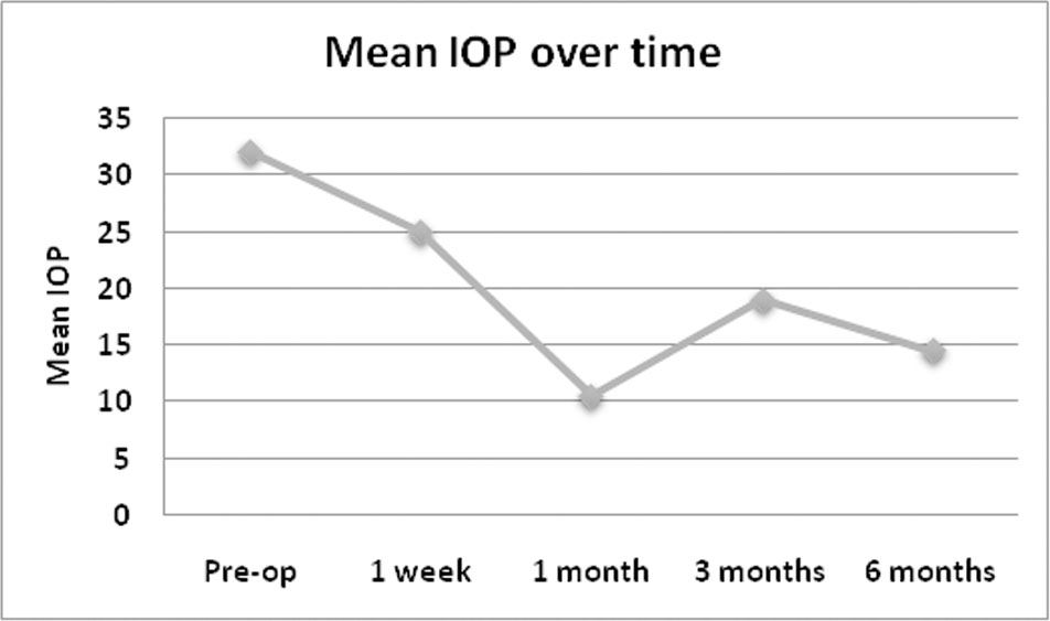
Fig 3. Shows the IOP trend over time. A sharp decline in IOP is noticed by around 1 month, which usually corresponds to the tube opening after vicryl suture dissolves. Mean Preopeartive IOP was 32.07 (±11.14) mm Hg. It showed a statistically significant decline at 1 month- 10.61(±8.31) mm Hg, 3 months-19(±9.89) mm Hg and at 6 months -14.53(±4.66 ).
None of the patients developed any serious vision threatening complications. There is no difference between pre-op and post-op BCVA. (p=0.72, >0.05).
DISCUSSION
Trabeculectomy is still the gold standard anti-glaucoma surgery. Glaucoma drainage devices have their own role in the management of refractory glaucomas, whose management becomes difficult with trabeculectomy alone. These devices offer us good results even when the virgin conjunctiva is violated by prior ocular surgery. Over the years, there have been robust modifications in the drainage devices implantation. Our device, AADI-350 is a modification of Baerveldt implant.
The long tube course of these drainage devicesthrough the conjunctiva can lead to many problems including tube erosion and extrusion, increasing the chances of endophthalmitis.Conjunctival erosion usually occurs 1-3mm from the limbus.
Stewart et al. performed a meta-analysis of 38 studies and reported the incidence of tube exposure to be 2% in more than 3000 eyes within a mean follow-up period of 26 months. They reported that there was no difference between Ahmed, Baerveldt, and Molteno implants in terms of tube exposure incidence but that tube exposure might occur in any time within the first five years.
In order to avoid such complications, different methods of tube coverage have been adopted including glycerin preserved donor scleral graft, dehydrated
human dura mater, clear corneal graft, fascia lata, buccal mucosa, KeraSys, bovine and human pericardium, but none with universal success. Smith et al. showed that no material was more prone to melting than another, studying 64 eyes that underwent tube shunt surgery using processed pericardium, processed dura mater and donor sclera. The authors reported no statistical difference in rate of thinning between groups.
Ekici et al. reported 169 patients who were implanted with a 9-mm split-thickness half-moon γ-irradiation corneal allograft (VisionGraft). They reported an erosion rate of 2%. Gamma irradiated corneas provide the added advantage of reducing allograft rejection. The inherent clarity and tensile strength of corneal tissue helps in easy tube inspection, suture lysis and easy suturing during surgery.
These come at the risk of transmission of prion disease. Anatomically, these grafts alter the conjunctival corneal angle more, leading to tear film disturbances, dellen and making the eye more prone to infection. These not only increase the surgical time but also the cost of the procedure due to the dependency on eye banks. Procuring a patch graft on an emergency basis is subject to availability in nearby eye banks. With heterologous scleral grafts it is vital to conduct routine testing for all infections such as HIV 1 and 2, hepatitis B, hepatitis C, and syphilis.
Placing the tube under the scleral flap was tried initially, but there were complications like internal tube erosion and tube rotation leading to corneal touch.
Oscar-Donado et al. described a needle-generated scleral tunnel, 3-4 mm from the limbus with a bent 23-G needle in a set of 128 eyes and did not find any case of tube erosion or extrusion.
Kalamkar et al. used a modified scleral sleeve method for tube fixation along with a limbal based scleral flap for tube coverage. Of the 16 eyes studied, there were no tube related complications in any patient.
Ollila et al. reported no tube exposure in any of 92 eyes in which they performed Molteno implantation by long scleral tunnel technique, in a mean follow-up period of 22 months, whereas they observed tube exposure in 15 (4.5%) of 332 eyes that underwent classical surgery.
Tube microvibrations have been proven to induce more inflammation and fibrosis leading to bleb failure. This is more so in patch grafts due to the sheer thickness of the graft, altered conjunctival-corneal angle, leading to improper fit, tear film disturbances and constant lid rubbing. They can lead to more mechanical tension on overlying conjunctiva and even ischemia of conjunctival blood vessels, leading to necrosis and tube exposure. Many techniques have been described to manage tube exposure including direct closure of the defect, conjunctival autograft and sclera patch graft with double layered amniotic membrane graft.
Tube exposure leads to the dreaded complication, endophthalmitis. The reported rate of endophthalmitis following glaucoma drainage device implantation ranges from 0.8% to 6.3% with a mean of 2%. Byun et al. explored the risk factors for tube exposure and found the number of previous surgical procedures to be a risk factor, whereas Souza et al. did not.Koval and associatesfound Hispanic race, neovascular glaucoma, prior trabeculectomy, and combined surgicalprocedures to be significantly associated with tube exposure.
The exposure rate for all inferiorly placed devices (AGV) was 12.8% compared to 5.4% forsuperior devices. Exposed inferior implants were found to be at a higher risk of infection than exposed superior implants (41.7% Vs 8.1%). We implanted all the devices in the superotemporal quadrant as it gives good coverage by the upper eyelid. None of our patients developed tube exposure or endophthalmitis.
None of the eyes had corneal decompensation.This was true even in eyes with a follow-up of less than six months.
This is the first study to analyse the safety and efficacy of the tunnel-track technique in AADI-350 tube insertion. Previously published reports have established the advantage of a similar technique in Ahmed glaucoma valve surgery. Both IOP control and reduced tube-related complications make this an effective alternative to other conventional methods of tube coverage.
The infinity knot using 9-0 nylon sutures placed at the point of initial scleral tunnel 4 mm from limbus reduces the chances of tube wriggling. This snug-fit of the tube could explain the reduced rate of tube-related complications in our series.
LIMITATION
Since it is a small retrospective cohort of 13 patients, future studies with larger sample size are required to reaffirm the findings. Also tube exposure from scleral erosion is predominantly a late complication and can occur even years after implantation. Another drawback of this study is the relative short follow-up time.Whether this technique also reduces the micromovementsof the implant, needs to be evaluated further.
CONCLUSION
Our data suggests that the scleral tunnel-track technique of AADI-350 tube insertion is a safe and efficacious technique for prevention of tube related complications. It also reduces the cost, effective surgical time and the dependency on eye banks.
Leave a Comment