

Dr. Diva Kant Misra, M15899, Dr. Harsha Bhattacharjee, Dr. Dipankar Das
Introduction
Immunohistochemistry (IHC) is a vital molecular pathology study, which helps to diagnose various pathologies with more accuracy. This present study documents the uveitic conditions in enucleated eyeballs with IHCs carried out at a tertiary eye care institute in northeast India.
AIM
To study molecular pathologic (Immunohistochemistry) interpretations on enucleated uveitic eye
Methods
Histopathological study and IHCs of the enucleated eyeballs with uveitic conditions were studied from 2010-2016. Selections of pathological cases were made on the result of histological diagnosis. It was a technology transfer project from a tertiary eye care institute based in Chennai, India to the ocular pathology laboratory in a tertiary eye care institute of Northeast India funded by Indian Council of Medical Research. All the specimens in the cohort were subjected for IHC using kit method for different antibodies as per indications.
The study was approved by the Institutional Ethics Committee and written informed consent was obtained from all patients. The design of the study was prospective and laboratory based.After initial diagnosis using haematoxylin and eosin (H & E) stains, samples were subjected to IHCs using number markers as per indications.
Paraffin embedded tissues were sectioned at 2-4 μm on the coated slides with the help of LEICA RM 2145 microtome (Germany). The tissues in the slides were kept for one hour at 60°C followed by deparaffinization with xylene, graded alcohol and distilled water.
The IHC was carried out using kits available commercially (BioGenex, USA). Buffer washing was used with tris-buffered saline (×1), pH 7.6 followed by peroxidase block (100 μl) for 10 min at room temperature. After subjecting to second buffer, slides were drowned for a hydrophobic blockade in the region of the tissue using a tissue tek pen. Power block was prepared using 100 μl of commercially available kit solution for 30 min and antibody (primary) incubation for overnight at 2-8°C. The third buffer was used by washing with the addition of super enhancer (100 μl) for 1.5 hours at room temperature. Again, buffer wash was done with the addition of polymer horseradish peroxidase (100 μl) for another 1.5 hours at room temperature and a drop of diaminobenzidine (DAB) chromogen in one ml of stable DAB buffer was added for five minutes at room temperature. Further washing and H and E counterstaining for 45 sec to one minute was carried out. Final washing, clearing and mounting of slides were done, and slides were seen and documented under a compound microscope (ZEISS, Axioskop 40 with AxioCam MRc, Germany).
Results
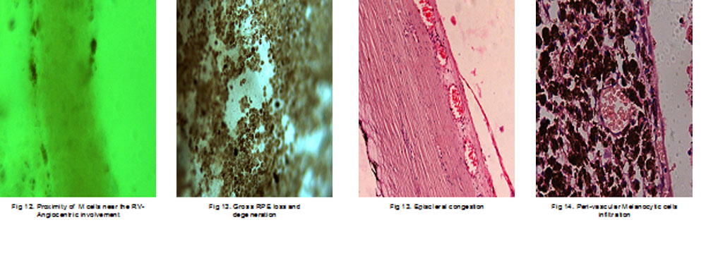
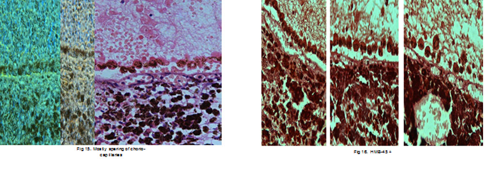
Three cases of sympathetic ophthalmia (SO), two cases of BDUMP, one case of IgG4RD mimicking as intraocular tumour were studied in the cohort. IHCs were done with various markers to confirm the histopathological findings. Appropriate controls were taken for retinal and uveal areas for the lesions. Sparring of choriocapillaris in all of SO cases. Angio-centric involvement in BDUMP was unique finding that were documented and supported by IHCs
Case: 1
A 23-year-old Indian female presented to a tertiary institute of Northeast India with the chief complaints of dimness of vision, pain and redness in the right eye (OD) for 3 months. The patient was previously treated elsewhere with two posterior sub-Tenon injections of triamcinolone acetonide given 6 weeks apart. She was also on topical steroids and cycloplegic. She did not report any benefit from the previous treatment. There was no history of trauma to the eye or elsewhere. Other medical, surgical, and family histories were unremarkable.
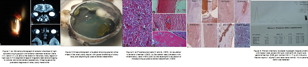
On examination, ocular alignment was normal with a visual acuity of counting finger at 3 m in the OD. There was minimal sclerouveitis noted in the inferior-temporal quadrant of OD. On slit-lamp examination, OD showed 360° posterior synechiae with anterior chamber flare and cells (+++) with 1 mm hypopyon [Figure 1]a. There was a suspected mass in the inferior-temporal quadrant behind the iris, pushing it anteriorly. The left eye examination was unremarkable.
B-scan ultrasound (B-scan USG) of OD was suggestive of ciliary body mass with adjoining exudative retinal detachment (ERD). USG bio-microscopy findings were consistent with the findings of B-scan USG. The patient was advised with complete blood count with peripheral blood smear which was within normal limit. Syphilis screening (venereal disease research laboratory) was nonreactive with normal thyroid function test and negative Mantoux test (5TU) reading. Based on the clinical examination and baseline investigations, we came to the differential diagnosis of ciliary body melanoma or similar tumors. Magnetic resonance imaging suggested the possibility of ciliary body melanoma in OD [Figure 1]b and c. After obtaining informed consent, enucleation was carried out in OD, and eyeball specimen was sent to ocular pathology laboratory for histopathological analysis. In grossing, there was a grayish-white-colored tumor seen in the inferior-temporal part of the ciliary body measuring (10.99 × 7.98) mm. Adjoining the grayish-white ciliary body mass, there was an ERD noted
Microscopic description of enucleated eyeball showed peripheral corneal vascularization. Few inflammatory cells were seen in the anterior chamber [Figure 3]b. Part of the iris tissue showed inflammation. An eosinophilic mass was seen in the ciliary body with diffuse plasmacytoid cells, with reactive lymphoid follicle and other inflammatory cells [Figure 3]a. Episcleral and scleral tissue were inflamed and infiltrated by various inflammatory cells [Figure 3]c and d. Occasional vascularization of the sclera was noted [Figure 3]c. Cut end of the optic nerve did not show any tumor involvement. Immunohistochemistry (IHC) was advised to explore the possibility of various inflammatory lesions. The steps for IHC were carried by kit methods available commercially
IHC for both Kappa and Lambda was positive. Cluster differentiation (CD20, CD3, and CD45) was focal-positive. CD138 was positive in the specimen and HMB45 was negative. IHC for IgG4 was positive in occasional cells which also expressed CD138 (plasma cells). All positive controls showed appropriate positive immunostaining. Negative control slide did not show immunostaining. IgG4-positive plasma cells were seen in high-power field in the documented slides. Nearly, 30% of all the CD138 positive plasma cells were found to be IgG4-positive [Figure 4].
Protein chemistry revealed hypergammaglobulinemia with higher levels of total protein and gamma fraction. No myeloma band or Bence Jones protein was detected [Figure 5]. Serum IgG4 levels were also elevated. A hematologist was consulted, and bone marrow examination revealed mild anemia, for which she was given supportive treatment. In the postoperative period, the patient was given a course of oral steroids. On follow-up at 6 weeks, she was given prosthesis in OD, and the patient was doing well till her last review.
Case 2 :
A 13-year-old boy presented with severe loss of vision in the right eye (RE) over the last month, not associated with penetrating injury or any ocular surgery. Best-corrected visual acuity (BCVA) was light perception in RE and 20/20 in the left eye (LE). RE showed relative afferent pupillary defect and vitreous cells. IOP of both eyes was 20 mmHg. The patient was diagnosed as Coats’ disease with exudative retinal detachment (RD) in RE. There were no pathologic findings in LE. Over a period of 18 months, he developed neovascular glaucoma in RE with an IOP of 45 mmHg. His BCVA in LE was 20/20 and an IOP of 20 mmHg. The patient was treated with DLCP to superior and inferior half (120° each) of RE ciliary body. Twenty-five diode laser burns of 2000 ms duration and using a power of 2000 mW were applied.
Six weeks after cyclophotocoagulation, he experienced photophobia and sudden deterioration of vision in his LE and mild pain in RE. BCVA in LE was 20/63 for distance and near vision was N18. IOP of RE was 21 and in LE 15 mmHg. Slit lamp examination revealed anterior chamber cells, flare and keratic precipitates in both eyes, and a dense cataract precluding fundal view in RE. Fundus examination of LE revealed disc hyperemia and yellow-white subretinal infiltrates that partly obscured choroidal vasculature [Figure 7]a. Ultrasound B-scan of RE showed total RD with increased choroidal thickness, and LE showed shallow RD with increased retinochoroidal thickness. Ocular coherence tomography showed retinal elevation due to the accumulation of subretinal fluid in LE [Figure 6]c. Fluorescein angiography of LE revealed multifocal punctate hyperfluorescent spots in the early venous phase which then continued to late leakage [Figure 7]b.
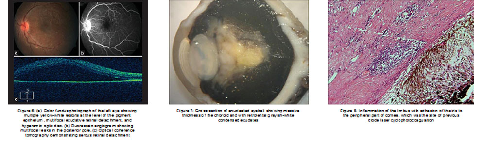
A diagnosis of SO was made, and the patient was treated with intravenous methylprednisolone (500 mg/day for 3 consecutive days) followed by oral prednisone. Over the next 20 days, intraocular inflammation in both the eyes reduced and visual acuity returned to 20/20 in the LE. Fundus examination revealed resolution of subretinal infiltrate and exudation. Although RE became phthisical, the patient had complaints of repeated pain and discomfort. The patient preferred removal of the phthisical eyeball for cosmetic reasons as well as the pain and discomfort. Enucleation with silicone ball implant was done after obtaining informed consent, and specimen was sent for histopathological examination. LE remained unremarkable with BCVA of 20/20.
Gross examination of vertical section of RE ball showed small whitish calcified areas with detached retina, vitreous hemorrhage, and thickened sclera posteriorly [Figure 7]. Microscopic examination under hematoxylin and eosin stain revealed corneal edema with neovascularization. The limbal area showed congestion with inflammatory cell infiltrate [Figure 8]. The anterior chamber was deep with angle closure at one end. Iris neovascularization with ectropion uvea was noted. Lens was partially cataractous with membranous structure in front of it. Ciliary body showed atrophic changes. There was exudative RD with fibrotic retinal structure. Retinal telangiectatic vessels with numerous cholesterol clefts in the exudation with focal osseous changes noted [Figure 9]a,[Figure 9]b,[Figure 9]c,[Figure 9]d. There was diffuse choroidal involvement with lymphocytic infiltration sparing the choriocapillaris [Figure 10]. Immunohistochemistry showed CD 20 (B-cell) and CD 3 (T-cell) positivity [Figure 11]a and [Figure 11]b.
He is presently under the follow-up with gastroenterologist for UC and is on systemic immunosuppressive. Two cases had pathological evidence.Sparing of chorio-capillaries were characteristic findings
 Conclusion:
Histopathological study and IHCs of the enucleated eyeballs with uveitic conditions were studied from 2010-2016. Selections of pathological cases were made on the result of histological diagnosis.All the specimens in the cohort were subjected for IHC using kit method for different antibodies as per indications. Three cases of sympathetic ophthalmia (SO), two cases of BDUMP, one case of ciliary body medulloepitheli
oma mimicking as intraocular inflammation were studied in the cohort. IHCs were done with various markers to confirm the histopathological findings. Appropriate controls were taken for retinal and uveal areas for the lesions. Foreign body documentation in uveal tissue in one of the SO cases, sparring of choriocapillaris in all of SO cases. Angio-centric involvement in BDUMP was unique finding that were documented and supported by IHCs.
Leave a Comment