

RANGACHARI SESSION VIDEO
Dr.Santanu Das, Dr.Raghuraj S Hegde,Dr.Sundeep
ABSTRACT:
PURPOSE-To determine when and why to use titanium or porous polyethylene (PPE) implant in orbital fracture repair.
METHODS-30 patients with confirmed orbital fractures on CT were included. They were divided into 3 groups, type1fractures (single wall fracture, displacement≤ 1cm2) type2 (>1 wall fracture, displacement1-2cm2) and type3 (orbit plus other facial bone fractures, displacement>2cm2).All type3 and type2 fractures (20) were repaired using titanium mesh.5 patients with Type1fractures were treated with PPE. Surgeries were done within first 10 days of admission.
RESULTS-23 patients with large fractures underwent orbital reconstruction with titanium mesh recovered well with no complications.7patients with small orbital floor fractures were treated with PPE had complete resolution of symptoms.
CONCLUSION-From our study we conclude that both type of implants are excellent for reconstructing fractures. Titanium is used in large defects whereas PPE is used in small floor fractures.
Keywords: orbital implants, titanium combined implant, porous polyethylene, orbital fractures, methodology.
INTRODUCTION-Orbital fractures are common and constitute approximately 18 to 50% of all craniomaxillofacial traumas depending upon region.[1] Their management is not only difficult but also challenging as one has to not only correct the functional defects like enophthalmos, diplopia, muscle entrapment and extraocular muscle movement restrictions but also needs to restore the normal orbital contour, volume, vision as well as the facial aesthetics by not giving rise to any iatrogenic ectropion and entropion.[1,2,4,5] There has been a debate not only about the indications and timings of surgery but also regarding the best possible implant available for reconstructing the orbit.[2,3,6,7,8]
An ideal orbital implant should have the following properties:[1,6]
- The ability to bend into the proper anatomical shape
- Radio opacity
- Permanent stability
- Biocompatibility
Reconstructing the orbit is challenging because in case of a fracture the bony walls are comminuted and there might be missing bone fragments. Therefore it is very important to reconstruct the missing bone.[2] Although there are a lot of materials available to reconstruct the orbit, in this prospective study we will not only describe when and where to use titanium and porous polyethylene implants but also which one is better in which conditions.
MATERIALS AND METHODS:
30 patients who came to the emergency and ophthalmology outpatient department from April2017 to May2018 with confirmed orbital fractures on CT- scan were taken up for the study. 7 were female whereas 23 were male.
A thorough ophthalmic evaluation was done for each of them which comprised of visual acquity testing for both distance and near, colour vision testing, anterior segment evaluation, eliciting pupillary reflexes, diplopia charting, extraocular movement examination and posterior segment evaluation using indirect Ophthalmoscopy and B-Scan.
CT-Scan orbits 1mm cuts, bone windows, axial coronal and saggital views with 3D-reconstruction was done for all the patients following which they were divided into 3 groups that is patients with type-1, type-2 and type-3 fractures.
Type-1 fracture- single wall fracture, displacement≤ 1cm2
Type-2 fracture- >1 wall fracture, displacement1-2cm2
Type-3 fracture- orbit plus other facial bone fractures and displacement>2cm2
Following admission all the patients were put on one broad spectrum antibiotic, Metronidazole and Amikacin along with one analgesic and intravenous steroids to reduce the inflammation and periorbital swelling. Patients with associated head injury were put on intravenous Mannitol and one antiepileptic. Thrice daily random blood sugar monitoring was done. In patients with RAPD intravenous methyl prednisolone 1 gram was given for 3 days. All the surgeries were done within the first 10 days of accident under general anaesthesia and were done by a single surgeon. Before the surgery titanium implants were autoclaved whereas porous polyethylene implants were kept in the formalin chamber overnight before usage.
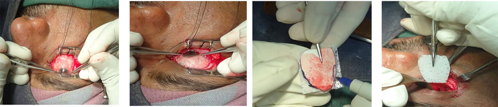
All the surgeries were done through the transconjunctival route using the swinging eyelid technique. We used either a titanium combined orbital implant or a porous polyethylene sheet (Biopore) depending upon the type of fracture. For all type-1 fracture patients PPE was used whereas for type-2 and type-3 fracture patients combined titanium orbital implant was used. Forced duction test (FDT) was done for every patient before starting the surgery as well as once the surgery was complete to rule out any mechanical restriction and iatrogenic entrapment. Titanium implant was placed over the palatine bone and fixed to the inferior orbital rim using 6mm*1.5mm screws while PPE was placed in the orbital cavity and no glue was used to fix it.
FIGURE- 1A,B,C,D
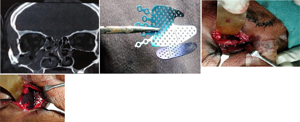
FIGURE- 2A,B,C,D
In the post op period all the patients were put on broad spectrum antibiotics, oral steroids to reduce the swelling and inflammation, analgesics and one antibiotic steroid eye drop. For all patients in the post-operative period regular dressing along with visual acquity testing, assessment of extraocular movements, diplopia charting and anterior segments evaluations were done. Post operative CT-scan was done for all patients to ascertain whether the implant, extraocular muscles and orbital contents are in proper orientation or not.
Following discharge they were followed up at 1week, 1month, 3months and at 6months.
RESULTS:
In our study 23(76.6%) patients were male and 7(23.3%) patients were female and the most common cause of orbital fractures amongst our patients being road traffic accident (RTA). 28 patients had RTA(93.3%), 2(6.6%) patients gave history of self fall. 2 wheeler accident was the most common mode of RTA in our patients.
TABLE-1
7 patients had type1, 8 had type2 and 15 had type 3 fractures in our study.
In our study both the implants gave us equally good results. There was no incidence of post operative infections which led to removal of the orbital implant, no incidence of implant extrusion, migration or malpositioning of the implant or orbital adherence in the immediate post-operative period as well as after 6 months of follow up.
In the group treated with PPE implant only one patient out of 7 had persistent diplopia in the post operative period because of nerve palsy.
In the group treated with titanium implant only 2 patients out of 23 had persistent diplopia even at 6 months follow up because of nerve palsy and not because of any implant related cause.
Enophthalmos was corrected in all the patients treated with titanium as well as PPE implants with satisfactory restoration of facial aesthetics in all of them.
In our study 5 patients had traumatic optic neuropathy at the time of presentation, for them intravenous methyl prednisolone was administered before they were take up for surgery. For 3 patients vision improved to 6/12 at 6 months post operative whereas remaining two patients had vision of 6/60 at 6 months post op. For the remaining 24 patients pre-op vision was restored in the post operative period and there was no implant related complications which lead to a drop in vision.
2 patients with persistent diplopia underwent strabismus surgery 6 months post primary surgery following which their diplopia in the primary gaze resolved.
DISCUSSION-
Materials for orbital fracture reconstruction have been classified as:[1]
- Autologous
- Allogenic
TABLE-2
Although there are a lot of materials available for reconstruction of orbit in this prospective study we have used only the titanium combined orbital implant and the porous polyethylene sheets.
After proving its competence in dental implants, bone screws and prosthetics titanium implants have become very popular amongst surgeons for the reconstruction of orbit.[2]
FIGURE-3A,B,C,D
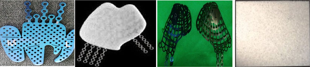
Titanium is rigid and malleable and is perfect for the reconstruction of large defects where rigidity and strength is required to maintain the contour of the orbit.[2,3,18,22,26] Also one of its properties is to osteointegrate which was proved endoscopically by Schubert et al who found that there was incorporation of soft tissue into the orbital implant approximately 1 month after the surgery. After 2 months it was found that the whole implant has been covered by a mucosal type epithelium.[21]
Gear et al in their study also proved that titanium implants are very good for reconstructing large defects and maintained adequate and satisfactory reduction in defects greater than 2cm which is similar to the results we obtained in our study.[26]
In our study no patients had post-operative infection in the immediate post operative period as well as at 6 months follow up which is in accordance with the results published by Sargent and Fulks who reconstructed 57 orbits with vitallium which is an alloy of titanium.[16,19,22] Mackenzie et al who reconstructed 51 orbits with titanium implant reported only 1 case of enophthalmos with no infection which proves that titanium has very good biocompatibility and incidence of infection is very rare which is similar to the results we obtained in our study.[16,19,27]
Some of the other advantages of titanium implant are that it is readily available, contouring of the implant that is making the postero-medial bulge of the orbit is easy, it is radio-opaque so it can be easily seen in post-operative CT-scan imaging.[3,5,7,16,17] There are no donor site related complications like infection, hematoma formation, increased post operative recovery time, bony defect at donor site and an additional surface scar. Some of the other complications associated with autologous grafts are that it increases operative time and sometimes bending the bone is difficult and might even break if tried to bend it beyond its capacity. All these complications not encountered with titanium. Lastly it is cheaper than composite implants and resorbable implants.[3,16]
Titanium is also corrosion resistant which is reported by Ling Xiao et al in her review article.[1,3,19]
In our study we have used PPE implants to reconstruct small defects in the orbit which is similar to what Ling Xiao et al suggested in her review article.[1,2,3,11] It is non-absorbable as well as malleable and connective tissue can grow into the pores which give good biocompatibility and is said to have greater biocompatibility than titanium as suggested by Ling Xiao et al.[1,2,3,11]
Romano et al used 128 PPE implants and reported only 1 case of post operative infection which is similar to the results we obtained in our study where we used PPE implants in 7 patients and none of them reported any infection at 6 months follow up.[26]
In the study conducted by Nam et al 214 orbital floor fractures were repaired using PPE sheets however they haven’t mentioned the type of defect small or large, reported 12 cases with complication which shows that the rate of complications with PPE implants are very minimal.[28] In or study we used PPE implants to correct small defects and therefore didn’t encounter any patients with post-operative persistent enophthalmos or any infection or any other problems like implant extrusion or migration even though we didn’t use any glue to fix the implant.
In a study conducted by Potter and Ellis they came to the conclusion that PPE implants can be successfully used to repair defects less than 2cm in diameter which is similar to what we have done in our study where we have used PPE implants in patients with type1fractures.[12]
In the study conducted by Sai Krishna Degala and Soumadip Dey there were no incidence of inflammatory reaction or implant extrusion in the 12 patients taken up for study which is similar to our results.[11] Although Mustafa et al reported post operative infections in 15.38% of their patients treated with PPE implants but in our study we didn’t encounter any infection if the proper preoperative and postoperative antibiotic protocol is followed.[25]
Although TotirM et al in their review article mentioned that PPE implant’s smooth edges and porous nature are its advantages over titanium but PPE implants are not radio-opaque therefore it can’t be seen in post operative CT scan pictures and also because of its lack of rigidity can’t be used for correction of large defects and also it is costly when compared to titanium.[3,10]
In our study only 1 patient out of 7 treated with PPE implants had persistent diplopia and restriction of ocular movements due to nerve paresis. Whereas only 2 patients out of 23 treated with titanium implant had persistent diplopia in the post operative period again due to nerve paresis and due to lower eyelid scarring. Lee et al, Otzturk et al, Lin et al using Medpor reported diplopia incidence to be 3.5%, 2.6% and 4.5% respectively.[13,14]
CONCLUSION:
In our prospective study both the implants gave us excellent results as there were no patients who came to us with complications like implant extrusion, migration or any inflammatory reactions because of the implant. Only1out of 7 patients treated with PPE had persistent diplopia and motility restriction because of nerve paresis whereas only 2 out of 23 patients treated with titanium had post operative diplopia again because of nerve palsy. Rest all our patients had satisfactory resolution of enophthalmos, diplopia, ocular motility along with restoration of facial aesthetics. Both the implants are good as they have very low rate of postoperative infection as suggested in literature and also in our study where there were none. Titanium implant has certain advantages like it has rigidity and provides excellent stability and good reduction of fractures where defects are more than 2cm and it is radio-opaque. On the other hand PPE implants are smooth porous which allows connective tissue to grow over it and because it is smooth there are less or no chances of orbital adherence. It is excellent for orbital fractures with defect < 1.5 cm.
REFERENCES:
- Ye LX, Sun XM, Zhang YG, Zhang Y. Materials to facilitate orbital reconstruction and soft tissue filling in posttraumatic orbital deformities. Plast Aesthet Res. 2016; 3:86-91.
- D Mok, L Lessard, C Cordoba, PG Harris, A Nikolis. A review of materials currently used in orbital floor reconstruction. Can J Plast Surg. 2004; 12(3):134-140.
- Totir M, Ciuluvica R, Dinu I, Careba I, Gradinaru S. Biomaterials for orbital fractures repair. Journal of Medicine and Life Volume 7, Special Issue 4, 2014.
- Xiaohui Han, Shunhai Yang, Zhizhang Dong, Wangqiang Feng. Reconstruction of orbital blowout fractures using MEDPOR titanium mesh implants. Int J Clin Exp Med 2016; 9(6):12411-12415.
- Jennings R Boyette, John D Pemberton, Juliana Bonilla-Velez. Management of orbital fractures: challenges and solutions. Clinical ophthalmology. 2015; 9:2127-2137.
- Dubois L, Steenen SA, Gooris PJ, Bos RR, Becking AG. Controversies in orbital reconstruction-III. Biomaterials for orbital reconstruction: a review with clinical recommendations. Int J Oral Maxillofac Surg. 2016;45:41-50.
- Gunarajah DR, Samman N. Biomaterials for repair of orbital floor blowout fractures: a systematic review. J Oral Maxillofac Surg.2013; 71:550–570.
- Emily M. Bratton, Vikram D. Durairaj. Orbital implants for fracture repair. Current Opinion in Ophthalmology.2011; 22(5):400–406.
- Mansour Khorasani, Pejman Janbaz, Farshid Rayati. Maxillofacial reconstruction with Medpor porous polyethylene implant: a case series study. J Korean Assoc Oral Maxillofac Surg. 2018; 44:128-135.
- Ali Mubin Aral, MD, Selahattin O zmen, MD, Safak Uygur, MD, Basar Kaya, MD, Neslihan Coskun, MSc, Suna O meroglu, MD, and Koray Kılıc, MD. Comparison of Resorbable Mesh (Poly L-Lactide/Glycolic Acid) and Porous Polyethylene in Orbital Floor Fractures in an Experimental Model. Plastic Surgery. 2017; Vol. 25(3) 163-170.
- Degala Sai Krishna, Dey Soumadip. Reconstruction of Orbital Floor Fractures with Porous Polyethylene Implants: A Prospective Study. J. Maxillofac. Oral Surg. (July–Sept 2016); 15(3):300–307.
- Potter JK, Ellis E. (2004) Biomaterials for reconstruction of the internal orbit. J Oral Maxillofac Surg 62:1280–1297.
- Lee S et al. (2005) Porous high-density polyethylene for orbital reconstruction. Arch Otolaryngol Head Neck Surg 131:446–450.
- Otzturk S et al. Long-term outcomes of ultra-thin porous polyethylene implants used for reconstruction of orbital floor defects. J Craniofac Surg.2005; 16:973–977.
- Buchel P, Rahal A, Seto I, Lizuka T. Reconstruction of orbital floor fracture with polyglactin 910/polydioxanone patch (Ethisorb). A retrospective study. J Oral Maxillofac Surg. 2005; 63:646–650.
- Bogdan BANICA; Patricia ENE; Daniela VRANCEANU; Razvan ENE. Titanium Preformed Implants in Orbital Floor Reconstruction – Case Presentation, Review of Literature. MAEDICA – a Journal of Clinical Medicine. 2013; 8(1): 34-39.
- Ellis III E, Tan Y. Assessment of Internal Orbital Reconstructions for Pure Blowout Fractures: Cranial Bone Grafts versus Titanium Mesh. J Oral Maxillofac Surg. 2003; 61:442-453.
- Gear, Andrew J. L. MD; Lokeh, Adam MD; Aldridge, Jeffrey H. MD; Migliori, Mark R. MD; Benjamin, Charles I. MD; Schubert, Warren MD. Safety of Titanium Mesh for Orbital Reconstruction. Annals of Plastic Surgery. January 2002; Volume 48: Issue 1: p 1-9.
- Scolozzi P, Momjian A, Heuberger J, Andersen E, Broome M, Terzic A, Jaques B. Accuracy and predictability in use of AO three dimensionally preformed titanium mesh plates for posttraumatic orbital reconstruction: a pilot study. J Craniofac Surg. 2009 Jul; 20(4):1108-13.
- Garibaldi DC, Iliff NT. Use of porous polyethylene with embedded titanium in orbital reconstruction: a review of 106 patients. Ophthal Plast Reconstr Surg.2007; 23(6):439–444
- Schubert W, Gear AJ, Lee C, Hilger PA, Haus E, Migliori MR, Mann DA, Benjamin CI. Incorporation of titanium mesh in orbital and midface reconstruction. Plast Reconstr Surg 2002; 110:1022-30.
- Sargent LA, Fulks KD. Reconstruction of internal orbit fractures with Vitallium mesh. Plast Reconstr Surg 1991; 88:31-8.
- Patel PJ, Rees HC, Olver JM. Fibrovascularization of Porous Polyethylene Orbital Floor Implants in Humans. Arch Ophthalmol. 2003; 121(3):400–403.
- Villarreal PM, Monje F, Morillo AJ, Junquera LM, González C, Barbón JJ. Porous polyethylene implants in orbital floor reconstruction. Plast Reconstr Surg.2002; 109:877–885.
- Mustafa V, Haluk Vayvada, Enver Aydin, Adnan Menderes, Atay Atabey. (2007) Repair of fractures of the orbital floor with porous polyethylene implants. Br J Oral Maxillofac Surg. 45:640–644.
- Romano JJ, Iliff NT, Manson PN. Use of Medpor porous polyethylene implants in 140 patients with facial fractures. J Craniofac Surg. 1993; 4:142-7.
- Mackenzie DJ, Arora B, Hansen J. Orbital floor repair with titanium mesh screen. J Carniomaxillofac Trauma. 1999; 5:9-18.
- Nam SB, Bae YC, Moon JS, Kang YS. Analysis of the postoperative outcome in 405 cases of orbital fracture using 2 synthetic orbital implants. Ann Plast Surg. 2006; 56:263-7.
TABLE-1
| TYPE OF FRACTURE | NUMBER OF PATIENTS | IMPLANT USED |
| TYPE-1 FRACTURE | 7(23.3%) | POROUS POLYETHYLENE IMPLANT |
| TYPE-2 FRACTURE | 8(26.6%) | COMBINED TITANIUM IMPLANT |
| TYPE-3 FRACTURE | 15(30%) | COMBINED TITANIUM IMPLANT |
TABLE-2
| AUTOLOGOUS | ALLOGENIC | ALLOPLASTIC |
| BONE | IRRADIATED BONE | NON-METALLIC PERMANENT |
| CARTILAGE | LYOPHILIZED DURA | SILASTIC SHEETS |
| FASCIA LATA | LYOPHILIZED CARTILAGE | BIOACTIVE GLASS |
| PERIOSTEUM | FASCIA LATA | MARLEX MESH |
| BOVINE BONE | POROUS POLYETHYLENE | |
| TEFLON | ||
| METALLIC- PERMANENT | ||
| TITANIUM | ||
| VITALLIUM | ||
| RESORBABLE MATERIAL | ||
| POLYDIAXANONE | ||
| POLYGLACTIN 910 | ||
| POLYLACTIC/POLYGLYCOLIC ACID POLYMER |
Figure-1: (A) coronal view of ct scan showing displaced orbital floor fracture; (B) posteromedial bulge of the combined titanium orbital implant; (c) fractured inferior orbital rim being fixed with orbital plate and screw; (D) titanium implant being placed in the orbital cavity and fixed into the inferior orbital rim with screws.
Figure-2: (A) and (B) forced duction test being done at the beginning of the surgery; (C) porous polyethylene implant being cut as per the size of the orbital cavity; (D) porous polyethylene implant being placed in the orbital cavity.
Leave a Comment