

Dr.Jayati Pandey, Dr.Mohan Shalini,Dr.Rajnath Singh Kushawaha,Prof.Gupta Ramesh Chandra
AIM: To compare Optic Nerve Sheath Diameter (ONSD) in Primary Open Angle Glaucoma (POAG), Primary Angle Closure Glaucoma (PACG) and Normal Tension Glaucoma (NTG).
MATERIAL AND METHOD: Patients with POAG (n=38), PACG (n=32), NTG (n=18) and Controls (n=48) underwent B-scan ultrasound and Computed Tomography Scan (CT scan) measurement of ONSD. Intraocular pressure (IOP) was measured in all groups and was correlated with ONSD.
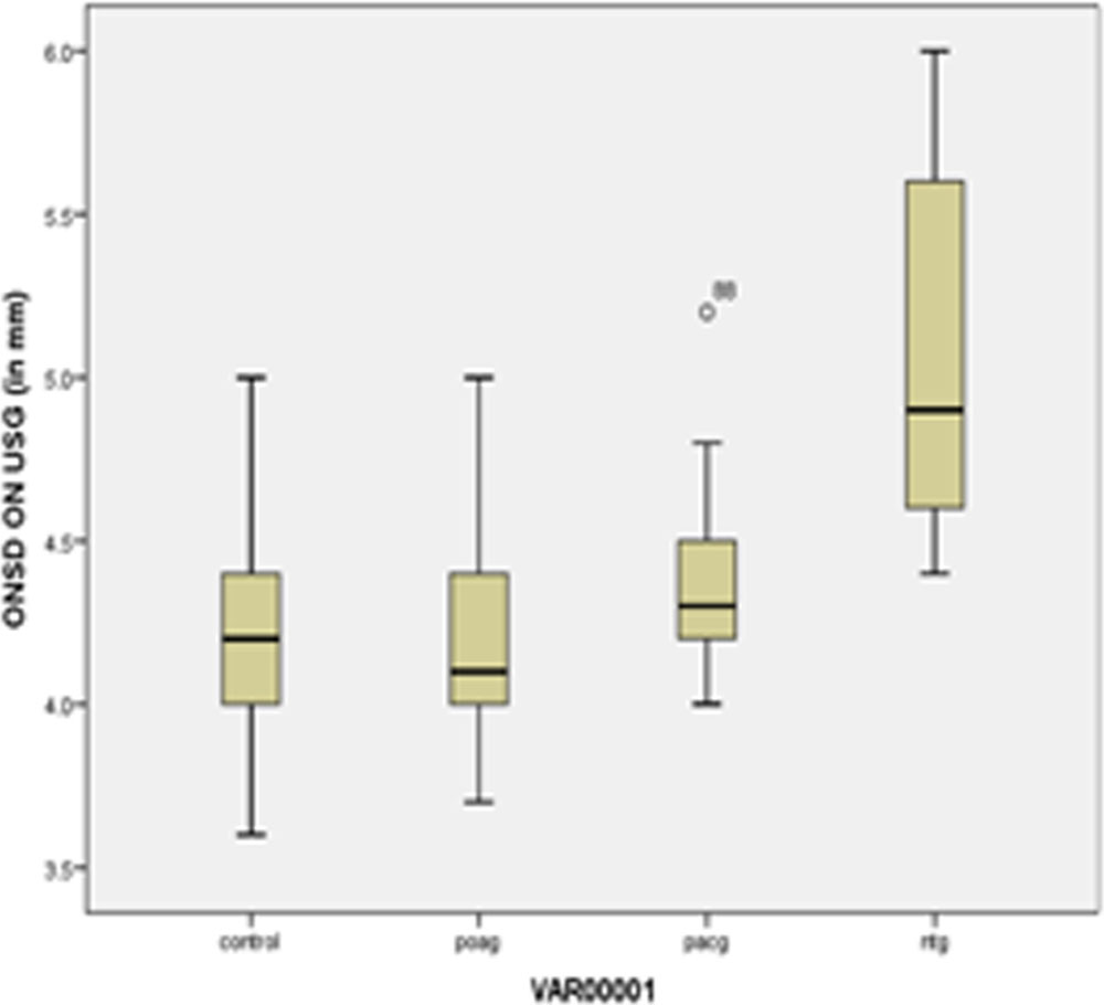
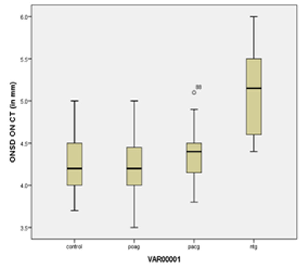
RESULT: ONSD was significantly (p=<0.001) increased in NTG patients (mean=5.0mm ±0.48SD) compared with POAG (mean=4.20mm±0.32), PACG (mean=4.33mm±0.27) and control (mean=4.21mm±0.31). ONSD showed correlation with IOP in PACG group (r=0.392, p=0.02) while it did not in other groups.
CONCLUSION: ONSD in a group of NTG patients were significantly increased compared with POAG, PACG and controls indicating the role of translaminar cribriform pressure gradient in NTG patients.Indirect measurement of ICP by assessment of ONSD may provide further insight into retrolaminar pressure component and pathophysiology of glaucoma.
INTRODUCTION
Glaucoma is the leading cause of irreversible blindness affecting more than 60 million people worldwide[1]. Glaucoma is a chronic progressive optic neuropathy that is recognised by the appearance of characteristic cupping of optic disc associated with corresponding visual field defects. The disease is characterised by progressive loss of retinal ganglion cells & their axons associated with tissue remodelling of the optic nerve head.
A sustained increase in intraocular pressure(IOP) may be due to increased formation of aquous humor, difficulty in exit or raised pressure in the episcleral veins. Of these first & last rarely occurs and it follows that raised intraocular pressure is essentially due to an increased resistance to its drainage through angle of anterior chamber.
The optic nerve is derived from an out-pouching of the diencephalon (optic stalk) during embryonic development. As a consequence, the fibres of the optic nerve are covered with myelin produced by oligodendrocytes, rather than Schwann cells of the peripheral nervous system, and are encased within the meninges. The optic nerve is ensheathed in all three meningeal layers (dura, arachnoid and pia mater). Its diameter increases from about 1.6 mm within the eye to 3.5 mm in the orbit to 4.5 mm within the cranial space. The optic nerve component lengths are 1 mm in the globe, 24 mm in the orbit, 9 mm in the optic canal, and 16 mm in the cranial space before joining the optic chiasm.[2]
The optic nerve sheath is an anatomical extension of the duramater and so the subarachnoid space around the optic nerve is continuous with the intracranial subarachnoid space. The optic nerve is separated from its sheath by a fluid layer of cerebral spinal fluid (CSF), which is in continuity with the rest of the central nervous system. The lamina cribrosa at the bottom of the optic nerve head acts as a pressure barrier between the intraocular space and the retrobulbar space of the optic nerve. Increase in intra cranial pressure (ICP) lead to an increase in the volume of the fluid layer, thereby increasing optic nerve sheath diameter (ONSD) as measured in the retrobulbar portion of the optic nerve trajectory. Dilatation of the optic nerve sheath has been shown to be a much earlier manifestation of ICP rise.[3,4] The forces of IOP and CSF pressure meet at lamina cribrosa , a modified extension of peripapillary sclera flaps , composed of collagen and non collagen components.[5] The lamina cribrosa functions as barrier between the anterior force of IOP and posterior force of CSF-p within the orbit also known as Translaminar Cribriform Pressure Dfference(TLCPD)
TLCPD = IOP – CSF-p
The TLCPD depends on the IOP and the retrobulbar CSF pressure. The ability of lamina cribrosa to withstand pressure gradient without deformity is dependent on its thickness, the rigidity of extracellular matrix and peripheral sclera tension. The lamina cribrosa’s ability to maintain shape is important in protecting structures that pass through it. Increased TLCPD could cause bowing of the lamina cribrosa. Such deformity may damage optic nerve ganglion cells via mechanical compression or ischaemia as the vessels pass through the lamina cribrosa [6]. CSF-P and IOP have equivalent effects on TLCPD and optic disc surface movement. It was also found that CSF-P affects axonal transport of the optic nerve, which might have an effect on glaucoma aetiology and retinal venous outflow [7].
The ideal method to measure CSF pressure is lumbar puncture but it is an invasive modality with increased risk with various life threatening complications. It has been found that raised CSF pressure causes increased ONSD and vice versa [3, 4]. Therefore, measuring ONSD can be surrogate method to measure the CSF pressure and can throw some light in pathophysiology of various types of glaucoma. The optic nerve sheath is fairly easy to visualize by ultrasonography by insonation across the orbit in the axial plane. A-mode ultrasonography was used to view the optic nerve sheath more than four decades ago; B-mode scanning was performed subsequently to assess intraocular lesions[8]. Evolution of ultrasound technology and the development of high frequency (> 7.5 MHz) linear probes with improved spatial resolution have enabled excellent views of the optic nerve sheath. The ONSD, measured at a fixed distance behind the retina has been evaluated to diagnose and measure intracranial hypertension in traumatic brain injury and intracranial haemorrhage[9,10]. Ultrasound-based assessment of the ONSD is a validated method for indirect measurement of the ICP [10,11,12]. ONSD is much easier to measure on Computed Tomography scan (CT scan) than with sonography due to the good reproducibility of CT and the lack of a learning curve.
OBJECTIVES : The objective of the study is to study Optic Nerve Sheath Diameter in patients of POAG , PACG and NTG and to correlate it with IOP
MATERIALS AND METHODS
STUDY DESIGN
It is a hospital based cross sectional study.
Three cohorts of individuals over 18 years old were recruited for the study from department of Ophthalmology LLR hospital Kanpur: patients with POAG (n=38),PACG (n=32), NTG(n=18) and healthy control (n=42). Glaucoma patients were defined on basis of intraocular pressure , having characteristic optic disc damage and visual field loss as described previously in the literature [13,14]. The healthy volunteers were screened by experienced ophthalmologists. Those with a family history of glaucoma, or an increased or asymmetrical cup/disc ratio or any other optic disc structural change (notching, disc hemorrhage), or an IOP above 21mmHg, were excluded as possible glaucoma suspects. Patients with a history of ocular trauma or eye disease (except glaucoma) that could not be accounted for by refractive error were excluded. Patients on antiglaucoma drugs or with any known neurological disorder was also an exclusion criterion.
The study was approved by the ethical review committee (Institutional Review Board) of our own institution and was conducted in accordance with Good Clinical Practice within the tenets of the Helsinki’s agreement. Each patient/subject was required to sign an informed consent statement before being enrolled into the study and prior to any study measurements being taken.
MEASURING DEVICES
IOP was measured with the Goldmann applanation tonometer (GAT). Central corneal thickness (CCT) was measured using a pachymeter (Pachscan, Sonomed, and U.S.A). Angle of anterior chamber was assessed by 2 mirror goniolens. Visual fields were assessed by Humphrey’s Perimeter (Zeiss, Germany ). Disc photograph was taken by Fundus camera (Zeiss Visucam Lite, Germany). Measurement of the ONSD was performed with a B-scan ultrasound probe (Sonomax, Montreal, Canada) and CT scan (Siemens, Germany).
EXPERIMENTAL DESIGN
During the study visit, the following examinations were be performed in the same order : BCVA using the Snellen’s chart placed in the same location at the same distance from the patient under the same illumination for all subjects, IOP measurement by Applanation tonometer, Angle of anterior chamber by goniolens, Disc photo by Fundus Camera, Visual field by Humphrey’s perimeter, and finally ONSD measurements. Latter was performed by an observer masked to the patient diagnosis. The patient was made to lie in the supine position with the head in a neutral position and both eyes closed and in primary gaze position. After application of coupling gel the insonation depth was set to 5–8 cm, the transducer was softly placed over the upper eyelid in an axial plane. This sonographic section provides a transverse view of the globe and the structures of the retrobulbar area . The ONSD was calculated perpendicular to the vertical axis of the scanning plane 3mm behind the globe, where the optic nerve sheath structure is more prone to expansion due to increase in ICP[15] probably due to a decrease in sheath thickness in that retrobulbar segment of the optic nerve[4,16] . Only one eye per patient was included in the study. The eye with greater glaucomatous damage was selected in the glaucoma patients.
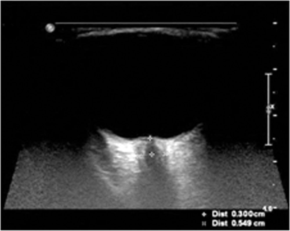
Brain CT scan was performed with a series of millimetre slices (one slice every 0.6 mm). As for ultrasound, ONSD was measured at a distance of 3mm behind the eyeball, immediately below the sclera[9,17] . ONSD was measured transversely as a section through the centre of the optic nerve.
STATISTICAL ANALYSIS
Statistical analysis was performed using SPSS 21.0 (IBM SPSS Inc., Chicago, IL, USA) for Windows statistical package. The Mann-Whitney test was used to compare between two variables. The Kruskal-Wallis test was used to compare variables between four diagnostic groups. Spearman’s correlation coefficient was used to study association between variables. Probabilities are two-tailed and considered statistically significant if p<0.05.
RESULTS
| VARIABLE | POAG | PACG | NTG | CONTROL |
| Number | 38 | 32 | 18 | 48 |
| AGE(in years) | 50.50±6.49 | 50.94±7.35 | 56.4±10.4 | 50.23±6.48 |
| VA (log MAR) | 0.44±0.32 | 1.76±0.26 | 1.13±0.55 | 0.06±0.09 |
| Mean IOP (in mmHg) | 27.29±3.45 | 49.75±11.13 | 16.72±3.84 | 15.19±2.51 |
| CCT( in µm) | 527.60±10.26 | 543.19±8.24 | 504.83±14.27 | 520.1±13.66 |
| MD | -6.17±2.14 | -11.35±2.02 | -5.11±0.94 | 0.39±0.65 |
| ONSD on USG (in mm) | 4.20±0.32 | 4.33±0.27 | 5.0±0.48 | 4.21±0.31 |
| ONSD on CT (in mm) | 4.23±0.33 | 4.35±0.26 | 5.01±0.5 | 4.20±0.27 |
VA – Visual Acuity ; IOP – Intra ocular pressure ; CCT – Central Corneal Thickness ; MD – Mean Deviation ; ONSD – Optic Nerve Sheath Diameter
In our study a total of 136 patients were evaluated out of which most were in 41-50 years age group (54.17%) whereas 41.67% were in 51-60 years and 4.16% in > 60 years age group. Overall prevalence in males were 61.76% and in females were 38.24%. Males outnumbered females in all the groups except in PACG group where females outnumbered males (62.5% females; 37.5% males). In our study ONSD was significantly increased in Normal Tension Glaucoma (5.0±0.48; p=0.00) compared to other groups and no significant correlation was found between IOP and ONSD in NTG group and POAG group(r=-0.209, p= 0.406; r=0.141, p= 0.398 respectively) while in PACG group there was mild positive correlation between IOP and ONSD which was significant(r=0.392, p=0.02).
DISCUSSION
The present study was conducted to evaluate the relevance of studying ONSD in glaucoma patients, and to study whether this indirect measurement of ICP correlates with IOP in these patients.
In the present study the ONSD in Caucasian NTG patients measured significantly (P=0.00) larger diameters compared to controls without ON diseases. The findings of larger ONSDs in NTG patients are in accordance with measurements in a study from Jaggi et al (7.9±0.9mm) [18] and A Pircher et al (6.4±0.9mm) [19] who showed that ONSD in NTG patients was significantly increased (p<0.001) and contradictory to Abegao Pinto et al[20] who did not find any significant difference in ONSD among NTG , POAG and Control (p=0.08) which may be due to difference in the methodology used or difference in head position scanning between these two groups.
The TLCPD is not the only factor affecting optic nerve head but it is also the thickness of the wall separating these compartments i.e. lamina cribrosa. An abnormally thin sclera has been seen in NTG patients [21] which results in increased stress on optic nerve in these patients.
Two mechanisms explain the enlarged ONSDs in NTG patients. First a localized CSF-Pressure elevation behind the globe due to impaired CSF outflow might lead to increased radial stress to the optic nerve sheath and thereby stretching the optic nerve sheath. Second, an accumulation of proteins of different biological functions [22].
In our study no significant correlation was found between IOP and ONSD in NTG group and POAG group(r=-0.209, p= 0.406; r=0.141, p= 0.398 respectively) while in PACG group there was mild positive correlation between IOP and ONSD which was significant(r=0.392, p=0.02) whereas study conducted by Abegao Pinto L et al[20] showed that OSND did correlate with IOP in NTG patients(r=0.53; p<0.001) but not in POAG patients and healthy controls(p=0.46 , P=0.86). The difference in Pinto et al study from our study may be because in all the patients IOP and ONSD was not measured on same day or same point of time, diurnal variation of IOP was not taken in to consideration. Since no study has been conducted regarding ONSD in PACG group hence, result cannot be compared.
LIMITATIONS OF STUDY
Our work has several limitations. As a full neurological examination was not performed in these patients, a direct measurement of intracranial pressure was not included thus intracranial pressure was presumed to be normal by taking history so there is possibility of contamination of the experimental groups by individuals with a yet undiagnosed neurological clinical condition. The sample size in NTG group was very small compared to other groups so further studies with larger sample size is required to confirm our result. In some patients IOP and ONSD could not be measured on same day or same point of time , hence changes due to diurnal variation in IOP could give different result.
CONCLUSION
It is the trans-lamina cribrosa pressure difference (and not the transcorneal pressure difference, i.e. the so called intraocular pressure) which is of importance for the physiology and pathophysiology of glaucoma.Indirect measurement of ICP by assessment of ONSD may provide further insight into retrolaminar pressure component and pathophysiology of glaucoma.ONSD measurement can be used as a useful tool for diagnosing NTG in early stage as ONSD greater than 5 mm might be indicator of greater glaucomatous damage in NTG patients. ONSD measurement through USG-B scan and CT scan being a non invasive procedure can be used as an alternate method of measuring CSF-pressure in glaucoma patients as well as in other neurological diseases.
BIBLIOGRAPHY
- Quigley HA, Broman AT : The number of people with glaucoma worldwide in 2010 and 2020. Br J Ophthalmol. 2006; 90:262 –267.
- Vilensky, Joel, Robertson, Wendy, Suarez-Quian, Carlos : The Clinical Anatomy of the Cranial Nerves : The Nerves of “On Olympus Towering Top”. Ames, Iowa: Wiley-Blackwell. 2015; ISBN 978-1118492017
- Helmke, Hansen HC : Fundamentals of transorbital sonographic evaluation of optic nerve sheath expansion under intracranial hypertension. Experimental study. Pediatr Radiol. 1966; 26:7015
- Hayreh SS: The sheath of the optic nerve. Ophthalmologica 1984; 189:54–63
- Gamero GE, Fechtner RD. The optic nerve in glaucoma. In: Choplin NT, Lundy DC, eds. Atlas of glaucoma. 2nd edn. Andover: Informa, 2007:59–74.
- . Guidoboni G, Harris A, Carichino L, et al. Effect of Lamina cribrosa deformation on the Hemodynamics of the Central Retinal Artery: a mathematical model. MathmBiosci Eng In press.
- Morgan WH, Yu DY, Balaratnasingam C. The role of cerebrospinal fluid pressure in glaucoma pathophysiology: the dark side of the optic disc. J Glaucoma 2008; 17:408–13.
- Gangemi M, Cennamo G, Maiuri F, D’Andrea F : Echographic measurement of the optic nerve in patients with intracranial hypertension. Neurochirurgia (Stuttg) 1987; 30:53–5.
- Geeraerts T, Merceron S, Benhamou D, Vigué B, Duranteau J : Non-invasive assessment of intracranial pressure using ocular sonography in neurocritical care patients. Intensive Care Med. 2008; 34:2062–2067
- Moretti R, Pizzi B, Cassini F, Vivaldi N : Reliability of opticnerve ultrasound for the evaluation of patients with spontaneous intracranial hemorrhage. Neurocrit Care. 2009; 11:406–410
- Soldatos T, Chatzimichail K, Papathanasiou M: Optic nerveSonography: a new window for the non-invasive evaluation ofIntracranial pressure in brain injury. Emerg Med J. 2009; 26:630–634
- Bäuerle J, Lochner P, Kaps M, Nedelmann M: Intra andInterobserver reliability of sonographic assessment of the opticNerve sheath diameter in healthy adults. J Neuroimaging Dec 1 2010
- Jampel H (1997) Target pressure in glaucoma therapy. J Glaucoma:133–138
- Zeyen T (1999) Target pressures in glaucoma. Bull Soc BelgeOphtalmol 274:61–65
- . Liu D, Kahn M : Measurement and relationship of subarachnoid pressure of the optic nerve to intracranial pressures in fresh cadavers. Am J Ophthalmol 1993; 116:548–556
- Hayreh SS: Pathogenesis of oedema of the optic disc. Doc Ophthalmol 1968; 24:289–411
- Geeraerts T, Newcombe VFJ, Coles JP, Abate MG, Perkes IE, Hutchinson PJA, Outtrim JG, Chatfield DA, Menon DK : Use of T2-weighted magnetic resonance imaging of the optic nerve sheath to detect raised intracranial pressure. Crit Care. 2008; 1:R114.
- . Jaggi GP, Miller NR, Flammer J, Weinreb RN, Remonda L, Killer HE (2011) Optic nerve sheath diameter in normal-tension glaucoma patients. Br J Ophthalmol. 2012; 96:1 53-56
- A Pircher, M Montali, J Berberat, L Remonda and HE Killer. Relationship between the optic nerve sheath diameter and lumbar cerebrospinal fluid pressure in patients with normal tension glaucoma. Eye (2017) 31, 1365–1372; doi:10.1038/eye.2017.70
- Abegao Pinto L, Vandewalle E, Pronk et al. Intraocular pressure correlates with optic nerve sheath diameter in patients with normal tension glaucoma. Graefes Arch Clin Exp Ophthalmol. 2012; 250:1075
- Mohamed-Noor J, Bochmann F, Siddiqui MA, Atta HR, Leslie T, Maharajan P, Wong YM, Azuara-Blanco A (2009) Correlation between corneal and scleral thickness in glaucoma. J Glaucoma18:32–36
- Killer HE, Jaggi GP, Flammer J, Miller NR, Huber AR. The optic nerve: a new window into cerebrospinal fluid composition. Brain 2006; 129(Pt 4): 1027–1030
Leave a Comment