

Dr.Devesh Kumawat, K18457, Dr.Vinod Kumar
Abstract
Purpose: To assess the correlation between the morphological features and serology in eyes with macular coloboma (MC).
Methods: All cases of MC who presented to the retina clinic between January 2016 to December 2017 were enrolled. Color fundus and swept source optical coherence tomography (SS-OCT, Topcon Inc.) features were assessed in three groups based on the serum IgG results: Toxoplasma +, Cytomegalovirus (CMV) + and serology negative.
Results: A total of 49 eyes of 27 patients were recruited. The mean age was 24.8 ± 14.9 yrs (range 7 to 60 years). While the lesion size, presence of satellite lesions, choroidal excavation and lacunae (large choroidal vessels) on SS-OCT differed significantly among the groups; pigmentation, retinal fibrosis, shape, retinal vessel pattern, and choroidal vessel visibility did not vary significantly. CMV+ lesions were solitary, large and deeply excavated. Toxoplasma+ lesions were flat, medium sized and either solitary or with satellite lesions. Serology negative eyes had small solitary, deeply excavated lesions with choroidal lacunae.
Conclusion: Clinical and OCT features along with serology can help ascertain etiology of macular coloboma.
Keywords:
Choroidal excavation;
Congenital cytomegalovirus infection;
Macular coloboma;
Ocular toxoplasmosis;
Swept source optical coherence tomography
Introduction
Retino-choroidal colobomata is an uncommon ocular malformation reported to occur at a rate of 0.14% in the general population.[1] An atypical variant of coloboma is macular colobomata which is even more uncommon. Because of their central nature, the patients often present with poor vision and nystagmus (in case of congenital lesions). Their etiology remains an enigma and they are believed to result from either congenital developmental abnormality or inflammation due to intrauterine or postnatal infection.[2–5] Congenital macular coloboma, unlike typical retino-choroidal coloboma which are a consequence of an anomalous closure of the fetal fissure, form as a result of faulty differentiation of the arcuate bundles along the horizontal raphe during retinal development.[2,5,6] On the other hand, post inflammatory macular scars are a result of ocular toxoplasmosis or cytomegalovirus (CMV) infection.[2,3,5,7] Ocular toxoplasmosis is often secondary to congenital infection and tends to occur during the chronic phase of the disease.[8–10] It sometimes follows acquired infection associated with the ingestion of undercooked meat or water contaminated with cysts of Toxoplasma gondii. Congenital CMV infection can also manifest with chorioretinitis lesions and macular scarring.[11] In addition, retinal dystrophies may be associated with macular scarring/coloboma. These include Leber’s congenital amaurosis, progressive cone-rod dystrophy, and North Carolina macular dystrophy.[3,5,12]
Assessing the serological status of the patient and family history may be of great help in differentiating the etiology of these cases. Though fundus and optical coherence tomography (OCT) features of these entities have been defined and differentiated in the past, but are often indecisive.[6,7,13,14]. Therefore, we performed this study to determine if the morphological characteristics of macular coloboma vary with the serology results.
Methods
This is a retrospective study of consecutive patients with macular coloboma, who attended the retina clinic over last two years (January 2016 to December 2017) at a tertiary eye care center in North India. The study adhered to the tenets of declarations of Helsinki and to the institutional research guidelines. A review of the database of the retina clinic was performed to identify the cases. Exclusion criteria included the presence of active chorioretinitis, unavailability of retinal imaging data, preverbal children and the patients in whom serology was not performed or was positive for both Toxoplasma and CMV.
Detailed ophthalmic, family, personal and systemic history of Toxoplasma and CMV infection was obtained. All patients had undergone a complete ophthalmic examination including best corrected visual acuity assessment (Snellen chart), anterior segment examination using slit lamp and dilated fundus examination. The macular lesions were imaged using color fundus photographs and swept source optical coherence tomography (Triton, Topcon Inc. Oakland, New Jersey, USA). The clinical features of macular coloboma in terms of the size of the lesion (as compared to the size of the optic disc), shape, location, retinal pigment epithelium (RPE) changes, and overlying fibrosis if any were noted. Swept source OCT (SS-OCT) imaging protocol included twelve equidistant radial line scans through the lesion. The various scans were analyzed for the presence of retinal thinning, retinal cysts, RPE atrophy/ proliferation, choroidal atrophy, excavation, choroidal lacunae, and scleral backscatter.
Based on size, lesion were classified into small [≤ 1 disc diameter (DD)], medium [1 to 3 DD], and large [>3DD] lesions. Based on the pigmentation within the lesions, the lesions were categorized into no/ minimally, moderately (<50% area pigmented), and heavily (>50% area pigmented) pigmented. Shapes defined were circular/round, oval, irregular and ill-defined. The excavation was subcategorized as none/flat, shallow (<50% of choroidal thickness), and deep (>50% of choroidal thickness). Choroidal vessel visibility was subdivided into not visible, just or barely visible, and prominently visible vessels.
Electrochemiluminescence immunoassay (ECLIA) was performed and results were available for TORCH serology for all recruited cases (IgM, IgG for Toxoplasma and CMV). IgM and IgG were considered negative if <1 and positive if ≥1.
Data entry was performed and analyzed using SPSS 20 software. The qualitative/quantitative data were expressed as frequency as well as percentages. Morphological changes on clinical and OCT-based examination were analyzed if different among eyes with different serological results. Visual acuity and age were compared between the groups with Kruskal Wallis test. Categorical data were subjected to Pearson’s Chi-square test or Fischer’s exact test to compare the features in between the groups.
Results
A total of 49 eyes with macular coloboma of 27 patients (5 unilateral, 22 bilateral cases) were recruited for this study. Family history including parental consanguinity was non-contributory in all cases. None of the patients had a history of seizures or central nervous system problems. Two patients with positive CMV serology had a sensori-neural hearing loss.
The mean age of patients was 24.8 ± 14.9 years (range 7 to 60 years). The males and females constituted 51.8% and 48.1% of the patients respectively. The mean logMAR BCVA was 0.78 ± 0.38 (range 0 to 1.48). The detailed characteristics of the patients and macular coloboma are mentioned in Table.
Serology was positive in 69.4% (34/49) of the eyes. Out of 34 eyes, Toxoplasma serology was positive in 26 eyes (12 bilateral and 2 unilateral cases) and CMV was positive in 8 eyes (3 bilateral and 2 unilateral cases). In patients with unilateral macular coloboma (n=5), two patients were positive for Toxoplasma, two were positive for CMV and one was negative for both. Age was statistically different between these groups (p= 0.045) with Toxoplasma positive cases being younger than the rest.
Small, medium and large size lesions (Figure 1) constituted 22.4% (11/49), 59.2% (29/49) and 18.4% (9/49) of eyes respectively. The majority of eyes (81.6%, 40/49) had a singular macular lesion, while rest 9 eyes (18.4%) had multiple satellite lesions (Figure 1 Middle Left). 93.8% (46/49) eyes had lesion involving the fovea while rest of the 3 eyes had extrafoveal lesions (Figure 1 Bottom Right). The majority of eyes (46/49, 93.8%) had pigmented lesions, out of which 36.9% (17/46) had profound pigmentation (Figure 1 Top Right, Middle Left). Fibrosis within the lesion (Figure 1 Bottom Left), retinal vessel continuity over the lesion (Figure 1 Bottom Left), choroidal vessel visibility through the lesion (Figure 1 Middle Right), temporal disc drag (Figure 1 Bottom Right), scleral show (Figure 1 Top Left, Middle Right), and surrounding retinal pigmentary changes (Figure 1 Top Right) were noted in 40.8% (20/49), 59.2% (29/49), 47.9% (28/49), 16.3% (8/49), 44.9% (22/49), 20.4% (10/49) eyes respectively. Round, oval, irregular but well defined, and ill-defined lesions were noted in 32.7%, 30.6%, 16.3%, and 20.4% eyes respectively.
On SS-OCT, choroidal excavation (Figure 2), intraretinal cystic changes (Figure 2 Bottom Left), generalized retinal thinning, choroidal thinning, and choroidal lacunae were visible in 81.6 (40/49), 28.6% (14/49), 69.4% (34/49), 83.7% (41/49), and 32.3% (16/49) eyes respectively. Increased hyper-reflectivity from thickened RPE layer was visible in 59.1% (29/49) eyes, while in rest RPE layer was absent or discontinuous.
The morphological changes on clinical and OCT-based examination were further analyzed if they were significantly different among eyes with different serological results. Most of the features like CDVA, location, pigmentation within and outside lesion, fibrosis, shape, retinal vessel pattern, choroidal vessel visibility, disc drag, intraretinal cysts, retino-choroidal thinning, and RPE changes were not different significantly in between the groups. However, features like lesion size, the presence of satellite lesion, the presence of excavation, choroidal lacunae, and scleral show were statistically significant in differentiating between the types (Fischer’s exact p value= 0.007, 0.008, 0.003, 0.035, and 0.001 respectively).
All eyes of patients with CMV positive serology had medium to large lesions (3 medium, 5 large) (Figure 3 Top Left). Most of the Toxoplasma lesions were of medium size (73.1%, 19/26) (Figure 3 Bottom Left), while most of the serology negative lesions were small to medium (small 40%, medium 46.7%) (Figure 3 Middle Left). On subgroup analysis, size differentiated significantly between CMV positive eyes and rest of the eyes (vs. Toxoplasma positive p value=0.006; vs. serology negative p value= 0.021), while it failed to differentiate significantly between serology negative and Toxoplasma positive eyes (p value= 0.20). All 9 eyes with satellite lesions belonged to Toxoplasma group.
The majority of serology negative eyes (73.3%, 11/15) and all eight CMV positive eyes had deep excavated lesions (Figure 3 Top Right, Middle Right) while Toxoplasma positive eyes had mostly flat to shallow excavation (only 32%, 8/25 eyes had deep lesion) (Figure 3 Bottom Right). On subgroup analysis, excavation differentiated significantly between Toxoplasma positive eyes and rest of the eyes (vs. CMV positive p value=0.004; vs. serology negative p value= 0.023), while it failed to differentiate significantly between serology negative and CMV positive eyes (p value= 0.36).
Choroidal lacunae were visible in 53.3%, 13.6%, and 37.5% of seronegative (Figure 3e), Toxoplasma and CMV positive eyes respectively. On subgroup analysis, choroidal lacunae differentiated significantly between serology negative and Toxoplasma positive eyes (p value= 0.025), while it failed to differentiate significantly between CMV positive eyes and rest of the eyes (vs. Toxoplasma positive p value=0.30; vs. serology negative p value= 0.67).
The scleral show was noted in 53.3%, 23.1%, and 100% of seronegative, Toxoplasma and CMV positive eyes respectively. On subgroup analysis, it differentiated significantly between Toxoplasma positive eyes and rest of the eyes (vs. CMV positive p value<0.005; vs. serology negative p value= 0.049), while it just failed to differentiate significantly between serology negative and CMV positive eyes (p value= 0.052).
Due to a small number of patients with unilateral macular coloboma, laterality could not be compared statistically in between the groups.
Discussion
Congenital macular coloboma are atypical as they occur in an area different from the area of the embryonic cleft.[15] Congenital macular coloboma and its simulations have long been a topic of controversy, starting from the etiology to the pathogenesis and clinical features. The reasons behind grouping such abnormalities of the macular region under congenital category include absence of a certain history of onset, quite stationary nature throughout the life and young age at diagnosis in all such cases.[16]
Based on the ophthalmoscopic appearance, Ida Mann classified congenital macular colobomatous lesions into three types: common pigmented non ectatic lesion which arise late in gestation due to irritative insult (seventh-eighth month), non pigmented ectatic lesions occurring due to a destructive insult during fifth-sixth month of gestation when retinal vessels are forming, and rare lesions with abnormal vessels where insult occur prior to the third month of gestation.[16] However, in clinical practice, most of the cases are intermediate and the classification fails to identify the etiology of the lesion.
Various etiopathological theories have been put forward to account for these lesions.[16–18] First, there may be a failure of normal development of the eye different from the choroidal cleft closure defects. Second, the development may be initially normal to start with but later gets affected by any pathological process, such as infection in intrauterine life or soon after birth and retinal degenerations later in life.
An abnormality of the developmental mechanism has been proposed in cases with positive family history, systemic abnormalities, and bilateral symmetric excavated/ punched out lesions with sharp borders.[17] Unilateral lesions with definitive inheritance have been reported as well.[19] Dominantly inherited cases mostly occur alone while an autosomal recessive inheritance is seen in association with Retinitis pigmentosa, Leber’s amaurosis and idiopathic infantile hypercalciuria. Pigmentary changes of the retina as seen in tapetoretinal degeneration and Leber’s congenital amaurosis, again point to a noninfective developmental abnormality.[20–22] Dominant foveal dystrophy of North Carolina, progressive cone dystrophy, and central areolar choroidal dystrophy are among other simulators of congenital macular coloboma but tend to have other symptoms like photophobia and color vision difficulties.[23–26]
Existing literature suggests that unilateral macular scars with or without pigmentation in the absence of positive family history are secondary to chorioretinitis,
especially due to toxoplasmosis, Cytomegalovirus and rarely larval toxocariasis.[11,27,28] The earlier the infection occurs in gestation, more resemblance is to the developmental defects. Reported pathological features in post inflammatory scars secondary to these infections include a destruction of the choriocapillaris, prominent scar tissue, proliferation of pigment epithelium and disorganization of retinal layers.[16,17] Clinically the presence of pigmentation and fibrosis within the lesion, and absence of excavation have been known to be relative but not absolute factors in favor of post inflammatory scars when compared to developmental defects.[17]
Literature shows that serological results can help in clinching the diagnosis in such situations. Ocular toxoplasmosis (OT) can be the result of a congenital maternal to fetal transmission and infection or acquired disease due to acute infection after birth or otherwise a reactivation of latent disease.[8,27] Though the diagnosis of OT can be confirmed only based on the detection of Toxoplasma DNA (PCR) in aqueous/vitreous samples,[29] early and specific treatment against Toxoplasma following serological diagnosis has been shown to limit the size of the scars. Acquiring the aqueous/vitreous samples is an invasive procedure. Routinely serum IgM/ IgG levels are being performed at most places and can be of supporting value in the diagnosis of OT. CMV has also been reported to cause macular colobomatous lesions.[11,28]
We performed this study to answer certain controversies that revolve around the topic. We classified serology negative eyes into the developmental group to include all non-infective malformations or hereditary disorders. Rest of the eyes were divided into Toxoplasma positive and CMV positive eyes.
Firstly, do clinical features vary between these groups and which one is characteristic? We found that the previously reported features like pigmentation within and outside lesion, fibrosis, shape, retinal vessel pattern, choroidal vessel visibility, and disc drag did not vary significantly among the groups. Only lesion size, the presence of satellite lesion, and scleral show were significant factors. CMV lesions were solitary, large with a significant scleral show. A significant scleral show was seen in all CMV positive cases. Toxo positive lesions were either solitary or with satellite lesion, medium in size and had lack of scleral show. Satellite lesions occurred only in Toxoplasma positive cases. Serology negative eyes had small-medium solitary lesion but the scleral show was distributed equally.
Secondly, there is a vast amount of literature on OCT features of Toxoplasma scars where the diagnosis was clinical and serology-based. But we failed to observe any significance in parameters like the presence of intraretinal cysts, retino-choroidal thinning, and RPE changes. Excavation has previously been reported as a relative feature to differentiate among these eyes, but we found it to be very significant. All CMV eyes and majority of serology negative eyes had deep excavation leading to severe choroidal thinning, while the majority of Toxoplasma positive eyes had flat or shallow excavation. A new observation made was of large hyporeflective spaces in the choroid referred as ‘choroidal lacunae’ which may reflect abnormally large choroidal vessels. Most of such lesions were noted in serology negative eyes in which we believe an early insult in gestation severely affected the choriocapillaris and inner Sattler layers leaving behind abnormally large Haller vessels.
The study carries some limitations. First, ocular fluid was not used for serological diagnosis. Second, family history was ascertained only from the patients/ parents and rest members were not examined by which we may have misdiagnosed inherited cases of macular coloboma. Third, genetic and electrophysiological tests were not performed in cases with retinal pigmentary changes to rule out retino-choroidal dystrophies. Fourth, Toxoplasma and more often CMV may cause sub-clinical systemic infection.[30] So should mere seropositivity be considered indicative of OT/ ocular CMV infection if macular scars exist in these eyes, is indecisive.
To conclude, although most previously reported morphological features failed in differentiating between the etiologies of macular coloboma, some features like size of the lesion, presence of satellite lesion, excavation, scleral show and choroidal lacunae can help guide in reaching a diagnosis so that appropriate counselling of the patients/ parents regarding recurrence of disease, follow-up and family screening can be advised.
References
- JESBERG DO, SCHEPENS CL. Retinal detachment associated with coloboma of the choroid. Arch Ophthalmol 1961;65(2):163–73.
- Satorre J, López JM, Martinez J, Piñera P. Dominant macular colobomata. J Pediatr Ophthalmol Strabismus 1990;27(3):148–52.
- Heckenlively JR, Foxman SG, Parelhoff ES. Retinal dystrophy and macular coloboma. Doc Ophthalmol Adv Ophthalmol 1988;68(3–4):257–71.
- Sharma S, Naqvi A, Cruess AF. Bilateral macular colobomas. Can J Ophthalmol J Can Ophtalmol 1996;31(1):27–8.
- Chen MS, Yang CH, Huang JS. Bilateral macular coloboma and pigmented paravenous retinochoroidal atrophy. Br J Ophthalmol 1992;76(4):250–1.
- Oh JY, Yu YS, Hwang J-M, Park KH. Optical Coherence Tomographic Finding in a Case of Macular Coloboma. Korean J Ophthalmol KJO 2007;21(3):175–7.
- Garg S, Mets MB, Bearelly S, Mets R. Imaging of congenital toxoplasmosis macular scars with optical coherence tomography. Retina Phila Pa 2009;29(5):631–7.
- Mets MB, Holfels E, Boyer KM, Swisher CN, Roizen N, Stein L, et al. Eye manifestations of congenital toxoplasmosis. Am J Ophthalmol 1996;122(3):309–24.
- Holland GN. Ocular toxoplasmosis: a global reassessment. Part I: epidemiology and course of disease. Am J Ophthalmol 2003;136(6):973–88.
- Holland GN. Ocular toxoplasmosis: a global reassessment. Part II: disease manifestations and management. Am J Ophthalmol 2004;137(1):1–17.
- P KM, Lakshmi N. Microphthalmia and microcornea: In congenital cytomegalovirus. Indian J Ophthalmol 2009;57(4):323.
- Mathew DJ. Bilateral macular colobomata: Temporal dragging of optic disc. Indian J Ophthalmol 2015;63(4):348–50.
- Abe K, Shirane J, Sakamoto M, Tanabe F, Kuniyoshi K, Matsumoto C, et al. Optical coherence tomographic findings at the fixation point in a case of bilateral congenital macular coloboma. Clin Ophthalmol Auckl NZ 2014;8:1017–20.
- Goldenberg D, Goldstein M, Loewenstein A, Habot-Wilner Z. Vitreal, retinal, and choroidal findings in active and scarred toxoplasmosis lesions: a prospective study by spectral-domain optical coherence tomography. Graefes Arch Clin Exp Ophthalmol Albrecht Von Graefes Arch Klin Exp Ophthalmol 2013;251(8):2037–45.
- Duke-Elder S, Cook C. Congenital deformities. In: System of Ophthalmology. St. Louis: Mosby Company; 1963. page chapter 14.
- Mann IC. ON CERTAIN ABNORMAL CONDITIONS OF THE MACULAR REGION USUALLY CLASSED AS COLOBOMATA. Br J Ophthalmol 1927;11(3):99–116.
- Sorsby A. CONGENITAL COLOBOMA OF THE MACULA: TOGETHER WITH AN ACCOUNT OF THE FAMILIAL OCCURRENCE OF BILATERAL MACULAR COLOBOMA IN ASSOCIATION WITH APICAL DYSTROPHY OF HANDS AND FEET. Br J Ophthalmol 1935;19(2):65–90.
- Moore AT, Taylor DS, Harden A. Bilateral macular dysplasia (‘colobomata’) and congenital retinal dystrophy. Br J Ophthalmol 1985;69(9):691–9.
- Evans PJ. FAMILIAL MACULAR COLOBOMATA. Br J Ophthalmol 1937;21(9):503–6.
- Phillips CI, Griffiths DL. Macular coloboma and skeletal abnormality. Br J Ophthalmol 1969;53(5):346–9.
- Freedman J, Gombos GM. Bilateral macular coloboma, keratoconus, and retinitis pigmentosa. Ann Ophthalmol 1971;3(6):664-665 p.
- Leighton DA, Harris R. Retinal aplasia in association with macular coloboma, keratoconus and cataract. Clin Genet 1973;4(3):270–4.
- Brown J, Kimura AE, Gorin MB. Clinical and electroretinographic findings of female carriers and affected males in a progressive X-linked cone–rod dystrophy (COD-1) pedigree. Ophthalmology 2000;107(6):1104–10.
- Ma K, Yang X, Han C, Zhang N, Xu J, Liu S, et al. Clinical features and linkage analysis for a Chinese family with autosomal dominant central areolar choroidal dystrophy. Chin Med J (Engl) 2009;122(22):2686–90.
- Frank HR, Iii MBL, Williams RJ, Sidbury JB. A New Dominant Progressive Foveal Dystrophy. Am J Ophthalmol 1974;78(6):903–16.
- Krill AE, Archer D. Classification of the choroidal atrophies. Am J Ophthalmol 1971;72(3):562–85.
- Perkins ES. Ocular toxoplasmosis. Br J Ophthalmol 1973;57(1):1–17.
- Malm G, Engman M-L. Congenital cytomegalovirus infections. Semin Fetal Neonatal Med 2007;12(3):154–9.
- Villard O, Cimon B, L’Ollivier C, Fricker-Hidalgo H, Godineau N, Houze S, et al. Serological diagnosis of Toxoplasma gondii infection: Recommendations from the French National Reference Center for Toxoplasmosis. Diagn Microbiol Infect Dis 2016;84(1):22–33.
- Singh S, Munawwar A, Rao S, Mehta S, Hazarika NK. Serologic Prevalence of Toxoplasma gondii in Indian Women of Child Bearing Age and Effects of Social and Environmental Factors. PLoS Negl Trop Dis 2014;8(3):e2737.
Figures and legends
Fig. 1 Varied clinical fundus presentations of macular coloboma: a. The right eye of a 23-year-old male with serology negative for both Toxoplasma and CMV. A solitary small moderately pigmented excavated lesion with an underlying scleral show is noted. b. The right eye of a 23-year-old male with serology negative for both Toxoplasma and CMV. A solitary medium sized foveal profoundly pigmented excavated lesion with surrounding retinal pigmentary changes is noted (more prominent outside the inferotemporal arcade). c. The left eye of a 13-year-old male with serology positive for Toxoplasma. A profoundly pigmented medium sized relatively flat lesion at the fovea is noted with adjacent multiple small satellite lesions. d. The left eye of a 38-year-old male with serology negative for both Toxoplasma and CMV. A medium sized foveal minimally pigmented and excavated lesion with prominently visible large choroidal vessels and sclera is seen. e. A seven-year-old female with serology positive for Toxoplasma. The macular lesion in the right eye is a single mildly pigmented and excavated lesion with retinal vessels continuing through the lesion. A central area of retinal fibrosis is also noted. f. The right eye of a 13-year-old female with serology positive for CMV. A large extra-foveal mildly pigmented and excavated lesion with prominent large choroidal vessels and associated temporal disc drag is seen.
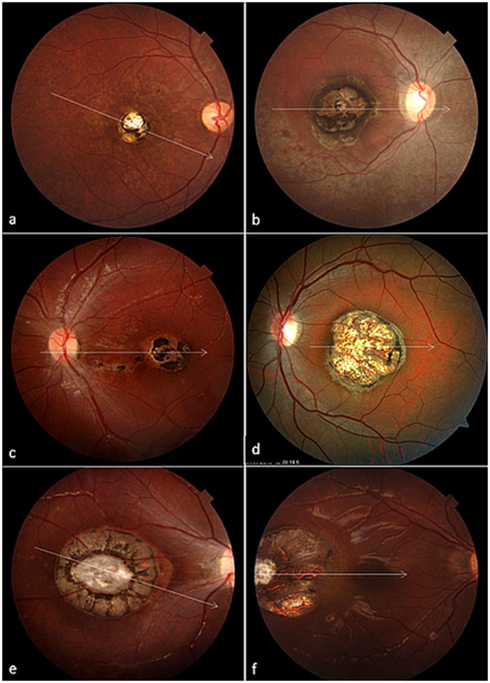
Fig. 2 Radial optical coherence tomography (OCT) line scan along the white axis shown in respective color fundus photographs in Figure 1. a. OCT shows a sharply defined deep macular retino-choroidal excavation with overhanging retinal layers. Immediately adjacent to it, choroidal lacuna can be visualized (white arrow). b and d. Deep excavated lesions with disrupted RPE layer (white arrow) and choroid and continuous dysplastic retinal layers with cavitations (white arrowheads). c. A relatively flat macular lesion with severe retinal thinning and hyperreflective RPE layer. e. A relatively shallow macular excavation with a central area of retinal hyperreflectivity corroborating with the glial tissue seen in figure 1e. Outer retinal cavitations (white arrowheads) are visible just adjacent to the central fibrotic area. f. A large deeply excavated lesion temporal to the fovea, with severe retino-choroidal atrophy.
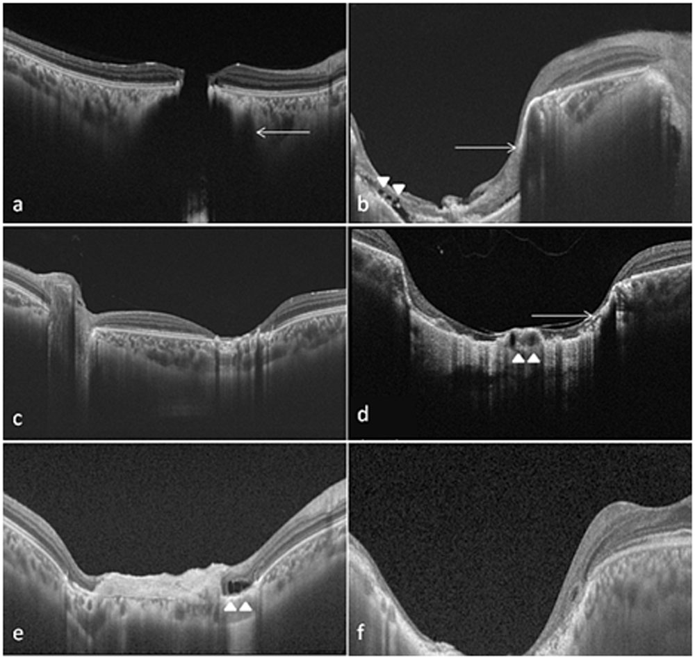
Fig. 3 Characteristic color fundus photographs and OCT line scans of macular coloboma with different serological results. a and d: A 30-year-old female with serology positive for CMV had a large deeply excavated relatively non-pigmented macular lesion with prominent visible choroidal vessels, scleral show, and severe retinal atrophy. b and e: A 29-year-old female with serology negative for both CMV and Toxoplasma had a medium sized deeply excavated moderately pigmented lesion with prominent large choroidal lacuna (white arrow). c and f: A 20-year-old male with serology positive for Toxoplasma had a medium sized ill-defined foveal pigmented relatively flat lesion with adjacent multiple small pigmented satellite lesions.
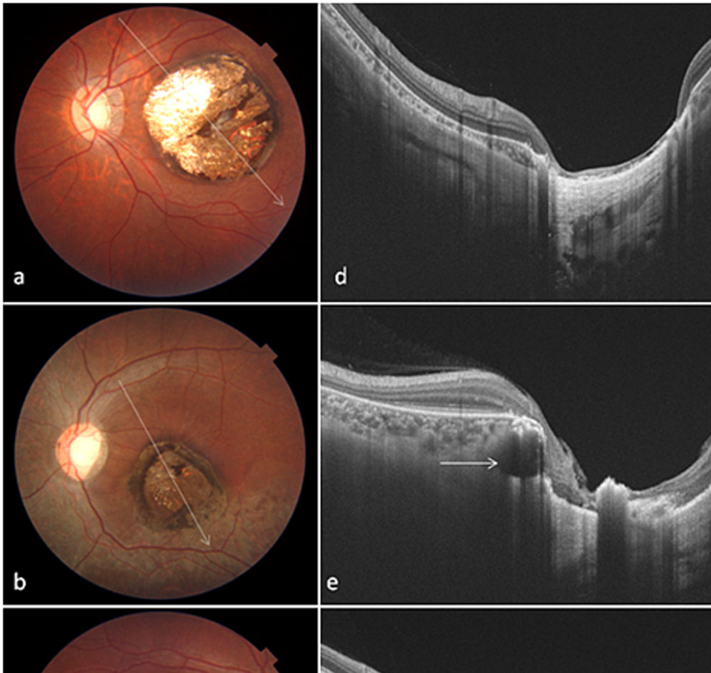
Table 1. Clinical and optical coherence tomography based characteristics in macular colobomatous lesions grouped on the basis of serological results.
| Feature | Serology negative | Toxoplasma IgG positive | CMV IgG positive | Total | P value |
| Number of eyes | 15 | 26 | 8 | 49 | – |
| Age (years)– Median, Min-Max | 24 (19-60) | 17 (7-58) | 18 (14-29) | 20 (7-60) | 0.0451 |
| CDVA– Mean SD,
Median (min-max) |
.82 ± .27,
.78 (.3-1.48) |
.74 ± .38
.6 (.18-1.48) |
.85 ± .52
1 (0-1.48) |
.78 ± .38
.78 (0-1.48) |
0.561 |
| Size
Small Medium Large |
6 7 2 |
5 19 2 |
0 3 5 |
11 29 9 |
0.0072 |
| Satellite lesion
No Yes |
15 0 |
17 9 |
8 0 |
40 9 |
0.0082 |
| Location
Foveal Extrafoveal |
14 1 |
25 1 |
7 1 |
46 3 |
0.732 |
| Pigmentation
None Some Profound |
2 11 2 |
1 13 12 |
0 5 3 |
3 29 17 |
0.182 |
| Fibrosis
Absent Present |
10 5 |
12 14 |
7 1 |
29 20 |
0.0662 |
| Shape
Round Oval Irregular Ill-defined |
6 4 3 2 |
9 7 4 6 |
1 4 1 2 |
16 15 8 10 |
0.812 |
| Retinal vessels
Discontinuous Continuous |
7 8 |
11 15 |
2 6 |
20 29 |
0.622 |
| Excavation
Flat Shallow Deep |
3 1 11 |
6 12 8 |
0 0 8 |
9 13 27 |
0.0032 |
| Choroidal vessels
Not visible Visible Prominent |
7 4 4 |
12 7 7 |
2 3 3 |
21 14 14 |
0.872 |
| Disc drag
Absent Present |
13 2 |
23 3 |
5 3 |
41 8 |
0.232
|
| Intra-retinal cyst
Absent Present |
13 2 |
17 9 |
5 3 |
35 14 |
0.682 |
| Retinal layers
Intact inner layers Atrophic |
3 12 |
9 17 |
3 5 |
15 34 |
0.942 |
| Retinal Pigment Epithelium changes
Atrophic Hypertrophic |
7 8 |
9 17 |
4 4 |
20 29 |
0.132 |
| Choroidal thinning
None Some Profound |
1 4 10 |
7 9 10 |
0 3 5 |
8 16 25 |
0.0752 |
| Retinal pigmentation
Absent Present |
12 3 |
20 6 |
7 1 |
39 10 |
0.992 |
| Choroidal lacunae
Absent Present |
7 8 |
21 5 |
5 3 |
33 16 |
0.0352 |
| Scleral show
Absent Present |
7 8 |
20 6 |
0 8 |
27 22 |
0.0012 |
Footnote: 1Kruskal Wallis test; 2Fischer’s exact test
Leave a Comment