

Dr. Nidhi Gadkar, Dr. Samar Kumar Basak, Dr. Tuhin Chowdhury, Dr. Nibedita Das
Corneal vascularization can occur due to inflammation after infection, trauma, post keratoplasty and others.VEGF has a role in the pathogenesis of corneal neovascularization1. Neovascularization can cause leakage of lipids leading to secondary lipid keratopathy which can be visually and cosmetically compromising.
Regression of neovascularization can decrease the lipid deposition and hence improve the transparency of cornea and improve the visual acuity.
Corneal neovascularization is one of the most common risk factors contributing to corneal graft rejection.The risk of an immune reactionis approximately 50% in a vascularized cornea compared withapproximately 10% in an avascular cornea.2,3
PURPOSE:
To study the effect of intrastromal ranibizumab on corneal vascularization and lipid keratopathy.
METHODS:
20 Cases with corneal vascularization with or without secondary lipid keratopathy were included in the study. Patients with corneal vascularization post HSV keratitis were included if disease was inactive. Two doses of Ranibizumab 0.01 ml(Accentrix) was givenintrastromally under topical anaesthesia with 30 gauge needle. Site was decided preoperatively by slit lamp photography. If more than one quadrant involved injection was given in each quadrant. Second dose was repeated at 6 weeks interval. Regression of vessels and lipid keratopathy was assessed by comparison of the slit lamp photographs taken pre and post operatively.
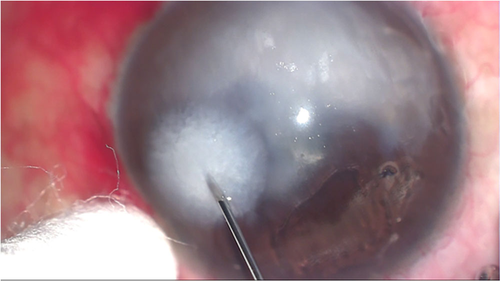
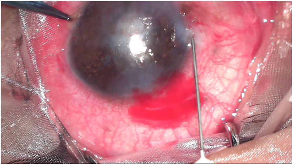
Final visual acuity was taken 6 weeks after the second injection.
Patients having dense opacities involving the pupil and stable vascularization underwent Optical penetrating keratoplasty(OPK)/ Deep Anterior lamellar keratoplasty (DALK).
RESULTS:
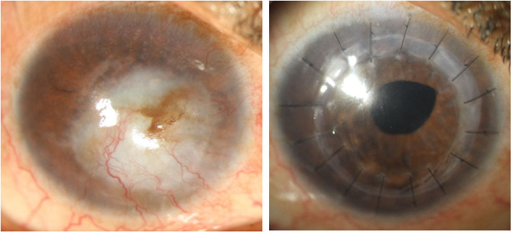
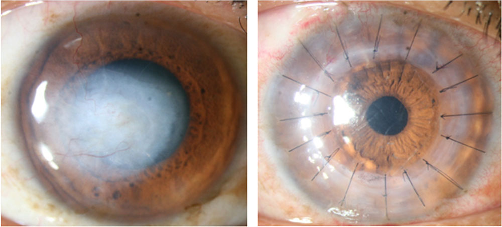
20 Cases were included. Mean age was 40 years (Range 20 to 70 years) Of the 20 cases 14(70%) were post infectious keratitis in which 11(55%) were due to HSV keratitis sequalae and 3 (15%) due to others, 1 was a failed vascularized graft, 1 healed fascicular ulcer, 4(20%) had vascularized corneal opacity in which the etiology was unknown. (Figure 1) All cases had improvement in visual acuity 6 weeks after the second dose, mean 0.24 Logmar units on follow up from 2 to 12 months. There was reduction in area of vascularization and vessel calibre. (Figure 2) All patients had regression of lipid keratopathy. (Figure 3,4) 5 patients underwent OPK and 1 patient underwent DALK. All patients are maintaining clear grafts with no episode of graft rejection on follow up ranging from 3 to 12 months after keratoplasty. (Figure 5,6) 5 patients having stable vascularization are planned for OPK. No intraoperative/ postoperative or systemic complications were noted. One patient had a recurrence of HSV keratitis after 18 months of second injection.
CONCLUSION:
Previous studies have been done on topical, subconjunctival Ranibizumab and Bevacizumab and intrastromal Bevacizumab5-10butfewer studies done on effect of intrastromal Ranibizumab11,12. Intrastromal injection allows greater exposure of the corneal vessels to the drug, as well as delivery of a known concentration of the drug. It is superior to topical administration where penetration of the drug through an intact epithelium is limited. There is also lesser likelihood of treatment failure due to lack of patient compliance. It is a safe treatment option in patients with inactive HSV keratitis and corneal vascularization with low risk for reactivation while few cases have been reported after intravitreal injection of anti VEGFs13-14 Patients having lipid keratopathy have better visual and cosmetic outcomes after injection. Reduction in vascularization pre operatively reduces an important risk factor for graft rejection post keratoplasty. The limitation of our study was lack of a long follow up to assess the long termstability of the regression of vascularization and lipid keratopathy.
REFERENCES
- Philipp W, Speicher L, Humpel C. Expression of vascular endothelial growth factor and its receptors in inflamed and vascularized human corneas. Invest Ophthalmol Vis Sci. 2000;41(9):2514–2522.
- Maguire MG, Stark WJ, Gottsch JD, et al. Risk factors for corneal graft failure and rejection in the collaborative corneal transplantation studies.Collaborative Corneal Transplantation Studies Research Group. Ophthalmology.1994;101:1536–1547.
- Maier P, Bohringer D, Reinhard T. Clear graft survival and immunereactions following emergency keratoplasty. Graefes Arch Clin ExpOphthalmol. 2007;245:351–359.
- Faraj LA, Said DG, Al-Aqaba M, et al. Clinical evaluation and characterisation of corneal vascularisation Br J Ophthalmol 2016;100:315–322.
- Oh JY, Kim MK, Wee WR. Subconjunctival and intracorneal bevacizumab injection for corneal neovascularization in lipid keratopathy. Cornea. 2009;28(9):1070-3.
- Vieira ACC,Hofling-Lima AL, Gomes JAP, et al. Intrastromal injection of bevacizumab in patients with corneal neovascularization. Arq Bras Oftalmol, 2012;75:277–279
- Ferrari, G., Dastjerdi, M. H., Okanobo, A.et al. Topical Ranibizumab as a Treatment of Corneal Neovascularization. Cornea,2012; 32(7), 992-997. doi:10.1097/ico.0b013e3182775f8d
- Hashemian MN, Zare MA, Rahimi F, Mohammadpour M. Deep intrastromal bevacizumab injection for management of corneal stromal vascularization after deep anterior lamellar keratoplasty, a novel technique. Cornea. 2011;30(2):215-8.
- Dastjerdi MH, Al-Arfaj KM, Nallasamy N, et al. Topical Bevacizumab in the Treatment of Corneal Neovascularization: Results of a Prospective, Open-label, Non-comparative Study. Archives of ophthalmology. 2009;127(4):381-389. doi:10.1001/archophthalmol.2009.18.
- Stevenson, W., Cheng, S., Dastjerdi, M. H. et al. Corneal Neovascularization and the Utility of Topical VEGF Inhibition: Ranibizumab (Lucentis) Vs Bevacizumab (Avastin). The Ocular Surface,2012; 10(2), 67-83. doi:10.1016/j.jtos.2012.01.005
- Kim, J., Seo, H.,Han, H. The Effect of Bevacizumab versus Ranibizumab in the Treatment of Corneal Neovascularization: A Preliminary Study. Korean Journal of Ophthalmology,2013;27(4), 235. doi:10.3341/kjo.2013.27.4.235
- J. Ahn, H. B. Hwang, S. K. Chung. Ranibizumab injection for corneal neovascularization refractory to bevacizumab treatment. Korean Journal of Ophthalmology,2014;28(2),177–80.
- Khalili, M. R., Mehdizadeh, M., Mehryar, M. Herpetic Epithelial Keratitis After Intravitreal Injection of Bevacizumab (Avastin). Cornea,2009;28(3), 360-361. doi:10.1097/ico.0b013e318189137a
- Derham, A. M., Chen, E., Bunya, V. Y. et al. Bilateral Herpetic Keratitis After Bilateral Intravitreal Bevacizumab for Exudative Macular Degeneration. Cornea,2017; 36(7), 878–879. doi:10.1097/ico.0000000000001233
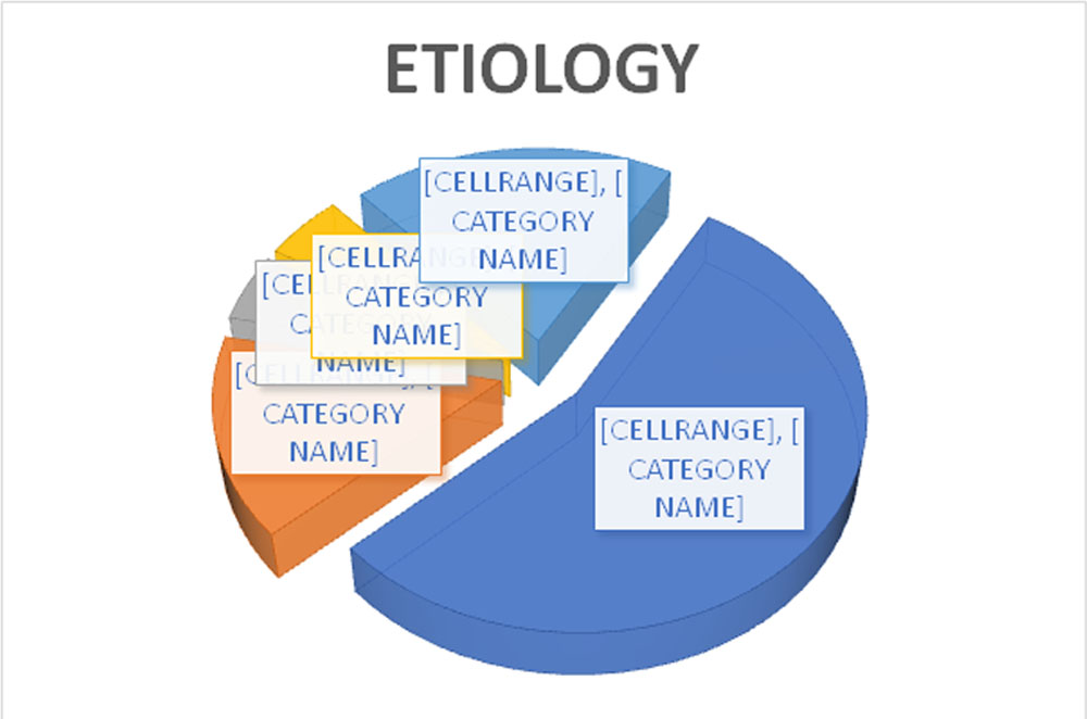
FIGURE 2
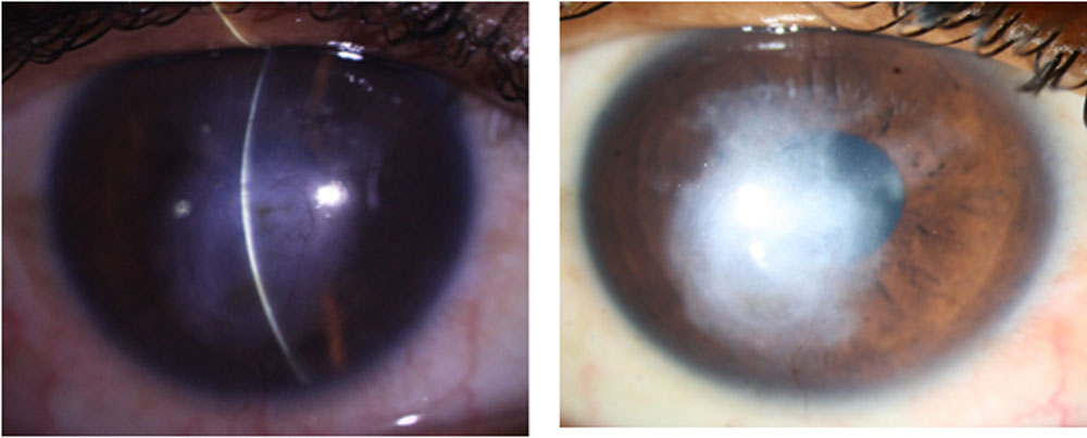
PRE INJECTION POST INJECTION
FIGURE 3
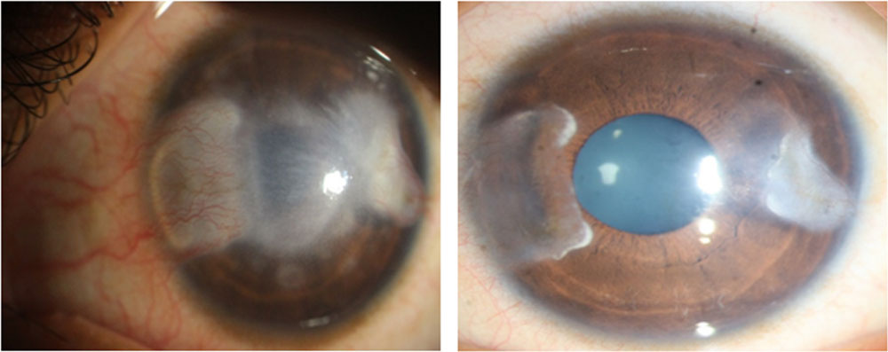
PRE INJECTION POST INJECTION
FIGURE 4
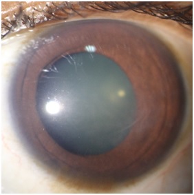
PRE INJECTION
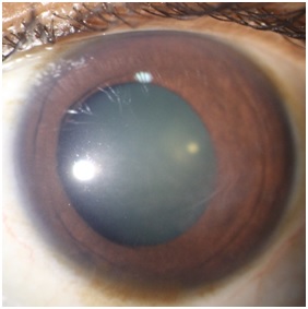 POST INJECTION
FIGURE 5,6
PRE INJECTION POST OPK
Leave a Comment