

INTRODUCTION
Optic neuritis is defined as inflammation ,infection or demyelination of optic nerve
Causes acute ,usually monooccular visual loss
Highly associated with multiple sclerosis
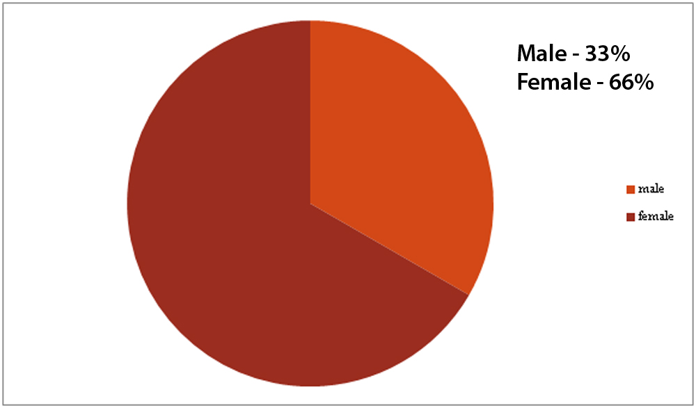
Female prepondorence(2/3)cases
Usually between 20-40 years of age
Typical –associated with multiple sclerosis,steroid dependent.
Atypical –not associated with multiple sclerosis,steroid dependent improvement.Causes of atypical optic neuritis includes connective tissue disorder SLE,vasculitis,sarcodoisis,or neuromyelitis optica.
Most common pathology –inflammatory demyelination of optic nerve
Perivascular cuffing,edema in myelinated nerve sheath and myelin breakdown
Inflammation of the retinal vascular endothelium can precede demyelination and sometimes retinal vein sheathing
Myelin loss exceeds axonal loss
There is acute onset of visual acuity loss,visual field loss,colour distoration,pain upon eye movement
Axonal loss occurs following retinal ganglionic cell neuronal loss occur
Macular ganglion cell layer gives rise to retina nerve fibre layer and affected by axonal inflammation
Previously RNFL was used to asses eye damage instesd of GCLbecause GCL could not analyse in vivo
Recent development of spectral domain optical coherent tomography (SD-OCT) can assess both peripapillary retinal nerve fibre layer and retnal GCL thinning in optic nerve injury
MATERIAL AND METHOD
Prospective study of 40 pts with first attack of unilateral optic neuritis who attented our hospital from July 2017 to Apr 2018. Age ,sex ,colour vision,VA,optic nerve findings on MRI,time from onset of visual symptoms to steriod t/t,Visual field(MD),average PRNFL & GCIPL thickness(SDOCT) were recorded
RESULT
Visual recovery usually occurs after optic neuritis after treatment of corticosteriod.but residual visual loss occur in patients with PRNFL and GCIPL thinning after optic neuritis
ANALYSIS
ANALYTICAL TABLE
Table showing patients without PRNFL and GCIPL thinning
| Vision before treatment | Vision after treatment | No of patient |
| CF | 6/18 | 15
|
| 6/60 | 6/12 | 10 |
Table showing patients with PRNFL and GCIPL thinning
| Vision before treatment | Vision after treatment | No of patients
|
| CF | 6/60 | 10
|
With residual symptoms abnormal optic nerve thickening
CONCLUSION
Retinal ganglion cell and axonal losses occured in optic neuritis cases showing severe initial VF loss. There is possibility of residual VF loss accompanying PRNFL and GCIPL thinning.
REFERENCES
Bradly’s text book of neurology
Acute optic neuritis.AAN neurology
Current options for treatment of optic neuritis;john et al
Jones SJ,Brusa A neurological evidencefor long term repair of MS lesion
Henderson AP,altman DR a study of retinal changes following optic neuritis
Leave a Comment