

Dr.Parthvi Shah,Dr.HARSHA CHETAN JANI,Dr.CHAITALI PATEL (PATHAK)
Abstract:
Animal rearing is a common practise in rural parts of India. India being an agricultural country,males and females across the country work in close proximity with cattle and are prone to various injuries through direct trauma via horn, tail or legs. Eyes are commonly injured in these circumstances. Here we present one such rare case where a bull’s horn injury to the eye resulted in a blow out fracture with displacement of the eyeballinto the ethmoidal sinus. Timely diagnosis and intervention plays a major role in reducing morbidity as will be highlighted in this case.
KEYWORDS:bull horn injury, blow out fracture , eyeball in ethmoidal sinus .
KEY MESSAGES :
Agricultural trauma is a common cause of monocular blindness in rural India.[1,2]Closed globe injury being more common than open globe injury.[2]Corneal ulcer, blunt trauma, penetrating injuries are often caused by animals.Rarely there are serious injuries to the eye to the extent that patient presents with an empty socket.
INTRODUCTION :
The case is being reported for its rarity as a variant of blow out fracture presenting with the eyeball displaced into the ethmoidal sinus. The patient thereafter regained vision in the affected eye and such a case has not been reported so far.
CASE REPORT :
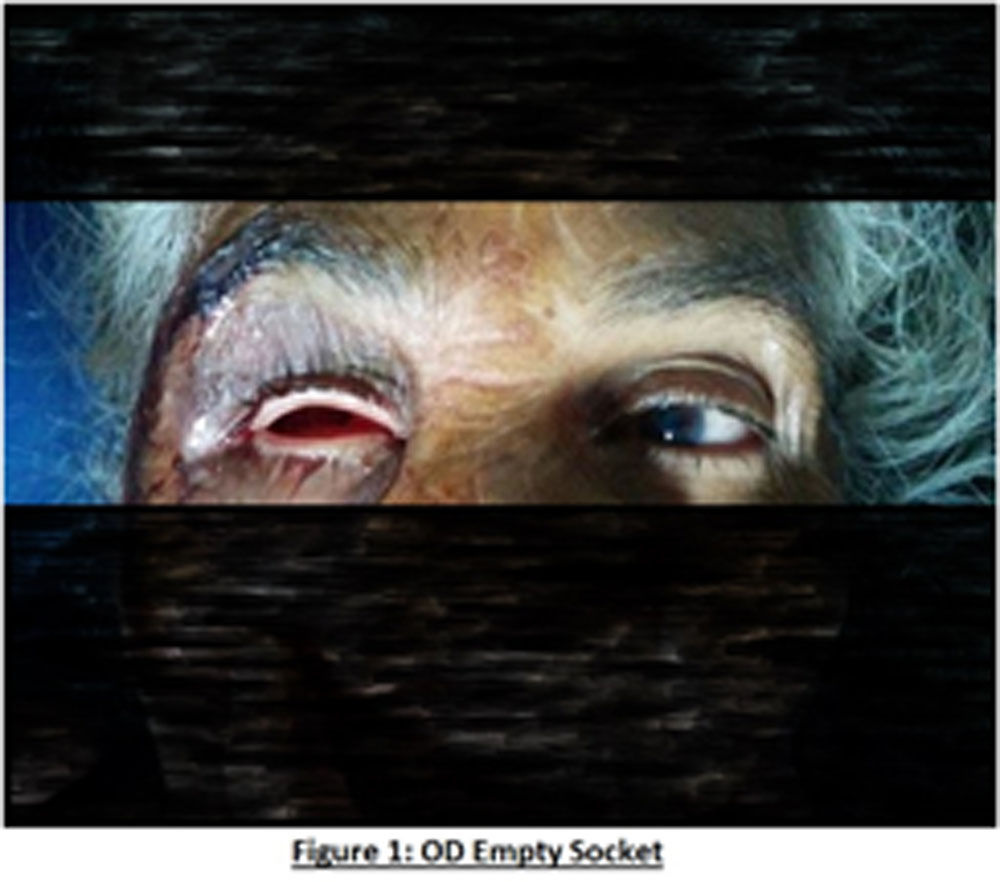
A 68 yr old female presented to the emergency department of a tertiary care hospital with sudden and painful dimness of vision in the right eye since one hour after being hit by abull’s horn in the right eye. Ophthalmological examination revealed a visual acuity of (?) Perception of Light and Counting Finger 2 meters in OD and OS respectively and empty socket with upper lid and lower lid edema and palpable fracture of inferior orbital rim.(figure1).
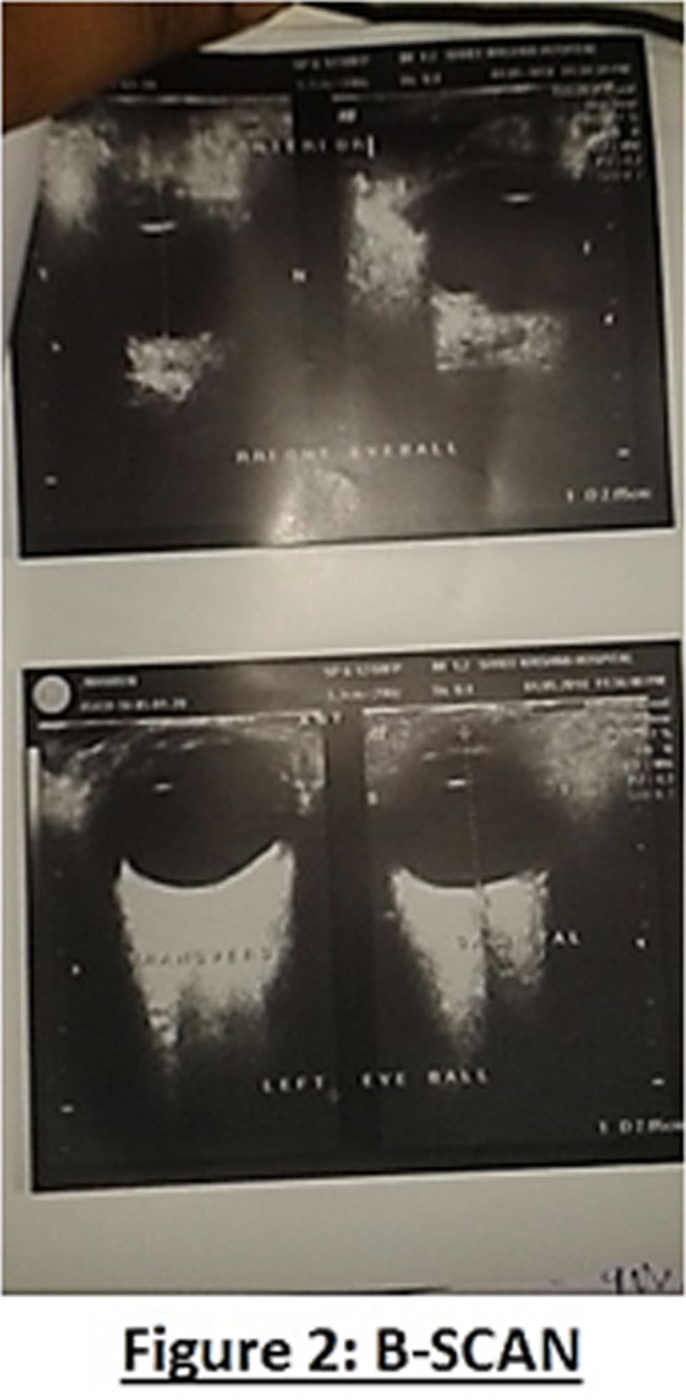
Patient was otherwise stable. Ultrasound revealed postero-infero and medial displacement of the right eyeball.(figure2)
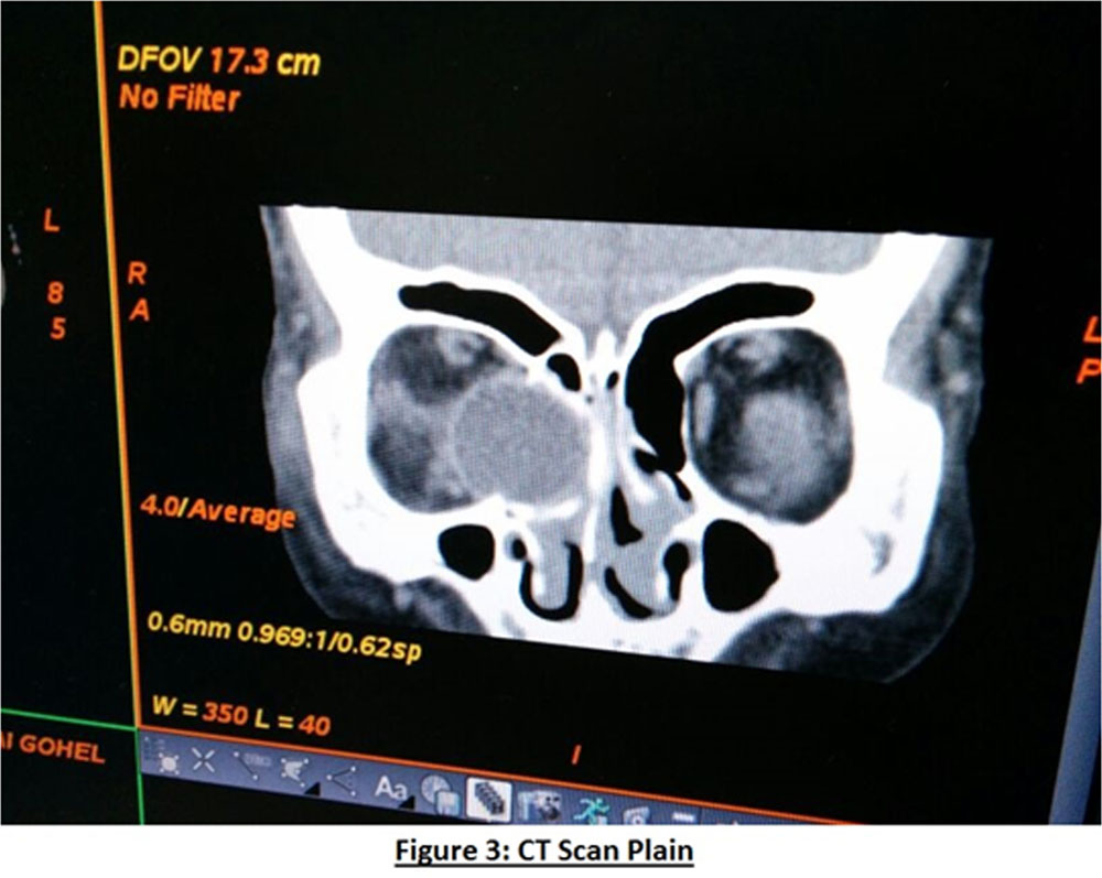
CT scan showed blow out fracture of the medial wall and floor of right orbit with the medial portion of right eyeball lying in the region of anterior ethmoidal air cells and anterior part of right nasal cavity.There was no visualization of the medial and inferior rectus muscles. The intra orbital part of the optic nerve was shown to be in contact with the displaced eyeball. (figure3)
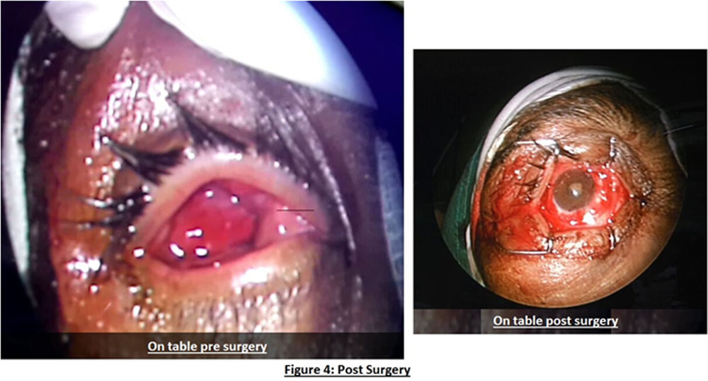
The case was managed surgically by combined efforts of ophthalmologists and otorhinolaryngologists in the following manner. Under endoscopic guidance the eyeball was repositioned into the orbital cavity and conjunctival sutures were taken to keep the eyeball in position. On table findings revealed an intact globe, clear cornea, multiple conjunctival tears, muscle lacerations, pupil 3mm dilated not RàL and immature catarct.(figure4).
Post surgery the patient was started on systemic antibiotics, steroids, analgesics with topical antibiotic and lubricating eye drops.
First post-operative day(figure 5) examination revealed a visual acuity of Counting Finger 1.5 meters for distance and N18 for near in OD, upper lid &lower lid edema, mechanical ptosis, and restriction of all ocular movements and a 15 degree exotropia of right eye.
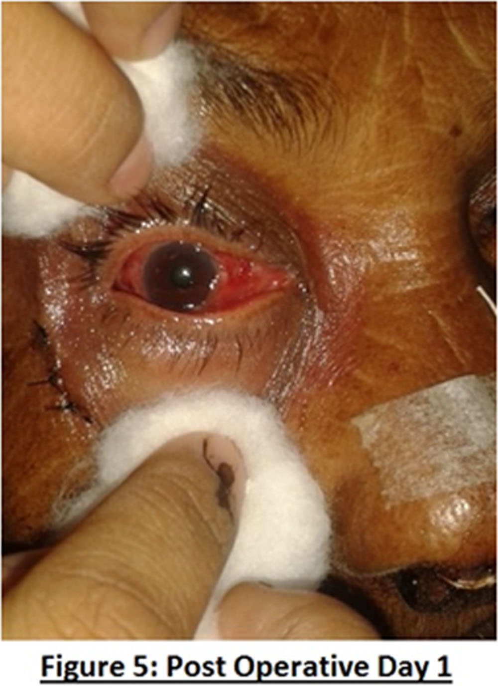
Slit lamp examination findings were as follows: Conjunctivalchemosis, Cornea clear, anterior chamber quiet, pupil 3mm sluggishly RàL , Nuclear sclerosis grade II/III and cortical cataract, intraocular pressure of 12 mm hg.
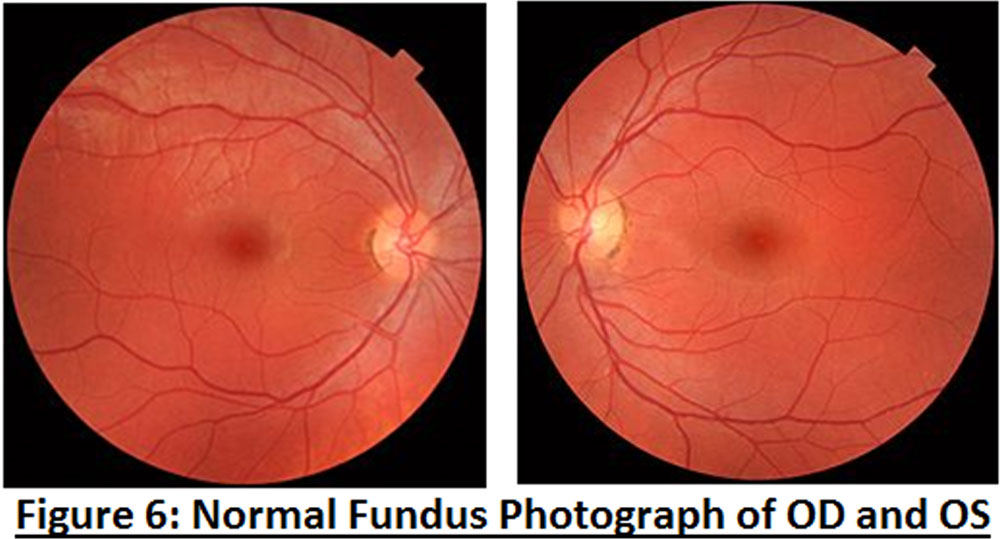
Dilated fundus examination revealed a normal disc and macula.(figure 6)
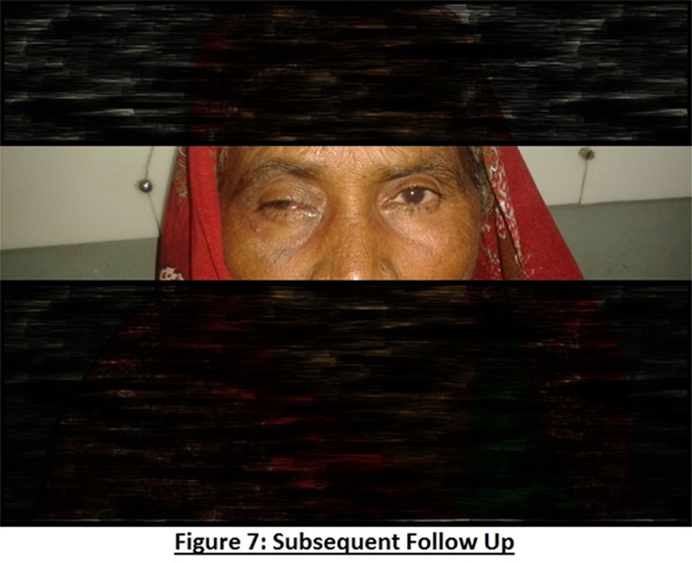
Subsequent follow ups showed an improvement in visual acuityupto 20/80 for distance and N18 for near in the operated eye. Examination revealed slight reduction in the ptosis .Exotropia of 15 degree with restriction of all ocular movements persisted.Pupil became central& reacting to light. (figure 7)
DISCUSSION:
Agricultural trauma being a common cause of ocular morbidity in our country, timely management is of utmost importance to prevent blindness.The above mentioned patient was brought to a tertiary care hospital well equipped with higher facilties like B scan, CT scan etc and a rapid diagnosis of the conditionwas made.This is probably the first case of its kind, to the best of our knowledge that even after such a grevious injury, the timely diagnosis and effective management saved the patients eye and vision.
REFERENCES:
1.Onyekwe LO, OhaegbulamSC. Penetrating orbito-cranial and ocular cow-horn injuries. Nigerian journal of clinical practice. 2007 July ;10(2):177-179.
2.Misra S, Nandwani R, Gogri P, Misra N.Clinical profile and visual outcome of ocular injuries in a rural area of western India. Australas Med J.2013 Nov 30;6(11):560-4.
Leave a Comment