

Dr.Ravindra Kumar Chowdhury, Dr.Sharmistha Behera,Dr.Jayashree Dora,Dr.Kanhei Charan Tudu
Abstract:
Present case series comprises 24 cases of lacrimal sac rhinosporidiosis over a period of two years at a tertiary care hospital of India. All the cases underwent dacryocystectomy with meticulous excision and copious irrigation with 5% povidone-iodine for 5 minutes. Clinicallydiagnosed cases were confirmed by histopathological correlation.Postoperative Dapsone therapy of 100 mg once or twice daily dose was given for three to six months. All the patients were followed up for a period of one year.Rhinosporidium seeberi, an aquatic protistan parasite was found to be the main causative agent. Males and females were affected equally. Children less than 10 years of age comprised 58.3%.The most common presentation was boggy swelling over lacrimal sac.All the cases were unilateral. In 66.6%, the lesion was limited within the sac. Recurrence was noted in 25% of cases. Low recurrence rate could possibly be due to early detection and standard management and limited follow-up period.
INTRODUCTION:
Rhinosporidiosis is a chronic and localized infection of mucous membrane caused by Rhinosporidium seeberi.[1]Rhinosporidium is anaquatic protistan parasite belonging to a novel group of fish parasites that infect fishand amphibians, located phylogenetically between the fungal animal divergence.[2]The common sites of affection are nose and nasopharynx (78%) followed byconjunctiva and associated structures including lacrimal apparatus(15%) and rarely skin and genitalia.[3]Among the ocular rhinosporidiosis (15%), the most common presentation is conjunctival polyp (77.6%).Twenty-six percent of ocular rhinosporidiosis occurs in lacrimal sac either withconjunctival or nasal involvement.[4]This aquatic protistan thrives in a hot tropical climate and endemiczones are located in southIndia and SriLanka.[5]The ocularmanifestation of rhinosporidiosis is rarely encountered outsidethe coastal areas of Kerala, Tamil Nadu, and Puducherry.However, sporadic occurrence has been reportedfromWestBengal,MadhyaPradesh,Rajasthan,Odisha,BiharandMaharashtra. The state of Odisha lies on the eastern coast of Indiaand comes wholly under the tropical zone. The climate in Odishais moderate. However, in western districts, namely Bolangir,Sambalpur and Sundergarh, extreme climatic conditions areexperienced. Summer temperature varies between 20 degreesand 45 degrees Celcius, while in winter it varies from 13 degrees to 32 degrees Celcius.[6]
We present the clinical and histological profile of 24 cases of rhinosporidiosis of lacrimal sac in a tertiary care hospital of Western Odisha,India.This is a first reported case series in this part of India as per the literature review.The history of pond bath,unilateral manifestation, and non-involvement of adjacent structure like nasal mucosa and conjunctiva in all of our cases were interesting to note.
MATERIALS AND METHODS:
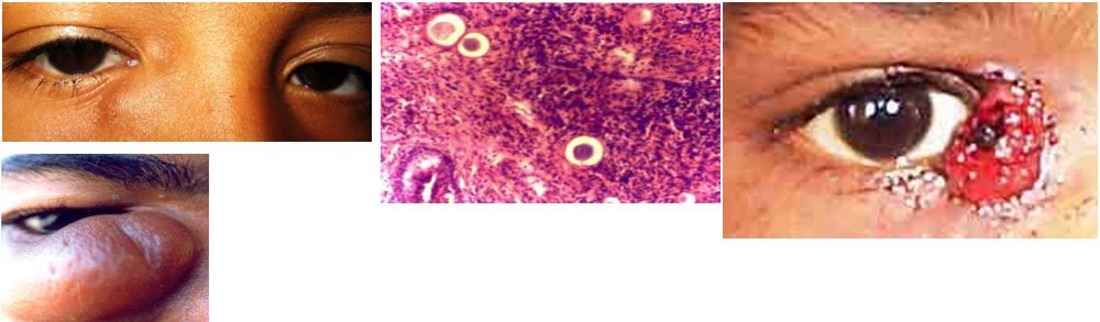
We studied 24 patients presenting with boggy swelling over lacrimal sac area retrospectively during May 2014 to May 2016 at a tertiary care hospital of Western Odisha. The study staff included four ophthalmologists and two pathologists. The inter-observer variation of our study was minimized by a standard clinical definition to suspect andastandard histological definition to confirm the lacrimal sac Rhinosporidium.In our study, all cases presenting with soft, fluctuant boggy swelling of the lacrimal sac were suspected to have rhinosporidiosis of the lacrimal sac [Figure – 1]. However, the diagnosis in all our cases was confirmed histologically by the presence of fibro-cellular elements covered by proliferating stratified epithelium containing innumerable sporangia of all sizes in the subepithelial layer [Figure – 2].
![[Figure – 1]](https://www.ijo.in/viewimage.asp?img=IndianJOphthalmol_2007_55_4_299_33045_1.jpg)
![[Figure – 2]](https://www.ijo.in/viewimage.asp?img=IndianJOphthalmol_2007_55_4_299_33045_2.jpg)
For each patient entered into the study, detailed clinical features, epidemiology (age, sex) and treatment were studied and analyzed.All the patients were referred to ENT specialist to look for any nasal involvement.The patients suspected of rhinosporidiosis of lacrimal sac clinically underwent dacryocystectomy (DCT). Care was taken to avoid spilling of spores during complete excision.After complete removal of the sac,5% povidone-iodine was applied for 5 minutes in all of our cases.Post-operatively all of our patients were given dapsone in a dose of 100mg once or twice daily for 3 to 6 months.Glucose-6 phosphate enzyme deficiency was excluded in all cases before prescribing Dapsone therapy.All the patients were followed up for one year.Special attention was given to collect information regarding the bathing habits of all patients.
OBSERVATION:
Rhinosporidiosis of lacrimal sac were uniocular in all 24 patients. The right and left eye were affected in almost equal numbers.The condition was more common in patients below 10 years of age [Table-1].The males and females were affected in equal numbers.
![[Table-1]](https://www.ijo.in/viewimage.asp?img=IndianJOphthalmol_2007_55_4_299_33045_3.jpg)
Rhinosporidiosis of the lacrimal sac presented with complains of boggy swelling of sac area(24 cases),epiphora(6 cases),epistaxis(4 cases) and polypoidal growth loaded with numerous grey white dots at lacrimal sac area(1 case).[Table-2] & [Figure-3]All of our patientshad a history of taking bath in pond water.The lesion was confined within the sac in 16 cases(66.6%) and outside the sac in 8 cases(33.3%).On opening, the sac pink vascularized growth with the finger-like extension was seen in all of them.Bleeding was remarkable during excision of the sac. Recurrence was noted during follow-up in six cases(25%).[Figure-4] The duration of follow-up was one year in our series.
Neither nasal involvement nor any conjunctival involvement was associated with any of our patients.
TABLE-1: Age & Sex Distribution of Lacrimal sac Rhinosporidiosis
| Age in years | No. Of Males | No. Of Females | Total |
| <10 | 8 | 6 | 14 |
| 11-20 | 1 | 3 | 4 |
| 21-30 | 2 | 1 | 3 |
| > 30 | 1 | 2 | 3 |
| Total | 12 | 12 | 24 |
TABLE-2:Clinical Presentations of Lacrimal sac Rhinosporidiosis
| Presentation | No.of cases | Percentage(n=24) |
| Boggy swelling over lacrimal sac area | 24 | 100 |
| Epiphora | 6 | 25.0 |
| Epistaxis | 4 | 16.6 |
| Polypoidal growth over sac area | 1 | 0.04 |
Fig-1: Rhinosporidiosis of the Lacrimal sac,
Fig 2 :Histopathology of Rhinosporidium Seeberi
Fig-3: Rhinosporidiosis of lacrimal sac with polypoidal growth
Fig 4:Recurrence of Lacrimal sac rhinosporidiosis
DISCUSSION:
Lacrimal sac rhinosporidiosis is usually suspected in patients presenting with boggy swelling over lacrimal sac region with a feeling of “bag of worms”.[7,8,9] In our study,such swelling over lacrimal sac area was found in 24 cases(100%) and epiphora in 6 cases(25%).This is in consistent with Nuruddin et al[10] who found 100% of patients had a feeling of boggy swelling over sac area and epiphora in 22.2%.The spread of Rhinosporidial infection is pericanalicular and perisacular.[11] Therefore only one-fourth of our patients presented with epiphora inspite of lacrimal sac involvement.
In the present study, the infection was seen most frequently in children less than 10 years of age. This is in contrast to the findings of Nuruddin et al[10] who had reported a higher prevalence in the 25 to 34 years of age group. This disparity may be accounted for by the higher number of children having the habit of bathing in pond water. Males and females were found to be affected equally in our study.
Rhinosporidiosis is a waterborne disease and the organism is suspected to be present in the stagnant water like ponds.However, it may also be transmitted via air and dust.[11,12]In our study all of the patients had a history of pond bath which is consistent with most of the studies.The probable route of entry of this organism to lacrimal sac is canaliculi and nasolacrimal duct.But none of our patients presented with conjuctival involvement or any nasalinvolvement.This is an interesting variation.
Radiography investigation like CT dacryocystography was recommended before surgery to delineate the involvement of lacrimal sac and nasolacrimal duct involvement by Pushker et al.[13] However our patients were not exposed to such investigations to avoid the harmful effect of radiation and dye.We assessed the involvement clinically.Care was taken when the nasolacrimal duct was involved with thorough curettage and cauterization to prevent recurrence.
The definitive treatment for Rhinosporidiosis of lacrimal sac is Dacryocystectomy(DCT) [4,7,8,9],It is recognized that surgical removal the lacrimal sac is frequently difficult due to severe bleeding during excision of lacrimal sac rhinosporidiosis.So recurrence of the lesion is frequent. In our series, recurrence was seen in only six cases(25%).The recurrence was more commonly seen when the extension was outside the sac. The result of this study does not agree with observation of earlier studies which say recurrence is inevitable [7,14]Low recurrence in our study may be due a limited period of follow-up. Further confirmation may be done through a larger case series for a longer follow up period.
The main cause of recurrence of the lesion in lacrimal sac rhinosporidiosis is the inability to remove the spores of rhinosporidium completely.The intraoperative maneuver like 5% betadine soakage for 5 min after excision of the sac[15] ,electric cauterization of surrounding healthy area[16] and application of 1 to 5 ml of amphotericin B (0.15%)[17] have been tried by different authors with success.Arseculeratne et al have reported metabolic inactivation of endospores on exposuresto povidone iodine.[15]The postoperative dapsone therapy with 100 mg once/twice daily for three to six months has been found to prevent recurrence as it is known to arrest the maturation of spores and promote fibrosis in the stroma.[18] This drug should, however, be used after ruling out drug allergy and G6PD deficiency.[19]We believe that intraoperative maneuver with 5 % povidone-iodine and postoperative dapsone therapy have lessened the recurrence rate in our series.
REFERENCES:
1.Ashwarth JH. On Rhinosporidium seebri with special reference to its sporulation and affinities. Trans RSoc Edinb 1923;53:301-42.
2.Herr RA, Ajello L, Taylor JW, Arseculeratne SN, Mendoza L. Phylogenetic analysis ofRhinosporidium seeberi′s 18S Small-subunit Ribosomal DNA groups this pathogen among members ofthe Protoctistan Mesomycetozoa clade. J Clin Microbiol. 1999;37:2750.
3.Gonazalez G, Viada J, Escalona A, Na’quira N. Nasal rhinosporidiosis – four cases relate literature review.Int Arch Otorhinolaryngol 2007;11:428-9.
4.Shrestha SP, Hennig A, Pariya SL. Prevalence of rhinosporidiosis of eye and it adenexa in Nepal. Am JTrop Med Hyg 1998;59:231-4.
5.Sood NN, Rao SN. Rhinosporidium granuloma of conjunctiva. BrJ Ophthalmology 1967; 51:61-4.
6.Chowdhury RK, Behera S, Bhuyan D, Das G. Oculosporidiosis in a tertiary care hospital of westernOrissa, India: A case series. Indian J Ophthalmol. 2007;55:299–301.
7.Kuriakose ET. Oculosporidiosis: Rhinosporidiosis of theeye. Br J Ophthalmol 1963;47:346–349.
8.Mukherjee PK, Shukla IM, Deshpande M, Kher P.Rhinosporidiosis of lacrimal sac. Ind J Ophthalmol 1982;30:513.
9.Pushker N, Kashyap S, Bajaj MS, et al. Primary lacrimal sacrhinosporidiosis with grossly dilated sac and nasolacrimalduct. Ophthal Plast Reconstr Surg 2009;25:234–235.
10.NuruddinM1,MudharHS,OsmaniM,RoySR.Lacrimal sac rhinosporidiosis:Clinical Profile and surgical management by modifieddacryocystorhinostom.Orbit. 2014 Feb;33(1):29-32.
11.Watve JK, Mane RS, Mohite AA, Patil BC. Lacrimal sacrhinosporidiosis. Ind J Otolaryngol Head Neck Surg 2006;58:399–400.
12..Mukherjee PK. Rhinosporidiosis. In: Fraunfelder FT, RoyFH, eds. Current Ocular Therapy. 5th ed. Philadelphia, PA:WB Saunders Company; 2000. pp 66–67.
13.Pushker N, Kashyap S, Bajaj MS, et al. Primary lacrimal sacrhinosporidiosis with grossly dilated sac and nasolacrimalduct. Ophthal Plast Reconstr Surg 2009;25:234–235.
14.Mohapatra LN. Rhinosporidiosis. Warren KS, Mahmaud AA,editors. Tropical and Geographical Medicine. Mc Graw Hill: NewYork; 1990. p. 991-3.
15.Arseculeratne SN, Atapattu DN, Balasooriya P, Fernando R. The effects of biocides (antiseptics and disinfectants) on the endospores of Rhinosporidium seeberi. Ind J Med Microbiol 2006;24:85–91.
16 Ghorpade A, Gurumurthy J, Banerjee PK, Bhalla M, Ravindranath M. Oculosporidiosis presenting as an under -eye swelling. Indian J Dermatol Venereol Leprol. 2007;73:196–7.
17.Bhomaj S, Das JC, Chaudhari Z, Bansal RL, Sharma P. Rhinosporidiosis and peripheral keratitis. Ophthalmic Surg Lasers. 2001;32:338–40.
18.Ghorpade A, Gurumurthy J, Banerjee PK, Bhalla M, Ravindranath M. Oculosporidiosis presenting as an under -eye swelling. Indian J Dermatol Venereol Leprol 2007;73:196-7
19.Arseculeratne SN. Recent advances in rhinosporidiosis and rhinosporidium seeberi. Indian J Med Microbiol 2002:20:119-31.
Leave a Comment