

Dr.Ashish Jain, J20051, Dr.Navneet Mehrotra, Dr.Manish Nagpal
AIM: To compare the retinal vasculature features of OCT Angiography with FA in BRVO.
METHOD: 30 eyes with BRVO were retrospectively evaluated. All patients underwent FA and OCTA Panorama imaging (wide field image acquisition) for assessment of microvascular changes, including foveal avascular zone (FAZ), capillary non perfusion (CNP) areas and venous congestion.
RESULT: CNP areas, enlargement of the FAZ, collateral vessels were appreciated equally well on both FA & OCTA. OCTA provided precise details of microvascular changes like venous congestion in the deep capillary plexus (DCP), not visualized on FA. Vascular perfusion in the DCP detected by OCTA is a key parameter responsible for VA in patients with BRVO.
CONCLUSION: Superficial retinal microvasculature details in BRVO correlate well on FA and OCTA. In addition, OCTA reveals details of deep capillary plexus not identifiable on standard FA.
INTRODUCTION:
Retinal vein occlusion is the second most common retinal vascular disorder after diabetic retinopathy and is considered to be an important cause of visual loss.[1,2] Branch retinal vein occlusion (BRVO) is an acute cause of visual impairment secondary to thrombotic events, external compression, or vessel wall pathology. [3] Occlusion of the major veins of the retinal circulation leads to increased intraluminal pressure, hemorrhage, and edema. [4] Although the most common cause of decreased vision in BRVO is macular edema, thrombosis results in engorged veins frequently accompanied by variable amounts of retinal nonperfusion.
However, poor visual recovery has been reported despite complete resolution of the macular edema after treatment. [5] In such cases, the possible mechanisms accounting for persistent visual impairment, for example, potential abnormalities in the retinal microvasculature, cannot
be identified by conventional optical coherence tomography (OCT) and FA. Optical coherence tomography angiography (OCTA) is a new imaging modality that allows noninvasive visualization of retinal blood flow without use of exogenous dyes. [6] The layer-specific imaging capabilities of OCTA have the potential to simultaneously visualize both superficial and deep retinal capillaries by segmentation of each layer. [6]
The aim of this study was to assess retinal microvascular changes in BRVO using OCT-A and its comparison with FA.
METHODS:
The procedures used in this study conformed to the tenets of the Declaration of Helsinki. All patients were informed of the nature and possible consequences of the procedures, and signed informed consent was obtained from all patients.
A consecutive series of 32 patients who presented with BRVO in the outpatient department between Jan 2017 and Dec 2017 were enrolled in this observational study.
All patients underwent comprehensive ophthalmic evaluation including measurement of best-corrected visual acuity (BCVA), binocular indirect ophthalmoscopy, contact lens slitlamp biomicroscopy, fundus photography (TRC-50ex, Topcon, Japan), OCT (SPECTRALIS, Heidelberg, Germany) and FA (SPECTRALIS, Heidelberg, Germany).
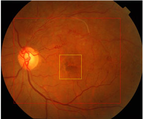
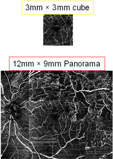
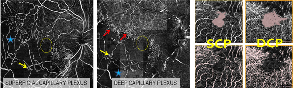
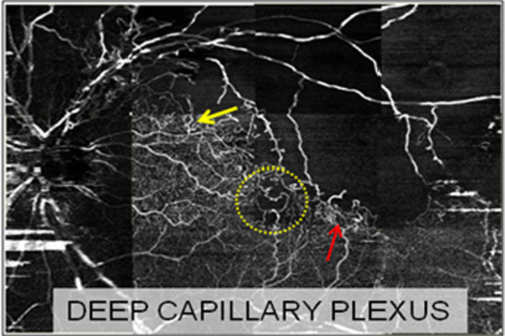
OCTA (Nidek RS 3000 advance) asessment involved scan of 3*3 and 12*9 panorama scan centered at the fovea. (fig a) assessment on OCTA involved Qalitative assessment (fig 2) like Capillary telengiactasia (yellow arrow), Capillary Non Perfusion (CNP) (blue star), Foveal Avascular Zone (FAZ) disruption (yellow dotted circle) and Venous congestion ( red arrow). Quantitative factors (fig 3) like FAZ (mm2) in Superficial and Deep Capillary Plexus and VD (mm2) in Superficial and Deep Capillary Plexus were assessed.
RESULTS:
| No of eyes | 32 |
| No of patients | 31 |
| Age, y, mean ± SD (range) | 53.4 ± 8.99 (21-68) |
| Sex, no (%) | |
| Male | 18(56) |
| Female | 14(44) |
| CFT mean ± SD (range) | 367 ± 153 (189 – 665) |
| VA mean ± SD (range) | 0.31 ± 0.23 (0 – 0.78) |
| Parameter | 32 eyes |
| SCP, no. (%) on 12 × 9 panorama (Fig d) | OCTA | FA |
| Capillary telengiactasia (yellow arrow) | 18 (54) | 20 (63) |
| CNP (red star) | 30 (93) | 26 (81) |
| Disrupted FAZ (dotted circle) | 16(50) | 14 (44) |
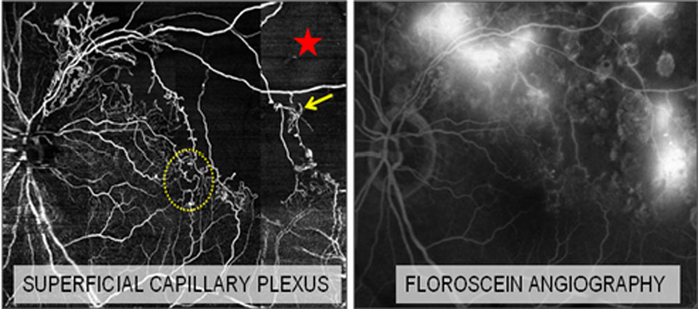
| Parameter (fig e) |
32 eyes |
||
| DCP, no. (%) on 12 × 9 panorama | OCTA | ||
| Capillary telengiactasia (yellow arrow) | 22(69) | ||
| Disrupted FAZ (dotted circle) | 17(53) | ||
| Venous congestion (red arrow) | 24(75) |
| Parameter | 32 eyes |
| Quantitative FAZ area within 3 × 3 mm2 | |
| SCP, mean ± SD (range) | 0.65 ± 0.54 (0.24 – 2.41) |
| DCP, mean ± SD (range) | 1.51 ± 0.61 (0.65 – 3.52) |
| Quantitative VD within 3 × 3 mm2 | |
| SCP, mean ± SD(range) | 1.39 ± 0.61 (0.87 – 2.01) |
| DCP, mean ± SD (range) | 2.11 ± 0.46 (0.85 – 2.97) |
| Linear regression | VA & DCP FAZ | P=0.0165 | SIGNIFICANT |
| VA & DCP VD | P=0.0108 | SIGNIFICANT | |
| VA & SCP FAZ | P=1.180 | NOT SIGNIFICANT | |
| VA & SCP VD | P=0.834 | NOT SIGNIFICANT | |
| VA & CFT | P=0.2445 | NOT SIGNIFICANT |
DISCUSSION:
Recent OCTA analysis has identified the significance of microvascular changes in the DCP in several retinal vascular diseases, including BRVO [7, 8] based on the depth-resolved imaging technique. Our results indicate that microvascular abnormalities, including capillary telangiectasia, microaneurysm, and disruption of the FAZ, are more common in the DCP than in the SCP, as previously reported. [9]
Because of the limited depth resolution, the DCP has not been evaluated previously in FA. The ability of OCTA to visualize the SCP and DCP separately and to quantify the area of perfusion offers additional information over FA, that is, that vascular perfusion of not only the SCP as detected by FA, but also the DCP, is important for VA.
The capillaries in the DCP seem to be important for nutritional and oxygen support of the synaptic connections responsible for transmission of visual signals. Because this area is located in the so-called watershed zone where the oxygen level is significantly lower than that in the inner and outer retinal layer and may be particularly vulnerable to ischemia, [10] hypoperfusion in the DCP may cause acute nutritional deficiency in the synaptic connections, resulting in decreased VA as shown in the present study. In our series, retinal hypoperfusion in the DCP was associated with retinal thinning and disruption of the photoreceptor layer. (fig f)
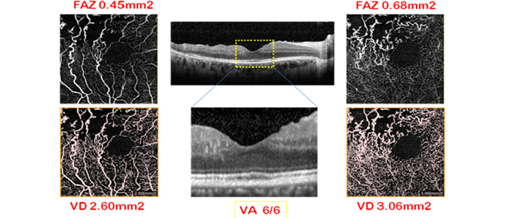
Fig f: retinal hypoperfusion in the DCP was associated with retinal thinning and disruption of the photoreceptor layer resulting in decreased VA
CONCLUSION:
Vascular perfusion in the DCP detected by OCTA is a key parameter responsible for VA in patients with BRVO. Significant association between vascular perfusion in the DCP and photoreceptor integrity, which has been reported to be associated with visual outcome OCTA is a potential clinical tool for BRVO, providing vascular details that have not been previously observed by standard fluorescein angiography.
REFERENCES:
- Klein R, Klein BE, Moss SE, Meuer SM. The epidemiology of retinal vein occlusion: the eaver dam eye study. Trans Am Ophthalmol Soc 2000;98:133–141.
- Rehak J, Rehak M. Branch retinal vein occlusion: pathogenesis, visual prognosis, and treatment modalities. Curr Eye Res 2008;33:111–131.
- Hayreh SS, Zimmerman MB, Podhajsky P. Incidence of various types of retinal vein occlusion and their recurrence and demographic characteristics. Am J Ophthalmol 1994;117: 429–441.
- Rogers SL, McIntosh RL, Lim L, et al. Natural history of branch retinal vein occlusion: an evidence-based systematic review. Ophthalmology 2010;117:1094–1101.
- McIntosh RL, Mohamed Q, Saw SM, Wong TY. Interventions for branch retinal vein occlusion: an evidence-based systematic review. Ophthalmology. 2007;114:835–854.
- Spaide RF, Klancnik JM Jr, Cooney MJ. Retinal vascular layers imaged by fluorescein angiography and optical coherence tomography angiography. JAMA Ophthalmol. 2015;133:45–50.
- Coscas F, Glacet-Bernard A, Miere A, et al. Optical coherence tomography angiography in retinal vein occlusion: evaluation of superficial and deep capillary plexa. Am J Ophthalmol. 2016;161:160–171.e2.
- Rispoli M, Savastano MC, Lumbroso B. Capillary network anomalies in branch retinal vein occlusion on optical coherence tomography angiography. Retina. 2015;35:2332– 2338
- Adhi M, Filho MA, Louzada RN, et al. Retinal capillary network and foveal avascular zone in eyes with vein occlusion and fellow eyes analyzed with optical coherence tomography angiography. Invest Ophthalmol Vis Sci. 2016;57:486–494.
- Wakabayashi T, Sato T, Hara-Ueno C, et al. Retinal microvasculature and visual acuity in eyes with branch retinal vein occlusion: imaging analysis by optical coherence tomography angiography. Invest Ophthalmol Vis Sci. 2017;58:2087–2094. DOI: 10.1167/iovs.16-21208
Leave a Comment