

Dr. ISHANI KAWATRA, Dr. Anuja Janakiraman, Prof. Manohar Babu Balasundaram, Dr. LAKSHEY DUDEJA
Introduction:
Argyria is generalized deposition of silver granules into skin and mucous membrane1 while ocular argyrosis is accumulation of silver in the region of eye.2Ocular argyrosis has been reported as an occupational hazard in silversmiths3, those working in mirror and battery manufacturing industries4, following silver clip disintegration after previous strabismus surgery, prolonged eye rubbing after contact with photographic developing solution3 and use of eye cosmetics5.
We are reporting clinical findings of two silversmith workers who had features of argyrosis. We have documented the systemic findings including pigmentation of the nails and skin of the limbs, serum silver levels and also noted the ocular findings including slit lamp photography, Humphrey’s visual field analysis, fundus fluorescein angiography, specular microscopy, optical coherence tomography of macula and cornea. In an attempt to prove presence of silver in the eye, when one patient was planned for cataract extraction, we sent the piece of lens matter for histopathological evaluation. This case report covers the systemic and ocular work-up of cases of argyrosis, confirming the presence of silver in both serum and ocular tissue.
Cases:
Two Indian males,case 1,56 years old and case 2, 50 years old, silversmith by occupation for more than 25 years, presented to out-patient department with complaints of gradually progressive loss of vision for distance and near (left eye more than right eye) for the last 3 months. First patientwas a known hypertensive and on treatment (tablet amlodipine 5mg) since 3 years. Second patient was a known diabetic and on treatment (tablet glimepiride) since 5 years. Both had no other systemic illness.
On examination, Case 1 had right eye uncorrected visual acuity of 6/18p improving to 6/9p with pinhole and left eye had uncorrected visual acuity of 5/60 improving to 6/60 with pinhole.Case 2 had visual acuity of 6/6 in right eye anduncorrected visual acuity of 6/18 improving to 6/9 with pinhole in left eye. Lid and adnexa of both patients were normal. In first patient, both eyes conjunctiva had grey-black pigmentation of medial bulbar conjunctiva, more prominent at the medial canthal region
(Figure 1).In both patients, cornea had diffuse confluent deposits at the level of descemet’s membrane (Figure 2) which was confirmed by anterior segment optical coherence tomography
(Figure 3). Lens showed cataractous changes in both the patients, case 1 hadnuclear sclerosis grade 2 with posterior sub capsular cataract in right eye and near mature cataract in left eye. Case 2 had posterior subcapsular cataract in both the eyes (left eye more than right eye). Intraocular pressure in both patients was normal. Fundus evaluation in first patientwas normal in right eye while details in left eye werenot clear due to dense cataract. B scan ultrasound in left eye was normal. Fundus evaluation in the second patient was normal in both the eyes.
Specular microscopy of both patients was within normal limits. On systemic evaluation, upper and lower limbs of both patientsshowed slate grey pigmentation along with pigmentation of nails (Figure 4). Pigmentation was more prominent in the first case. Both were advised to strictly use protective measures like gloves, mask to prevent systemic absorption of the metal at work place. Case 1 was advised to undergo left eye cataract extraction with posterior chamber intraocular lens implantation. Patient underwent small incision cataract extraction with PCIOL implantation,
following which best corrected visual acuity improved to 6/6. Lens matter was collected during cataract extraction and sent for histopathological evaluation. It showed evidence of diffuse deposits of silver which were confirmed using H & E (hematoxylin and eosin) and reticulin stain and were refractable in dark field illumination (Figure 5). Post cataract extraction, patient underwent gonioscopy, Humphrey’s 30-2 visual field analysis, macular OCT and FFA, which were within normal limits. All the ocular investigations in case 2 were also within normal limits.
Discussion:
Argyrosis is a condition caused by chronic exposure to silver. Silver is widely used in photography, metallurgy, medicine, water disinfection, making of jewellery, coins and mirrors.6Accumulation in eye and in the adjacent tissues can result from chronic exposure to silver compounds through ingestion, inhalation or skin contact. It may also occur due to local absorption following the use of silver containing eyedrops, certain eyelash and eyebrow dyes.7Outcomes of chronic silver exposure depend not only on the dose, duration and form of exposure but also on the individual characteristics like age, sex, nutritional status and general health.8In the cases presented,
patients had chronic exposure to silver through inhalation of fumes and skin contact while handling silver solutions with bare hands at workplace for more than 25 years.Systemic presence of silver was evident in the form of pigmentation of limbs and nails but this was not confirmed with tissue biopsy. Argyrosis can be diagnosed based on blood and urine tests.9Serum silver levels were raised in both the cases, case 1 had 2.3micrograms/ml and case 2 had 2.9micrograms/ml (normal <2 micrograms/ml).
This can be attributed to chronic exposure to silver as they did not use gloves and face shields because they found these deterrant to skills and fine dexterity. Regarding ocular involvement, silver salts deposition can cause discoloration around the eyes. Silver deposits can be noted in the elastic fibres of the connective tissue and basement membranes including the eyelids, conjunctiva, lacrimal sac, lens, ciliary body and Bruch’s membrane.
In cornea deposits have been noted within Bowman’s membrane, corneal stroma and Descemet’s membrane, causing its discoloration.10,11Slate grey pigmentation of corneal membrane was reported in a 50 year old silversmith.3 Prominent brown-black colored follicles like masses have also been noted in conjunctival fornices. Routine light microscopic examination identified abundant granular deposits which were later found to be silver particles deposited around the basement membrane of vascular and adnexal structures.
Small particles of silver can erode and deposit sub-epithelially causing discoloration.12Grey black discoloration of periorbital skin, lid margins and bulbar conjunctiva has also been reported. Our case 1 had conjunctival pigmentation which we presume may be due to silver deposits but it was not confirmed. Deposits were noted in our cases in deeper layers of cornea, Descemet’s membrane and this was confirmed by demonstrating the presence of hyper-reflective shadows in AS-OCT images. Argyrosis has also been associated with open angle glaucoma.3In our cases, patients had normal intra-ocular pressures, normal gonioscopy findings, normal Humphrey’s 30-2 visual fields and a non-glaucomatous disc.
Case reports due to ingestion of daily colloidal silver has revealed bilateral confluent perimaculardrusenoid deposits, localized at the level of or anterior to inner segment ellipsoid band and marked choroidal thinning. Fundus fluorescein angiography revealed a dark or silent choroid.13The cases presented had normal retina, normal fundus fluorescein angiography and a normal macular OCT. Electrophysiological tests have shown abnormal function of photoreceptors in the central and peripheral retina, with normal functioning of retinal pigment epithelium.8
We wanted to prove presence of silver in ocular tissue which has never been done in literature before. In an attempt to do this, when case1 underwent surgery for cataract extraction (MSICS) with posterior chamber intraocular lens implantation, we sent the lens material for histopathological evaluation. The presence of silver in the lens matter was confirmed by staining the tissue material with hematoxylin and eosin stain. Silver deposits were seen as refractable bodies in dark field microscopy. The presence of silver in ocular tissue with raised serum silver levels in patient 1 with history of chronic exposure to silver confirms the diagnosis of argyrosis in the patient.
Conclusion:
These cases stresses on the use of personal protective equipments to minimize occupational hazards. Presence of silver was confirmed in serum in both the patients and lens matter in one patient. To the best of our knowledge, the histopathological presence of silver has never been reported in lens matter inliterature before. Presence of silver in a cataractous lens can be debated as silver deposits in senile cataract or cataract induced by silver deposits.
References:
- Stafeeva K, Erlanger M, Velez-Montoya R, Olson JL. Ocular argyrosis secondary to long-term ingestion of silver nitrate salts. Clinical ophthalmology (Auckland, NZ). 2012;6:2033.
- Calvery ho, lightbodyhd, rones b. Effects of some silver salts on the eye(silver nitrate, silver ammonium nitrate, silver ammonium sulfate, silver ammonium lactate and a mixture of silver ammonium nitrate and silver ammonium sulfate). Arch ophthalmol. 1941;25(5):839–847. Doi:10.1001/archopht.1941.00870110091010.
- Kamath Y, Sinha A. Ocular argyrosis in a jeweller. BMJ case reports. 2013;2013.
- Pamela l. Drake, kyle j. Hazelwood; exposure-related health effects of silver and silver compounds: a review, the annals of occupational hygiene, volume 49, issue 7, 1 october 2005, pages 575–585, https://doi.org/10.1093/annhyg/mei019.
- Mark j.gallardomdaj. Bradleyrandlemanmdbkristina m.pricemdbdaniel a.johnsonmdasharonacostamdachans e.grossniklausmdbr. Doylestultingmd, phdb. Ocular argyrosis after long-term self-application of eyelash tint. American journal of ophthalmology. Volume 141, issue 1, january 2006, pages 198-200
- Pala G, Fronterré A, Scafa F, Scelsi M, Ceccuzzi R, Gentile E, Candura SM. Ocular argyrosis in a silver craftsman. Journal of occupational health. 2008;50(6):521-4.
- Gallardo MJ, Randleman JB, Price KM, Johnson DA, Acosta S, Grossniklaus HE, Stulting RD. Ocular argyrosis after long-term self-application of eyelash tint. American journal of ophthalmology. 2006 Jan 31;141(1):198-200.
- Sarnat-Kucharczyk M, Pojda-Wilczek D, Mrukwa-Kominek E. Diagnostic methods in ocular argyrosis: case report. DocumentaOphthalmologica. 2016 Oct 1;133(2):129-38.
- Wadhera A, Fung M. Systemic argyria associated with ingestion of colloidal silver. Dermatology Online Journal. 2005 Jan 1;11(1).
- Rungby J. Experimental argyrosis: ultrastructural localization of silver in rat eye. Experimental and molecular pathology. 1986 Aug 1;45(1):22-30.
- Flögel W, Widmeier S, Hotz P, Schärer L, Barthelmes D, Landau K, Thiel MA. Corneale und conjunctivaleBefundebeisystemischerArgyrose. KlinischeMonatsblätterfürAugenheilkunde. 2006 May;223(05):390-2.
- Tendler I, Pulitzer MP, Roggli V, Abramson DH, Marr BP. Ocular Argyrosis Mimicking Conjunctival Melanoma. Cornea. 2017 Jun 1;36(6):747-8.
- Rahimy E, Beardsley R, Ferrucci S, Ilsen P, Sarraf D. Optical Coherence Tomography Findings in Ocular Argyrosis. Ophthalmic Surgery, Lasers and Imaging Retina. 2013 Nov 25.
Figures:
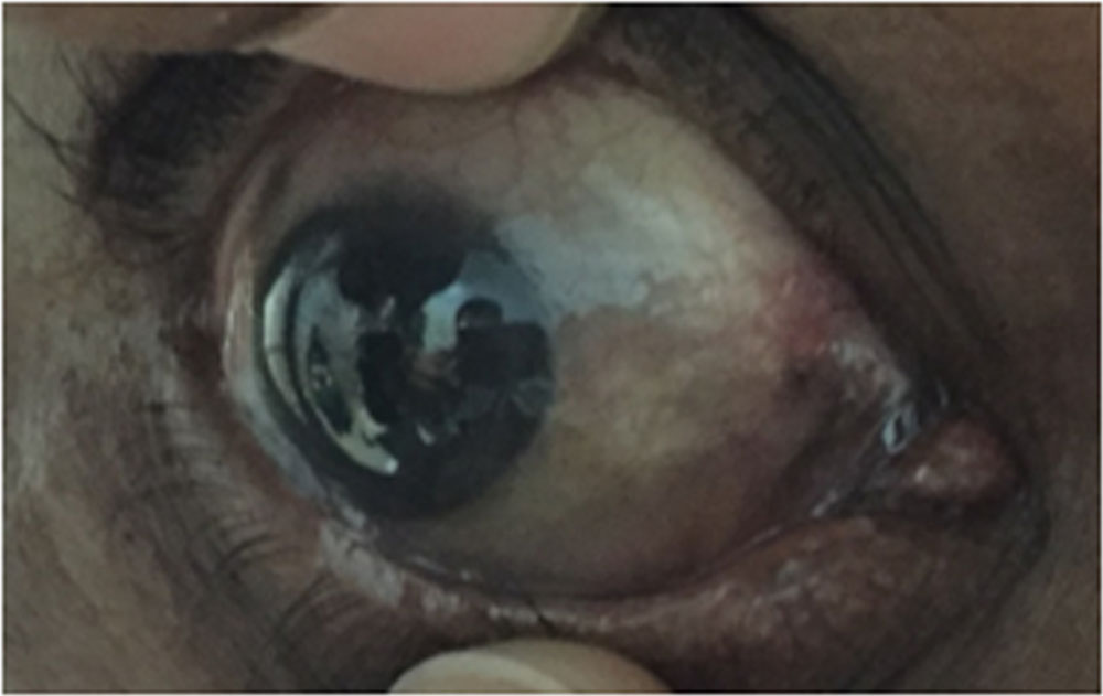
Figure 1: Slit lamp evaluation photograph showing pigmentation of bulbar conjunctiva and medial canthus in case 1
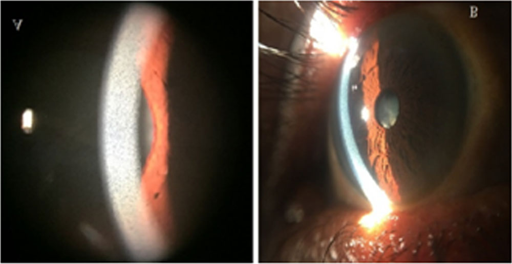
Figure 2: Slit lamp photograph (A) and anterior segment photograph (B) showing diffuse deposits in the deeper layer of cornea in case 1 and case 2
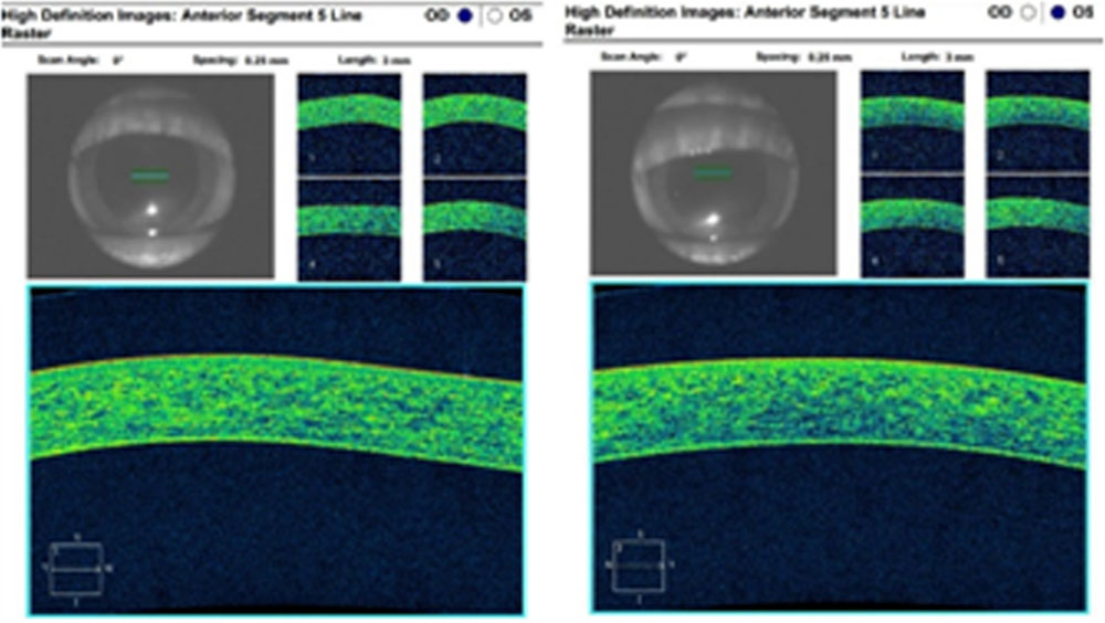
Figure 3: AS-OCT image of cornea (right eye and left eye) showing hyper-reflective shadows in the Descemet’s layer, suggestive of deposits in case 2
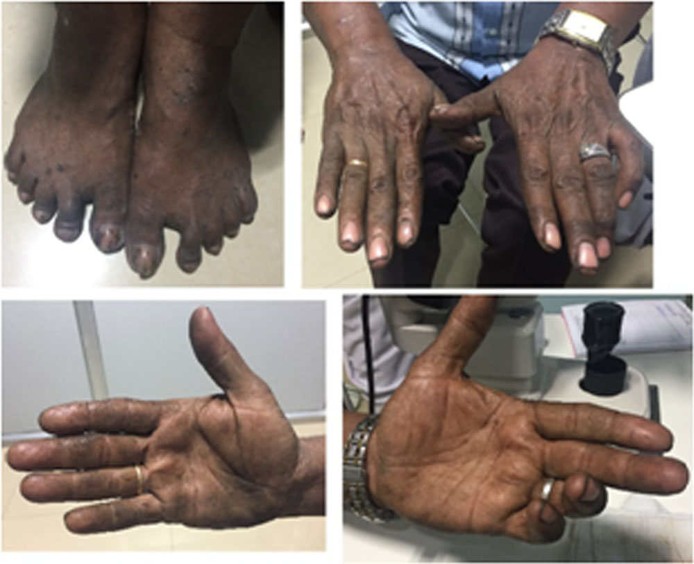
Figure 4: Picture showing pigmentation of limbs which is presumed to be due to deposition of silver in case 1.
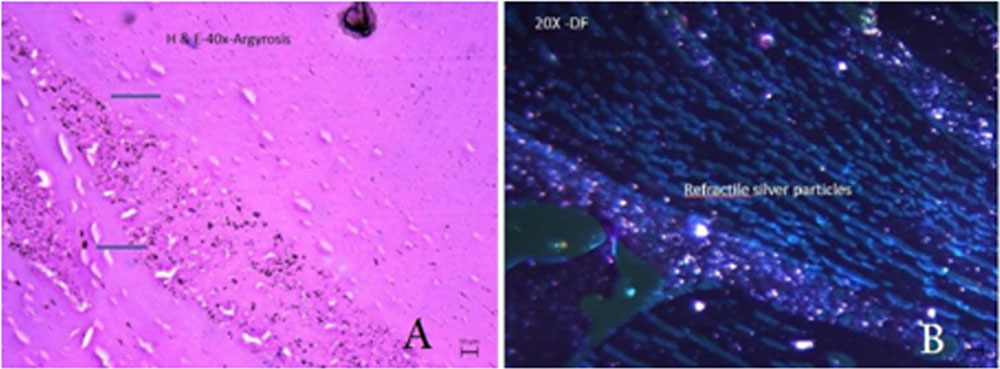
Figure 5: H and E (Hematoxylin and eosin)sections studied from the lens nucleus tissue of case 1 (A) shows diffuse deposition of black granules suggestive of silver particles which are refractable in dark field illumination (B).
Leave a Comment