

Mrs.RAFFAT ANJUM, Dr.Raina Usha Kaul,Prof.Goyal J L,Dr.PRATEEKSHA SHARMA
Purpose: To assess the visual and surgical outcome of anteriorcontinuous curvilinear capsulorhexis (ACCC) and posteriorcontinuous curvilinear capsulorhexis (PCCC)capsulorhexis by 25gauge(G) vitrectorhexis in pediatric cataract surgery with in the bag IOL implantation
Methods:A total of 15 eyes of children of age group 3 to 8 years with zonular cataract were selected for ACCC and PCCC by 25G vitrectomy cutter (vitrectorhexis) with IOL implantation. At 3 months postoperatively, best corrected visual acuity (BCVA) and visual axis opacification (VAO) were evaluated. Slitlamp photos of operated eyes were taken in retroillumination, the size and decentration of ACCC and PCCC were measuredfrom the center of IOL were measured with the help of python imaging library.
Results:At 3 months postoperatively, the mean BCVA was 0.56±0.07 ( visual acuity in decimals), mean average diameter (average of horizontal and vertical diameters) of ACCC diameter was 4.89±0.41milimeters (mm) and the mean average diameter (average of horizontal and vertical diameters) of PCCCwas 3.28±0.45 (mm). VAO was 0.00%.
Conclusion:25G vitrectorhexis gives adequate visual outcome with adequate sized ACCC and PCCC, with no extension of rhexis and no significant VAO.
Key words: 25 gauge vitrectomy cutter, anterior continuous curvilinear capsulorhexis (ACCC),posterior continuous curvilinear capsulorhexis (PCCC),visual axis opacification (VAO).
Paediatric cataract surgery consists of anterior continuous curvilinear capsulorhexis (ACCC), lens matter aspiration, posterior continuous curvilinear capsulorhexis (PCCC), limited anterior vitrectomy (LAV) and intraocular lens (IOL) implantation. Primary PCCC and anterior vitrectomy are preferred in younger children to avoid problems of posterior capsule opacification.
ACCC and PCCC are the most critical steps during paediatric cataract surgery. Various methods exist for the performance of these steps depending on the surgical situation as well as the surgeon’s preference and expertise. Two of the most commonly used methods are rhexis by manual forceps and by vitrectomy cutter. After nearly three decades of dominance of 20 gauge vitrectomy systems for vitrectorhexis, the 25 gauge vitrectomy systems have recently been introduced.1 The potential advantages of 25-gauge instruments are a smaller entry wound which allows the wound to be sutureless, easier manipulation in the narrow palpebral fissures and small eyes of children, lesser postoperative inflammation and shorter learning curve for anterior segement surgeons.1 To the best of our knowledge there is limited literature inpediatric cataract surgery with in the bag IOL implantation using 25-gauge vitrectomy system. This study aimed to assess the visual and surgical outcomes and complications of 25-gauge vitrectorhexis in pediatric cataract surgery with IOL implantation.
METHODS:
Study design:
This was a prospective interventionalconducted in the department of ophthalmology, in a tertiary hospital. The study was studied and reviewed by the institute’s ethical committee, and an informed written consent was taken from the parents of all the patients. The study included 15 eyes of children between the age group of 3-8 years with zonular cataractfor ACCC and PCCC by 25 gauge vitrectomy cutter (vitrectorhexis) with in the bag intraocular lens (IOL) implantation.
The exclusion criteria included presence of corneal opacity, active or chronic inflammation, uveitis, any lens subluxation, secondary glaucoma, any gross ocular pathology(like microphthalmos, aniridia, coloboma), presence of PHPV, retinal detachment and presence of any significant systemic diseases which made the child unfit for surgery.
Preoperatively, a complete ophthalmologic examination was done which included a detailed history regarding previous ocular and systemic complaints, uncorrected distance visual acuity (UDVA), best corrected visual acuity (BCVA), refraction under cycloplegia, intraocular pressure, slit lamp examination, keratometry, axial length, biometry and posterior segement examination.
Surgical technique
25 gauge vitrectomy system was used trans-corneally to perform anterior continuous curvilinear capsulorhexis (ACCC), lens matter aspiration, posterior continuous curvilinear capsulorhexis(PCCC) and anterior vitrectomy.The horizontal and vertical diameters of ACCC and PCCC were measured with the CRF with the rhexis ruler (MST DFH-0002) respectively. The MST CRF has calibrations of 1millimeter interval at its tip, so was easily used before and after rhexis for measurements. A single piece acrylic hydrophobic IOL (Acrysof SN60AT) was implanted in the bag in all the cases. All the steps including ACCC, lens aspiration, PCCC and anterior vitrectomy were done at the settings of 900 cuts per minute and aspiration of 300 mm Hg. The entry wound at the superior cornea was sutured with 10-0 vicryl suture.
Postoperative follow up was conducted at 1 day, 1 week, 1 month and 3 months post surgery.
Postoperatively, at 3 months visit, the implanted IOL was imaged on the slit lamp in retroillumination. These images were then analysed to measure the diameters of ACCC, PCCC and IOL with the help of Python imaging library (PIL). It is a free library for the Python programming language that aids opening, analysing and saving many different image file formats. With the help of PIL, the most approximate circles were drawn manually over the contours of ACCC, PCCC and IOL on the slit lamp image, and the diameters of these three circles were measured. These diameter values measured by PIL were in pixels. Knowing the diameter of IOL as 6.00mm (diameter of acrysof SN60AT), the pixel values were calculated in mm (millimeters) (figure no.) thus giving the measurement of ACCC and PCCC diameters in milimeters (Figure-19,20,21 and 22). Similarly, with the help of PIL, the center of IOL, ACCC and PCCC were located, and their corresponding distances from the IOL center were measured, thus giving the amount of decentration of ACCC and PCCC from the center of the IOL (Table no .15 and 17). Categorical variables were presented in number and percentage (%) and continuous variables were presented as mean ± standard deviation (SD) and median. Normality of data was tested by Kolmogorov-Smirnov test. If the normality was rejected then non parametric test was used.The outcome parameters were visual acuity BCVA, size of ACCC and PCCC, extension of rhexis and visual axis opacification.
Results:
The study included 15 eyes of pediatric patients between the age group of 3-8 years. There were no cases of corneal edema, unstable anterior chamber, wound leakage, irregular pupil or postoperative complications.At 3 months postoperatively, the mean BCVAwas 0.56 ± 0.07.
Intraoperatively, the mean average diameter (average of horizontal and vertical diameters) of ACCC was 4.98 ±0.41 (mm). The mean average diameter (average of horizontal and vertical diameters) of PCCC was 3.17 ± 0.47 (mm). At 3 months postoperatively, the mean averagediameter of ACCC was 4.89 ±0.41 (mm) and the mean average diameter of PCCC was 3.28 ± 0.45 (mm).Table 1 the mean average diameters of ACCC and PCCC intraoperatively and at 3 months postoperatively.
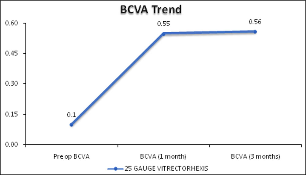
FIGURE 1 BCVA PREOPERATIVE, AT 1 MONTH POST-OP AND 3 MOTHS POST-OP (BCVA IN DECIMALS)
TABLE-1: MEAN AVERAGE ACCC AND PCCC DIAMETERS
| INTRAOPERATIVELY | 3 MONTHS POSTOPERATIVELY | |
| MEAN AVERAGE ACCC diameter
(in mm) |
4.98 ±0.41 | 4.89 ±0.41 |
| MEAN AVERAGE PCCC diameter(in mm) | 3.17 ± 0.47 | 3.28 ± 0.45
|
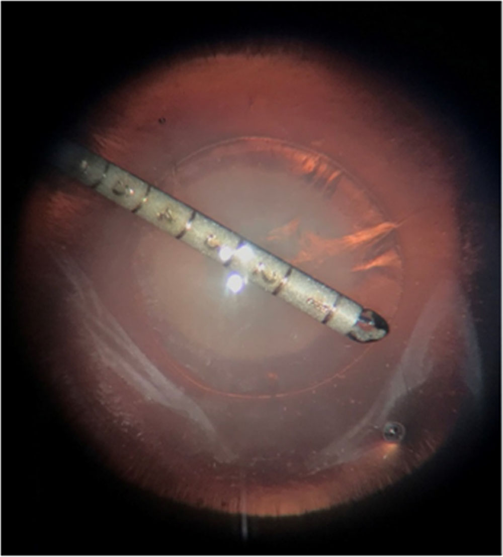
Figure 2 INTRAOPERATIVE MEASUREMENT OF ACCC
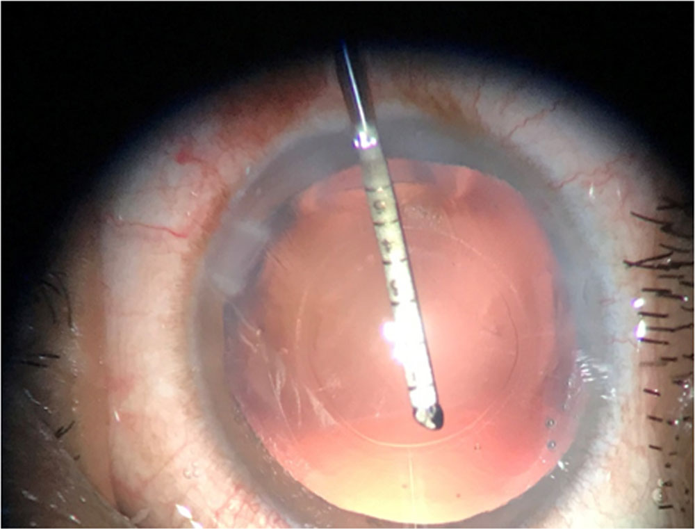
Figure3:INTRAOPERATIVE MEASUREMENT OF PCCC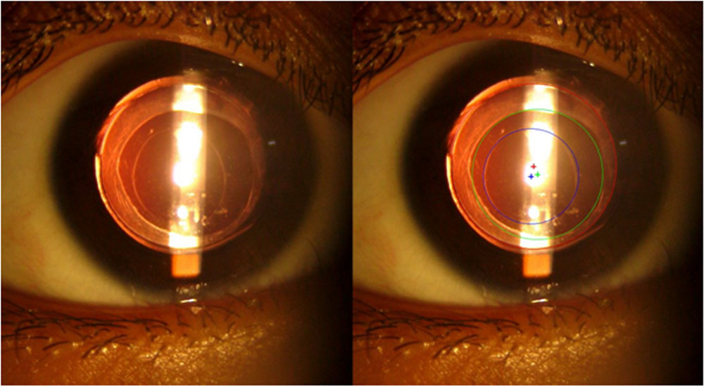
Figure4: POSTOPERATIVE MEASUREMENT OF ACCC AND PCCC IN THE MANUAL CAPSULORHEXIS GROUP ( DECENTRATION SHOWN BY THEIR RESPECTIVE CENTERS). Red-IOL, Green-ACCC, Blue-PCCC
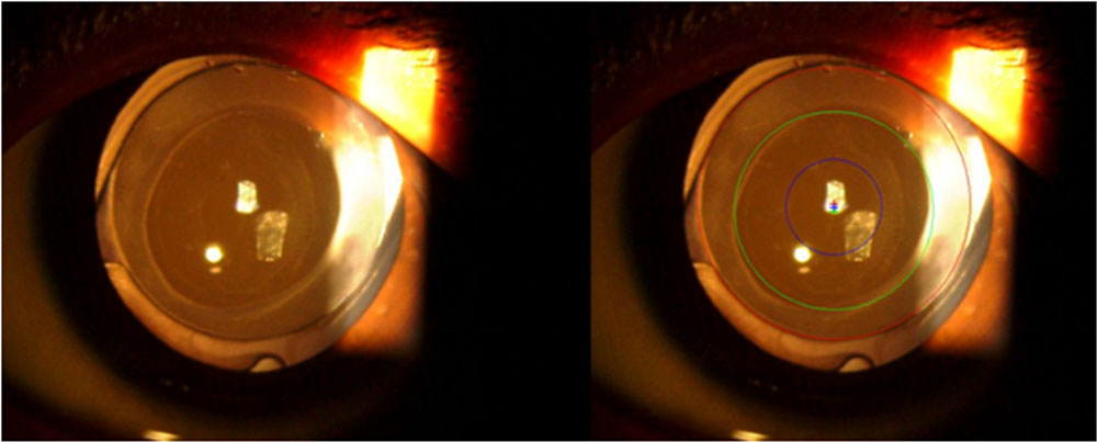
Figure5: POSTOPERATIVE MEASUREMENT OF ACCC AND PCCC IN THE 25G VITRECTORHEXIS GROUP (DECENTRATION SHOWN BY THEIR RESPECTIVE CENTERS). Red-IOL, Green-ACCC, Blue-PCCC.
DISCUSSION:
Surgical management of paediatric cataracts is a challenging task because of the elastic lens capsule, positive vitreous pressure, elevated inflammatory reaction due to irritation of the iris, and a higher rate of complications requiring resurgery.2,3,4 In children there is also a higher rate of posterior capsular opacification (PCO) as compared to adults. This visual axis opacification (VAO) is even more important in younger children as it may lead to amblyopia. Management of posterior capsule has been a challenging aspect of paediatric cataract surgery, especially when IOL implantation is involved.5The techniques introduced to prevent postoperative reopacification of the visual axis include primary posterior capsulorhexis with or without anterior vitrectomy, optic capture through posterior continuous curvilinear capsulorhexis (PCCC), and pars plana posterior capsulotomy combined with anterior vitrectomy.6
ACCC and PCCC are critical steps during paediatric cataract surgery. Two of the most common methods for ACCC and PCCC include using manual forceps and vitrectomy cutter. Manual capsulorhexis has been the gold standard in paediatric cataract surgery. It allows surgeons to perform capsulorhexis of a desirable size and integrity with greater ease and control.6 It offers a smooth regular edge with greater resistance to tearing and unwanted extension to the periphery. But manual capsulorhexis has disadvantages of a longer surgery time and difficulty in achievement of the appropriate size and shape of the rhexis with proper centration.6 The pediatric capsule is highly elastic, making it difficult to perform capsulorhexis with a bent needle or forceps specially in younger children.7
20-gauge vitrectomy instruments have been used most commonly but they necessitate for sclera incision suturing. Larger instruments also carry higher risk for ciliochoroidal detachment, vitreous incarceration and postoperative inflammatory response in children.6On the other hand, 25 G vitrectomy systems offer smaller wound size, rapid wound healing, preservation of conjunctiva, no need for sutures, ease of manipulation, intra-operative anterior chamber stability, no intra-operative corneal decompensation, and minimal post-operative inflammation, making these systems a better alternative.725 gauge vitrectomy system is being used for the management of paediatric cataract by pars plana and transcorneal routes.7,8
Anterior capsular fibrosis, posterior capsular fibrosis, visual axis opacification (VAO) and capsule contraction are common capsular complications that result from the high proliferative capacity of the lens epithelium and severe postoperative inflammation in pediatric cataract surgery. The efficacy of the CCC depends on its centration, location and size.The size of rhexis is very important so as to prevent PCO, as proposed in a study by Lin et al.7 It has been reported that the optimum CCC should be small enough to overlap 360 degrees of the IOL optic periphery to reduce posterior capsular opacification (PCO)9and an accurate CCC helps accomplish such 360 degree overlap10and this overlap may be more important than IOL design.11 A CCC that is bigger than the IOL optic has been associated with greater PCO and worse visual acuity than smaller capsulorhexis.12 In contrast, when CCC is too small, anterior capsule phimosis,13decreased vision, decreased visualization of the retina and decreased effectiveness in the properties of aspheric IOLs can occur. A study by Neuhann T. et al on adult cataract surgery proposed that a properly shaped and sized capsulorhexis enhances hydrodissection, cortical clean up, IOL fixation and centration
while decreasing the risk for PCO.14An ACCC diameter of 4.0-5.0 mm yielded optimal capsular outcomes, based on its relatively moderate contraction of anterior capsular opening, moderate enlargement of posterior capsular opening and lower percentage of posterior capsular opening opacification and visual axis opacification.7 For age-related cataracts, many studies have clarified that the optimal anterior capsulorhexis diameter is slightly smaller than the diameter of the simultaneously implanted IOL optic surface, with 0.50 mm-1.00 mm capsulorhexis edges covering the IOL optic surface.12 As opposed to the anterior capsular opening, for the initial posterior capsular opening, a 3.00 mm diameter capsulorhexis was well accepted and had a tendency to widen. In our study we measured the size of ACCC and PCCC intraoperatively and 3 months postperatively.
Kochagaway et al 15 reported extension of PCCC in 9.09% cases in vitrectorhexis group.In thatstudy PCCC was done after IOL implantation. In a retrospective study by Wilson ME et al16reported anterior capsular rupture in 5.3% cases in vitrectorhexis group. In our study, we implanted IOL after PCCC to see the strength of PCCC and found no extension of rhexis on IOL implantation.Wilson ME et al16 reported that vitrectorhexis for ACCC can be safely used upto 6 years of age. in our study, we performed both ACCC and PCCC with the vitrectomy cutter. we found that vitrectorhexis can be used safely for both ACCC and PCCC in paediatric cataract surgery in upto 8 years of children.
Hazirolan et al17did a study comparing manual capsulorhexis (ACCC and PCCC) with that of 23 gauge suturelessvitrectorhexis (ACCC, lens aspiration and PCCC) in pediatric cataract surgery. There was extension of PCCC in 8.33% cases in manual PCCC group and 10.4% extension in vitrectorhexis PCCC group and concluded that both manual capsulorhexis as well as vitrectorhexis to be comparable in terms of safety and efficacy for the achievement of anterior and posterior capsulorhexis.This study proved that in paediatric cataract surgery, sutureless23 gauge ACCC, lens extraction, PCCC and anterior vitrectomy by 23 gauge vitrectomy cutter is more reproducible, more predictable, faster and has a shorter learning curve. In our study, we found that 25 G ACCC and PCCC was fast, convenient, and safe as there was no extension of rthexis.
The incidence of postoperative visual axis opacification complication was reported as 23.5% in the Manual PCCC group (Hazirolan et al., 2004)17 while the incidence of PCO was 5 % in the Vitrectorhexis group (Xin Liu et al., 2016). In our study, no visual axis opacification was noted in any of the patient.
In our study, we found that the size of ACCC in 25 gauge vitrectorhexis group contracted over a period of 3 months postoperatively. The intraoperative mean average diameter of ACCC ( average of horizontal and vertical diameters) was 4.98 ± 0.41 (mm) and that at the end of 3 months was 4.89 ± 0.41 (mm).
Our study was conducted on a relatively small sample size of 15 eyes, with a short follow up of 3 months. We believe that a larger sample and a longer follow up will be more representative of changes in ACCC and PCCC sizes over time.
Conclusion:
The use of the 25 gauge vitrectomy system for the management of paediatric cataracts is both safe and effective with several advantages including the ability to maintain a stable and deep anterior chamber intra-operatively in spite of the high intra-vitreal pressure in children, no intra-operative corneal decompensation or iris prolapse. It allowed easier manipulations in paediatric eyes due to the smaller instrument size and higher instrument flexibility, minimal post-operative inflammatory response, preservation of the conjunctiva for future surgeries like trabeculectomy which may be required in patients operated for congenital cataracts who develop glaucoma. 25 gauge vitrectomy system allowed achievement of adequately sized ACCC and PCCC with convenience and without any extension of rhexis or VAO.
Financial support:
Nil
Conflicts of interest:
Nil
REFERENCES:
1.Wilson ME, Pandey SK, Thakur J. Paediatric cataract blindness in the developing world: surgical techniques and intraocular lenses in the new millennium. Br J Ophthalmol. 2003;87: 14-9
2.Wilson ME, Petersiem MW, Englert JA, Goldberg RL, Rosenbaum AL. Pseudophakia and polypseudophakia in the first year of life. J AAPOS 1999;5:238-45
3.Zetterstrom C, Lundvall A, Kugelberg M. Cataracts in children. J Cataract Refract Surg. 2005;31:824-40.
4.Lambert SR, Buckley EG, Piager DA, Medow NB, Wilson ME. Unilateral intraocular lens implantation during the first 6 months of life. J AAPOS. 1999;3:344-9.
5.Wilson ME. Management of aphakia in childhood. AAO Focal Points. Clinical Module for Ophthalmologists. Module Ophthalmol. 1999;17:1-15.
6.Huang Y. Xie L. Short-term outcomes of dry pars plana posterior capsulotomy and anterior vitrectomy in paediatric cataract surgery using 25-gauge instruments. Br J Ophthalmol. 2010; 94: 1024-7.
7.Lin H et al. Capsular Outcomes Differ with Capsulorhexis Sizes after Pediatric Cataract Surgery: A Randomized Controlled Trial. Sci. Rep. 2015; 5:16227.
8.Chee KYH, Lam GC. Management of congenital cataract in children younger than 1 year using a 25-gauge vitrectomy system. J Cataract Refract Surg. 2005;8:720-4.
9.Ravalico G, Tognetto D, Palomba M, Buratto P, Baccara F. Capsulorhexis size and posterior capsule opacification. J Cataract Refract Surg 1996; 22:98-103.
10.Dick, H.B, Pena-Aceves, A, Manns,M. and Krummenauer, F. New technology for sizing the continuous curvilinear capsulorhexis: prospective trial. J Cataract Refract Surg 34, 1136-44, 10.1016/j.jcrs.2008.03.025 (2008).
11.Smith SR, Daynes T, Hinckley M, Wallin TR, Olson RJ. The effect of lens edge design versus anterior capsule overlap on posterior capsule opacification. Am J Ophthalmol 2004; 138:521-6.
12.Hollick EJ, Spalton DJ, Meacock WR. The effect of capsulorhexis size on posterior capsule opacification: one-year results of a randomized prospective trial. Am J Ophthalmol 1999; 128:271-9.
13.Davison JA. Capsule contraction syndrome. J Cataract Refract Surg1993;19:582-9
14.Neuhann T. Theorie and Operationstechnik der Kapsulorhexis( Theory and surgical technic of capsulorhexis). KlinMonatsblAugenheilkd 1987; 190:542-5.
15.Kochgaway L, Biswas P, Paul A, Sinha S, Biswas R, MaityPuspen et al. Vitrectorhexis versus forceps posterior capsulorhexis in paediatric cataract surgery. Indian J Ophthalmol. 2013 Jul;61(7):361-64.
16.Wilson ME, Trivedi RH, Bartholomew LR, Pershing S. Comparison of anterior vitrectorhexis and CCC in pediatric cataract and IOL implantation surgery. A10-year analysis. J AAPOS. 2007; 11:443-6.
17.Lin A.A. and Buckley E.G. Update on pediatric cataract surgery and intraocular lens implantation. CurrOpinOphthalmol 2010; 21, 55-59, 10.
Leave a Comment