

Dr.Sowmya R, S11824
Abstract
Adie’s pupil , also called as Adie’s tonic pupil is the common cause of anisocoria or unequal pupils in clinics. It is used to denote the parasympathetic denervation of pupil with absent light responses and retained near reflex. We describe a case series of patients diagnosed as Adie’s pupil.
We retrospectively reviewed the case records of patients diagnosed with Adie’s pupil between january 2016 to december 2017 . Diagnosis was made based on light near dissociation of pupils, vermiform movements of iris, supersensitivity to diluted pilocarpine. Routine blood investigations , VDRL was ordered in all patients.
Out of the forty patients included, there were 23 males and 17 females .
It was bilateral in 6 and unilateral in 34 cases. Most cases were between 20-50 years. There were 3 cases < 20 years and 4 cases >50 years. Hypothyroidism and diabetes was noted in 2 cases each. Interestingly ESR was raised in 9 cases. Our series points towards common occurrence of Adie’s pupil and needs to be looked for during examination. Though the etiology seems not clear in our review but gives a clue to investigate further in view of raised ESR suggesting inflammation
Introduction
Tonic pupil is caused by the damage to the postganglionic parasympathetic fibres i.e., at level of ciliary ganglion or short ciliary nerves.1,2,3 Tonic pupils refers to pupil with following features: a)poor reaction to light with segmental iris palsy b)accommodative paresis which improves with time, c)with unusually strong and tonic near reaction with slow redilation after constriction to near stimuli, d)cholinergic supersensitivity . 2,4,5
Tonic pupils belong to one of three categories: local, neuropathic and Adie’s pupil 2,4,6
- Local tonic pupil is due to orbital or systemic conditions affecting ciliary ganglion or short ciliary nerves in isolation as in HZV, sarcoidosis , VKH, rheumatoid arthritis, blunt injury to globe or various intraocular surgeries .2,7
- Neuropathic tonic pupils are usually bilateral and occur as a part of widespread peripheral or systemic autonomic neuropathy. Various condition include Miller Fischer variant of gullian Barre syndrome, charcot marie tooth disease , shy Drager syndrome and many others. 1,2,8,9
- Adie’s syndrome refers to tonic pupil of idiopathic etiology . It is commonly seen in females ( around 70%) and in age group of 20-50 years . Unilateral affection is more common ( around 80% ) with 4% per year having the other pupil also involved 2,4,5
Symptoms are usually due to iridoplegia and cycloplegia like photophobia ,difficulty with dark adaptation , headache with near activities, blurred near vision.
The pathophysiology of an Adie pupil is acute denervation followed by appropriate and inappropriate reinnervation of the ciliary body and iris sphincter . Clinical features will vary depending on stage of reinnervation at time of examination. Accommodation paresis improves with reinnervation of cilary body. 2,10 Further the reinnervating fibres to ciliary body misinnervate the iris sphincter causing tonic response to near stimuli. 2,11,12
The denervated iris sphincter is supersensitive to topical parasympathomimetic solutions. Pilocarpine drops (0.125% or 1/8 th concentration) can be used to demonstrate this, as the normal pupil will constrict slightly, if at all. About 80% of patients with a tonic pupil show cholinergic denervation supersensitivity. 2,11,12
Materials and methods:
The case records of patients diagnosed with Adie ‘s pupil in a tertiary hospital between january 2016 to december 2017 were retrospectively reviewed.
Diagnosis of adie’s pupil was made based on following observations ; poor pupillary light response , tonic near response, vermiform movements of iris noted on slit lam examination , supersensitivity to diluted pilocarpine ( 0.5 ml of 2% pilocarpine dilated in 7.5ml of saline).
The following data like presenting symptoms, demographic data, results of diluted pilocarpine test, laterality of occurrence, investigation results, treatment given and follow up period were collected . Data of other systemic conditions recorded in case records were also noted .
Results :
The demographic profile, laterality , presenting symptoms, treatment data and investigation results were analysed .
Total of 40 patients were included. There were 23 males and 17 females contrary to description of being common in females.
Most cases were between 20-40 years (30/40cases) . There were 3 cases < 20 years and 4 cases >50 years.( Table 1 )
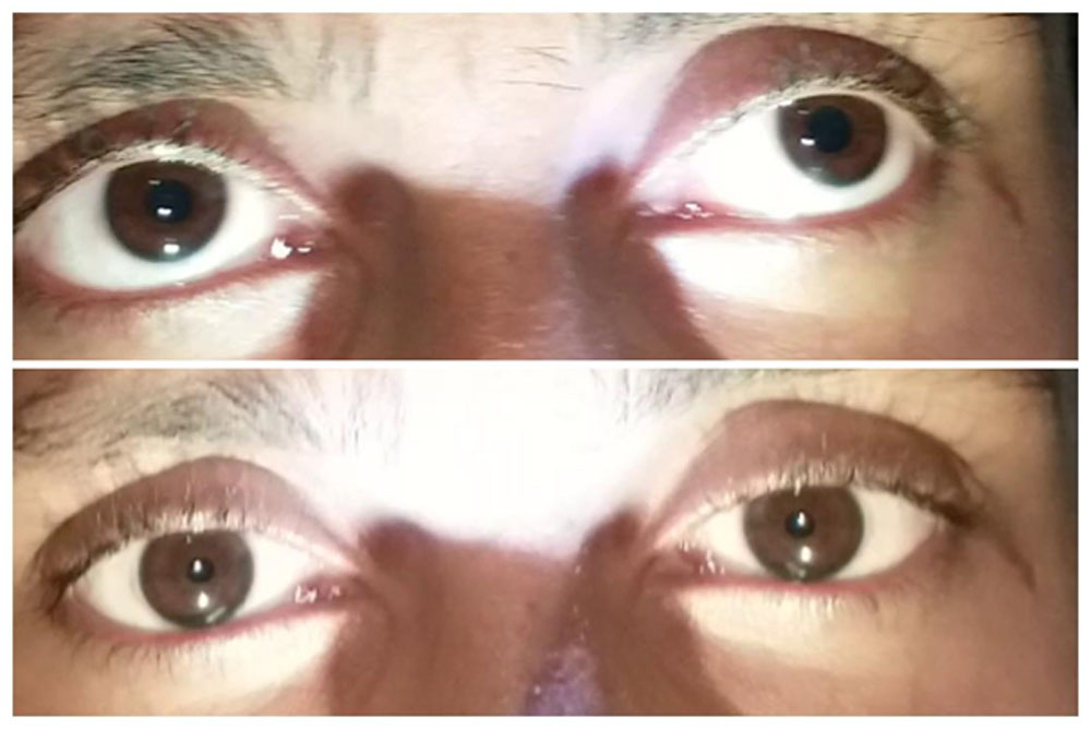
Unilateral involvement as seen in 34 cases(85%) with right eye being affected commonly than left eye ( 18:16 ) .( Fig 1) Bilateral involvement as noted in 6 cases , with asymmetrical involvement in all except one case.( Fig 2) Among the bilateral cases there were 3 males and females and most of them in range of 20-40 age group (3/6cases).
Considering the presenting complaints; difficulty with near tasks was the commonest (17 cases) , headaches in 7 cases , photophobia in 3 cases and 10 cases were noted as part of routine eye examination. Interestingly 3 cases presented with difference in pupillary size as noted by the patient themselves .
Routine investigation recorded was noted to be within normal range in all cases except raised ESR noted in 9 cases. Hypothyroidism and diabetes was noted in 2 cases each.
All patients showed supersensitivity to diluted pilocarpine except one patient , which remained the same at 1 month follow up .
Treatment data was analysed . Most (22/40 cases) of them given routine refractive correction, diluted pilocarpine was given in 2 cases( one bilateral involvement) and unilateral progressive lenses in 3 cases and rest needing no treatment.
On analyzing the follow up data,7 cases had less than 6 months follow up and 7cases were followed up for more than 1 year. interestingly there were no change in pupil size on follow up.
Discussion
In this retrospective study, we looked at demographic and clinical profile of patients with adie’s pupil. Age group affected was commonly between 20-40 years in our series ;similar to in literature .
The occurrence is reported to be commoner in females than males , 1,2,4 but our study showed males to be commonly affected.
Involvement was unilateral in 85% cases comparable to that reported . 1,2 Nearly 1/4th of cases were diagnosed as part of clinical examination suggesting the need for careful pupillary examination.
Our series points towards common occurrence of Adie’s pupil and needs to be looked for during examination. Though the etiology seems not clear in our review but gives a clue to investigate further in view of raised ESR suggesting inflammation .
References
- Kline LB, Bajandas FJ: Neuro-Ophthalmology Review Manual, Fifth Edition. Thorofare, NJ: Slack; 2004
- Miller NR, Biousse V, Newman NJ, Kerrison JB. Walsh and Hoyt’s Clinical Neuro-Ophthalmology: The Essentials, 2nd ed. Lippincott Williams & Wilkins, 2008:307-314
- Martinelli P. Holmes –Adie syndrome . Lancet 2000,356: 1760-1761.
- Thompson HS, Adie’s syndrome: some new observations. Trans Am Ophthal Soc 1977;75:587–626.
- Wilhelm H. Neuro-ophthalmology of pupillary function – practical guidelines. J Neurol. 1998;245(9):573-83.
- Thompson HS, Bell RA, Bourgon P. The natural history of Adie’s syndrome. In
Thompson HS, Daroff R, Frisan L et al, eds. Topics in Neuro-Ophthalmology. Baltimore, Williams & Wilkins, 1979:96-99.
- Kim JS, Yun CH, Moon CS. Bilateral tonic (Adie’s) pupils in Vogt-Koyanagi-Harada syndrome. J Neuroophthalmol. 2001;21:205–6
- Nitta T, Kase M, Shinmei Y, Yoshida K, Chin S, Ohno S. Mydriasis with light-near dissociation in Fisher’s Syndrome. Jpn J Ophthalmol. 2007;51:224–7
- Kajikawa S1, Ohi T, Fujita A, Kusunoki S. A Case of Anti-GQ1b Antibody Syndrome Associated with Pure Bilateral Adie’s Pupils. Brain Nerve.2016 Jan;68(1):93-6.
- Bell RA, Thompson HS. The symptoms of Adie’s syndrome. In Thompson HS, Daroff R, Frisan L, eds. Topics in Neuro-Ophthalmology. Baltimore, Williams & Wilkins, 1979:99-104
- Kardon RH , Corbett JJ, Thompson HS. Segmental denervation and reinnervation of the iris sphincter as shown by infrared videographic transillumination.1998;105(2):313–321.
- Thompson HS . Segmental palsy of the iris sphincter in Adie’s syndrome. Arch Ophthalmol.1978;96(9):1615–1620.
Figure legends
FIG 1. Upper picture shows (OD) Adie’s pupil. (OD) pupil constricts with near reaction in lower picture
FIG 2. Upper picture shows (OU) Adie’s pupil. (OU) pupil constricts with near reaction in lower picture
Table 1
| Age group | Males | Females | total |
| Less than 20 years | 2 | 1 | 3 |
| 21-30 years | 10 | 5 | 15 |
| 31-40 years | 7 | 8 | 15 |
| 41-50 years | 2 | 1 | 3 |
| Above 50 years | 2 | 2 | 4 |
| 23 | 17 | 40 |
Figure 1
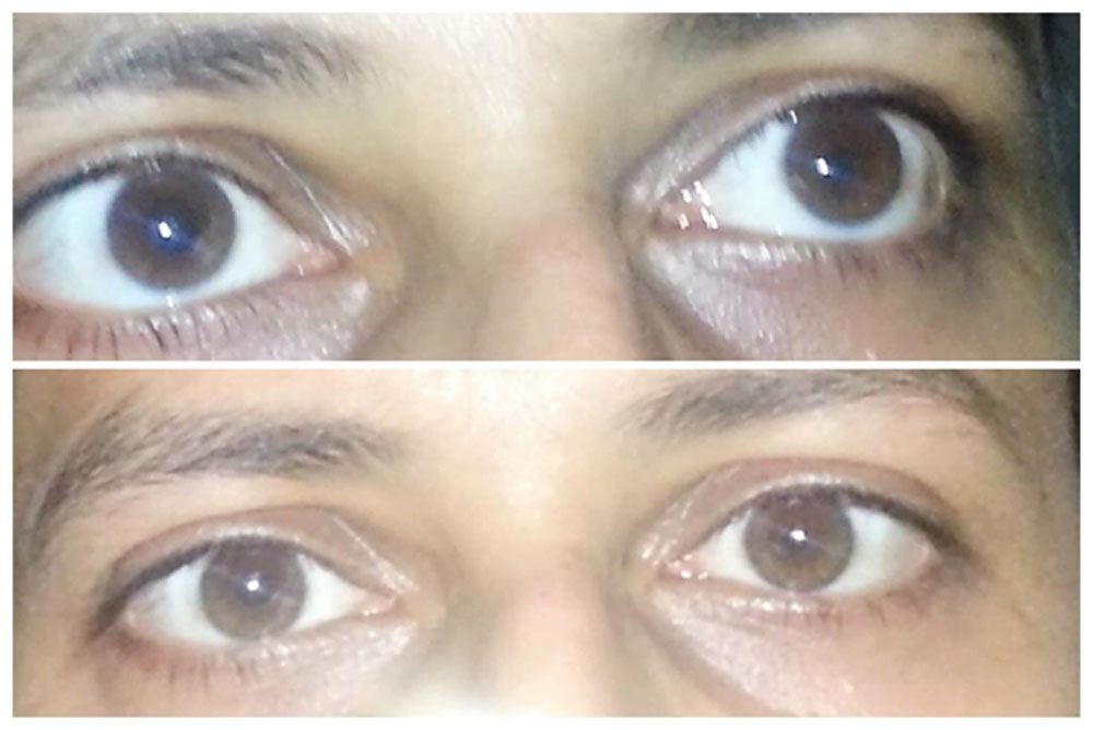
Figure 2
Leave a Comment