

Dr.Archana Rajamani,Dr.Archana Rajamani,Dr.Babu Rajendran
A NEW MODIF IED PHOTODYNAMIC THERTAPY (PDT) FOR RUBEOSIS IN NEOVASCULAR GLAUCOMA WITH GOOD RESULTS AND RISK BENEFIT AND COST BENEFIT RATIOS
ABSTRACT:
Dr ARCHANA RAJAMANI, Dr BABU RAJENDRAN
VIJAYA EYE FOUNDATION, VHENNAI 600026
Photodynamic therapy(PDT) uses a photo reactive drug along with red laser for the treatment of neovascularisation. However, in our experience, we have found that photodynamic therapy is a photochemical reaction and doesn’t need laser for new vessels on the choroid and retina. We tried this therapy for treatment of rubeosis after we got comparable results of this therapy with standard PDT. The IOP lowered after the One day of treatment and was stable till the patient was fit for surgery and the iris new vessels also showed regression post treatment. Conclusion:The Red Light onVisudyne (RLV) can be an effective the treatment of rubeosis.
INTRODUCTION
Photodynamic therapy was first introduced in mid to late 90s.
This procedure selectively occludes vascular endothelial cells. Vertiporphin binds to low density lipoproteins which are increased in abnormal endothelial cells. When this dye is exposed to the light in the red spectrum of wavelength 685 nm, it is activated from the ground state to the excited state. The dye in the excited state, after a series of biological reactions, releases nascent oxygen with free radicals. This causes activation of the clotting cascade and causes occlusion of new vessels1.
When photodynamic therapy was first introduced, it seemed to be the answer to all the retinal neovascularization. However, significant collateral damage like choroidal rip, bleedingand retinalscarring2 and the exorbitant cost brought down the popularity of the treatment which further declined with the advent of intravitreal anti–VEGF injections2
PDT was used for rubeosis by Muller VF et al3 in patients with neovascular glaucoma in the early 21ST
Century, but abandoned the procedure because of high cost of the dye and the laser and high rate of complications like bleeding.
We have used this protocol in patients with JFT (Fig 1), chronic recurrent central serous retinopathy(Fig
2) where patients have shown good response.
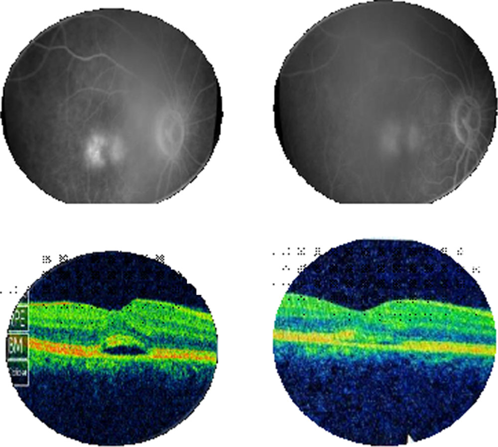
Fig 2: Oct picture showing flattening of the central serous retinopathy 24 hours post treatment with RLV
In an act of serendipity**, in one of our earlier PDT cases, the laser wasn’t working after we injected the dye in the patient and, out of sheer desperation, we used a bright white light source through a red safety filter that we had used with our tunable DYE laser and we found it created the expected reaction. In a second case soon after, we had a similar experience wherein the patient refused to sit at the slit lamp due to claustrophobia. We followed the same protocol with that patient as well and it worked again. On analysis we found that the filter was transmitting light in the wavelength of 620nm. We also found that the absorption spectrum of verteporphin had two peaks, one at 630 nm and another at 685 nm which is why 620nm worked.
We then created an independent non-laser Red light source using a LED in the 685 nm range and have
been using it in treating new vessels on the retina and choroid with results like published data using the red laser.
METHOD:
Our procedure with the RLV (Fig 3) is to inject the photosensitive drug intravenously, place the patient in a dark room and expose the patient to the special red light325 mW/cm2 for two minutes. No speculum, no contact lens, no topical anesthesia required and no collateral damage produced by the laser. The results obtained with RLV were comparable with standard PDT(Fig 4). In addition since the light fills the eye , the lesion with the dye will find the light ND hence focusing the light exactly on the lesion is not required..
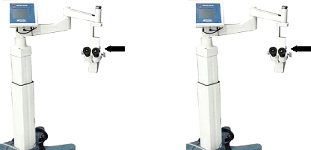
Fig 3 – our RLV machine. (patent pending) Manufactured by Appasamy* associates
Fig 4: results have been comparable with PDT with the Laser.
*APPASAMYASSOCIATES,20,SBIFFGICERSCOLONY,
1stSTREET,ARUMBAKKAM,CHENNAI-600106
SERENDIPITY** noun:
an aptitude for making desirable discoveries by accident.
The Neo Vascular Glaucoma Patient
This patient had a central retinal vein occlusion three months previous and came to us complaining of pain in that eye.
On day 0, his pressure was 25mmHg with no evidence of angle or iris neovascularization, we started
the patient on a combination eyedrops of latanoprost and timolol. His IOP was elevated to 30mmHg on day 7, when dorzolamide was added. The next day he came to the OPD with pain, redness and his IOP was recorded as 40mmHg. He was started on Acetazolamide oral tablets, and atropine eyedrops and steroid eyedrops as well. On day 9, his pressure was recorded as 42mmHg and Lumigan was added to the pre existing medications and still no NVI or NVA were noted. On day 10, when the physician hadn’t given the fitness yet due to uncontrolled blood sugars and pulmonary problems,, he returned with severe pain and vomiting with IOP of 60mmHg(Table 1), we thought of treating him with RLV as the iris new vessels were still abnormal vessels. We did anterior segment angiogram and then followed the standard protocol for PDT by injecting the photosensitive dye intravenously followed by exposure to RLV light for two minutes(Fig 5). The IOP dropped to 40mmHg post PDT and we saw regression of iris new vessels in iris angiogram.(Fig 6)
IOP MEDICATION & TREATMENT REMARKS DAY O 25 LATANOPROST+TIMOLOL
DAY 7 30 LATANOPROST+TIMOLOL+DORZOX NO RUBEOSIS NOTED DAYT 8 40 ADDED DIAMOX, ATROPINE, STEROID NO RUBEOSIS NOTED DAY 9 42 LATANOPROST+TIMOLOL+DORZOX+LUMIGAN
DAY10 60 RLV RUBEOSIS ++PAIN ++ PATIENT UNFIT FOR SURGERY
DAY 11 40 POST PDT+LATANOPROST+TIMOLOL NO PAIN-STOPPED DIAMOX DAY 25 40 PATIENT FIT FOR SURGHERY TRABACULECTECTOMY &
AVASTIN
DAY 30 20 POST OPERATIVE DROPS ONLY NO ANTIGLAUCOMA MEDICATION
Table 1: summarizing the IOP and medications of the patient.
RESULTS: The iris new vessels showed regression post treatment.
Fig.6: The picture on the left shows iris neovascularization pre treatment and picture on right shows the regression of new vessels 24 hours post treatment.
DISCUSSION :
The absorption spectrum of vertiporphin was approximately in the wavelength range of 600 to
685nmwhere our red filter light was 620nm which one of the peaks of the absorption spectrum of vertiporphin another peak being 685nm). This was when we thought that photodynamic therapy is a photochemical reaction like photosynthesis and doesn’t need light like a laser, since the light fills the entire eye and the new vessels with the dye will find the light, therefore no lens required to focus the light. Thus we came up with our Red Light on vertiporphin. This special light replaces the laser. When we replace the laser with the red light we also cut down the cost of the treatment along with reducing the complications of Laser like Choroidal rip, bleeding and scarring.
A search of literature to look for previous evidence of use of photodynamic therapy for anterior segment found that PDT was done for anterior segment neovascularization by Moller et al4 and by Maurizio et al5 but somehow the procedure didn’t become popular basically due to the high cost of the dye and the laser plus the collateral unintended damage caused by the laser. However, he did make one unexplained observation that after the PDT the new vessels never returned. This was the situation in our case as well.
In summary, this red light has its advantages. It is cost effective, no topical medications required, and the results are comparable to standard PDT with no chances of bleed and no need to measure GLD, since the lesion finds the light. In addition it has both good risk benefit and cost benefit ratios.
References :
1.DK Newman, Department of ophthalmology Addenbrookes hospital. Eye(2016) 30,202-210
2.Schmidt-Erfurth U, Chong V, Lowenstein A, Larsen M, Souied E, Schlingemann R, Eldem B, Monès J, Richard G, Bandello F. Guidelines for the neovascular age-related macular degeneration by the European Society of Retina Specialists(EURETINA). Br J Ophthalmol. 2014;98:1144-67
3.Pukhraj Rishi and Vishvesh Agarwal ,Sci J Med & Vis Res Found June 2015 | volume XXXIII | number 2 |
4.Müller VA1, Ruokonen P, Schellenbeck M, Hartmann C, Tetz Treatment of rubeosis iridis with photodynamic therapy with verteporfin–A new therapeutic and prophylactic option for patients with the risk of neovascular glaucoma Ophthalmic Res. 2003 Jan-Feb;35(1):60-4.
5. Maurizio Battaglia Parodi, Pierluigilacono: American journal of Ophthalmology 138(1). 2004;157-158
Leave a Comment