

Dr. ANJANA KARUNAKARAN,Dr. Rohit Shetty,Dr. Natasha Pahuja,Dr. Pooja Khamar
Introduction:
Keratoconus (KC) is a corneal ectatic disease which causes progressive focal thinning and irregular astigmatism leading to reduced visual acuity.2,3 The disease with its high predisposition in South Asia including Indian subcontinent may suggest an underlying genetic predisposition.2,4Keratoconus pathogenesis has been associated with mechanical factors such as eye rubbing and contact lens wear along with systemic conditions like connective tissue disorders, Down’s syndrome.5Atopy and genetic factors are also been reported to have a higher incidence of KC.4,6
The involvement of several inflammatory factors, enzymes, and cytokines such as matrix metalloproteinase 9 (MMP-9), IL-6, TNF-a, and Cathepsins that were found to be increased in tear samples of KC patients.7-12 KC has a genetic predisposition with increased incidence in familial and monozygotic twins.13,14,15 Most of the KC patients are sporadic but family history is reported in 6-10% of patients.16,17The modes of disease inheritance identified in KC families are dominant and recessive, but in autosomal dominant inheritance, it shows incomplete penetrance with variable phenotype.17 The etiopathogenesis of keratoconus and the triggering factors are still not fully elucidated.
Methods:
Patient details and clinical evaluations:
Patients were diagnosed for KC with the aid of retinoscopy, slit-lamp biomicroscopy, and corneal refraction measurements. Corneal topography of all the patients was obtained with Pentacam (OCULUS Optikger¨ate GmbH, Wetzlar, Germany) upon first visit at the clinic and used for diagnosis and grading of the KC patients.13,18 Keratoconus grades were determined from cumulative analysis of biomicroscopy data, slit-lamp, spherical and cylindrical refraction change, mean central keratometry measures, and corneal thickness as defined by the Amsler– Krumeich classification.19,20.
The blood from 3 KC subjects and their family members underwent genetic analysis 25 subjects with normal corneal topography, undergoing refractive correction with photorefractive keratectomy (PRK) were included as controls for the collection of corneal epithelium samples. Patients having history of using contact lenses or any type of anti-inflammatory systemic medications (e.g., antiallergic, anti-inflammatory drugs and steroids) were excludedalong with patients who had undergone any prior ocular surgical intervention (e.g., penetrating keratoplasty/ corneal collagen cross-linking and cataract surgery) for either eyes .
Isolation of DNA, genetic analysis and exome sequencing:
Total genomic DNA was isolated from five ml of peripheral blood using standard procedure of salt precipitation method1. Qubit quantified DNA was used for library preparation using the Illumina Nextera protocol as per the manufacturer’s instructions. These libraries were enriched and underwent whole-exome capture using BCM-HGSC core design (52 Mb; Roche NimbleGen, Inc., Madison, WI, USA), followed by sequencing on the Illumina HiSeq 2000 platform (Illumina, Inc., San Diego, CA, USA).
Isolation of RNA, cDNA synthesis and real-time PCR:
KC (n=23) subjects and age, gender matched controls (n=25) were taken for the gene expression analysis. Total RNA was isolated from patient epithelia obtained post operatively. Briefly, Trizol based RNA extraction was performed according to the manufacturer’s instructions (Invitrogen, Carlsbad, CA), followed by cDNA synthesis using Superscript III (Life Technologies, Carlsbad, CA). Real time PCR analysis was performed, which includes 40 amplification cycles with a denaturation at 95 °C for 15sec, annealing and extension at 58 °C for 30 sec using a CFX ConnectTM real-time PCR detection system (Bio-Rad, Philadelphia, PA). Human beta actin served as housekeeping reference gene.
Results:
Novel mutation in collagen gene (COLIV) was observed in KC subjects when compared with controls .The levels of COLIV was reduced in KC cohort (0.43 folds) compared to controls (1.05 folds) and is in association with collagen cross linking enzyme LOX. (Figure 1a, b)
Conclusion: This novel mutation in collagen gene first to be reported may be a causative factor in familial KC. Further, it suggests that matrix remodeling factor such as LOX play an important role in this process and the association with Collagen IV factors may trigger the disease pathogenesis and progressive thinning of the cornea.This study highlights the association of genetic(nature) and gene expression(nurture) may contribute to drive KC pathogenesis and can be harnessed for disease management.
LOX
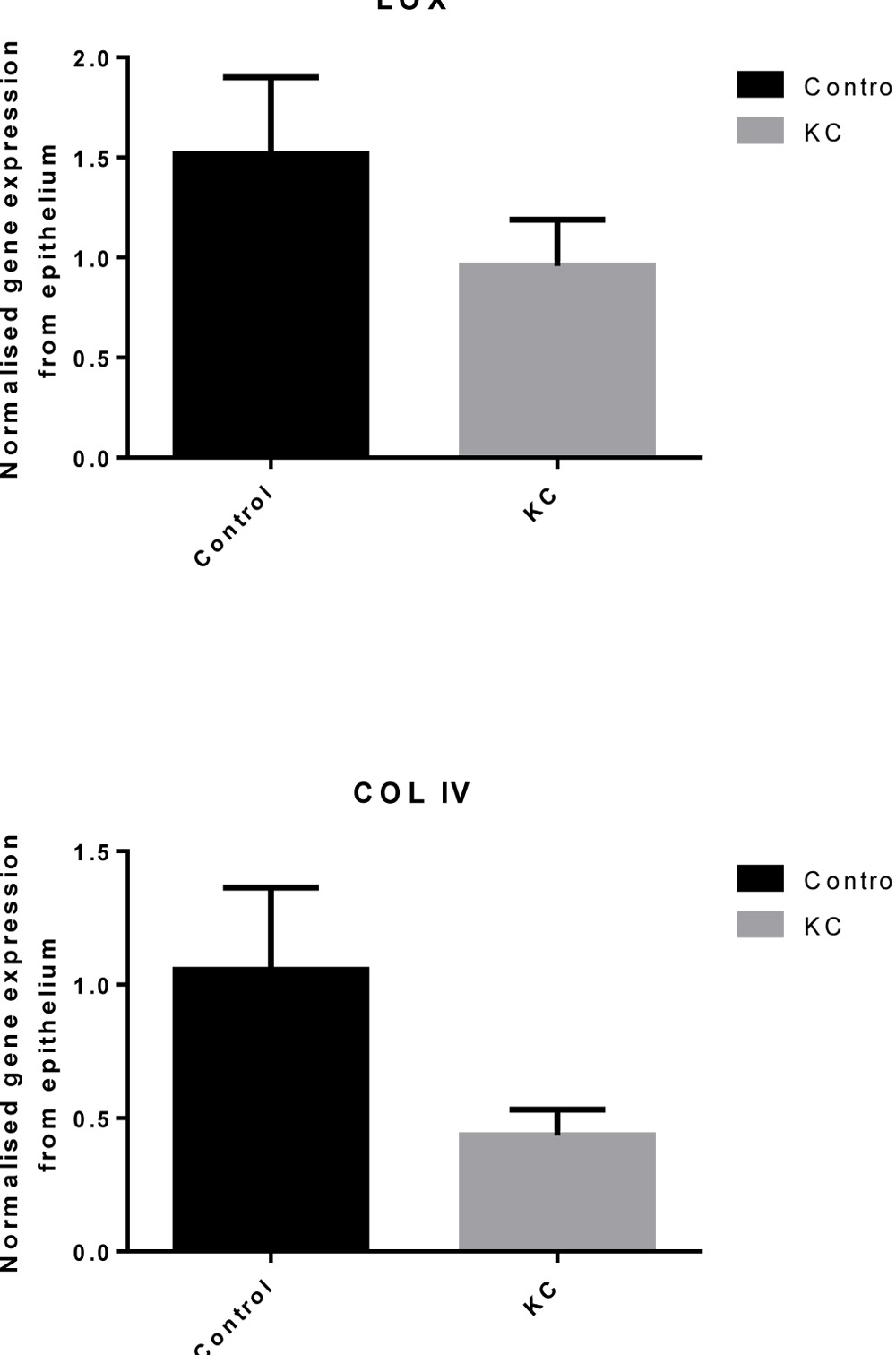
Figure 1 a, b: Represents patient epithelia derived normalized gene expression of COLIV and LOX.
References
- Miller SA, Dykes DD, Polesky HF. A simple salting out procedure for extracting DNA from human nucleated cells. Nucleic Acids Res. 1988;16:1215.
- Gordon-Shaag A, Millodot M, Shneor E, Liu Y. The genetic and environmental factors for keratoconus. Bio Med Res Int. 2015; 2015:795738.
- Zadnik K, Barr JT, Edrington TB, et al. Baseline findings in the Collaborative Longitudinal Evaluation of Keratoconus (CLEK) Study. Invest Ophthalmol Vis Sci. 1998;39:2537–2546.
- Jeyabalan N, Shetty R, Ghosh A, Anandula VR, Ghosh AS, Kumaramanickavel G. Genetic and genomic perspective to understand the molecular pathogenesis of keratoconus. Indian J Ophthalmol. 2013;61:384–388.
- Korb DR, Finnemore VM, Herman JP. Apical changes and scarring in keratoconus as related to contact lens fitting techniques. J Am Optometric Assoc. 1982;53:199–205.
- Krachmer JH, Feder RS, Belin MW. Keratoconus and related noninflammatory corneal thinning disorders. Survey Ophthalmol. 1984;28:293–322.
- Balasubramanian SA, Mohan S, Pye DC, Willcox MD. Proteases, proteolysis and inflammatory molecules in the tears of people with keratoconus. Acta Ophthalmol. 2012; 90:e303–e309.
- Lema I, Duran JA. Inflammatory molecules in the tears of patients with keratoconus. Ophthalmology. 2005;112:654– 659.
- Lema I, Sobrino T, Duran JA, Brea D, Diez-Feijoo E. Subclinical keratoconus and inflammatory molecules from tears. Br J Ophthalmol. 2009;93:820–824.
- Nemet AY, Vinker S, Bahar I, Kaiserman I. The association of keratoconus with immune disorders. Cornea. 2010;29:1261– 1264.
- Wisse RP, Kuiper JJ, Gans R, Imhof S, Radstake TR, Van der Lelij A. Cytokine expression in keratoconus and its corneal microenvironment: a systematic review. Ocular Surf. 2015; 13:272–283.
- Jun AS, Cope L, Speck C, et al. Subnormal cytokine profile in the tear fluid of keratoconus patients. PLoS One. 2011;6: e16437.
- Rabinowitz YS. Keratoconus. SurvOphthalmol. 1998;4:297-319.
- Parker J, Ko WW, Pavlopoulos G, Wolfe PJ, Rabinowitz YS, Feldman ST. Videokeratography of keratoconus in monozygotic twins. J Refract Surg. 1996;1:180-3.
- Edwards M, McGhee CN, Dean S. The genetics of keratoconus. Clin Experiment Ophthalmol. 2001;6:345-51.
- Hamilton JB. The Significance of Heredity in Ophthalmology. Preliminary Survey of Hereditary Eye Diseases in Tasmania. Br J Ophthalmol. 1938;2:83-108.
- Falls HF AA. Dominantly inherited keratoconus: report of a family. J Genet Hum 1969;317-24
- Rao SN, Raviv T, Majmudar PA, Epstein RJ. Role of Orbscan II in screening keratoconus suspects before refractive corneal surgery. Ophthalmology. 2002;109:1642–1646.
- Ishii R, Kamiya K, Igarashi A, Shimizu K, Utsumi Y, Kumanomido T. Correlation of corneal elevation with severity of keratoconus by means of anterior and posterior topographic analysis. Cornea. 2012;31:253–258.
- Mihaltz K, Kovacs I, Takacs A, Nagy ZZ. Evaluation of keratometric, pachymetric, and elevation parameters of keratoconic corneas with pentacam. Cornea. 2009;28:976– 980.
Leave a Comment