

Dr. Aniruddh soni, Dr. ANITHA VENUGOPAL, Dr. ADITYA GHORPADE
ABSTRACT
AIM:
To assess incidence, clinical features & treatment outcomes of Pediatric Keratoconus
METHODS:
A prospective observational survey in children aged 5 to 18 years with emphasis on visual impairment, history of VKC, topography & ultrasound pachymetry changes were noted. Children with progression were treated with CXL/DALK/PKP & were followed up till 6 months.
RESULTS:
70 eyes of 38 children with mean age of 13.26 +/- 2.2 years were included. Incidence in pediatric age was 0.26%. Common presenting complaint was poor vision. Bilateral KC was seen in 50 % of patients. History of VKC was present in 50% eyes.
Topographical parameters between eyes undergoing surgery and eyes not undergoing surgery were analysed pre operatively. 49 eyes underwent one of the three surgical procedures (CXL, DALK, PKP). Post operatively CXL group showed stabilization of UCVA, BCVA MRSE, SimK, Kmax, 3 mm zone, 5 mm Zone, SRAX & I-S difference values with reduction in CCT.
Post operatively DALK group showed improvement in UCVA, BCVA & CCT at end of 6th month followup. The Kmax & 3mm zone values was statistically reduced. The single eye operated for PKP, showed improvement in UCVA & BCVA, with reduction in MRSE post operatively at 6 months.
CONCLUSION:
KC is aggressive & rapidly progressive in children. VKC, atopic disease, and chronic eye rubbing must be controlled with topical medications. Regular followup and visual rehabilitation is paramount. CXL is an effective and alternative treatment to DALK for Pediatric KC.
Keywords :- Pediatric KC, VKC, CXL, DALK, PKP
BODY
Introduction
Keratoconus (KC) is a fairly common bilateral, non inflammatory, degenerative axial ectatic condition of the cornea in which the cornea assumes an irregular conical shape. These changes may result in visual impairment due to irregular astigmatism, progressive myopia, or corneal scarring. The disease most often occurs at puberty and progresses until the age of about 30 to 40 years before stabilizing. Keratoconus is a rare cause of amblyopia and visual impairment in children, as the development of visual function generally continues until the age of 8 to 11 years. Young age appears to be associated with more severe forms of keratoconus and faster disease progression, with an inverse correlation between age and severity. In addition, young age at diagnosis is linked to a greater risk of developing corneal opacity and requiring a corneal transplant
Methods
Children, aged 5 to 18 years were recruited from out patient department, from December 2016 till April 2017 and were followed up for a maximum of 6 months until October 2017. Ethical clearance was obtained from the Institutional Ethics Committee of Aravind Eye Hospital, Tirunelveli. The survey adhered to the tenets of the declaration of Helsinki. Demographic data of the children and a brief family history were obtained. The incidence of the disease was calculated in relation to number of pediatric patients (<18 years) & in relation to number of patients with keratoconus presenting to the hospital, during the recruitment A detailed ophthalmic examination was done including refraction, corneal topography & ultrasound pachymetry. Diagnosis of the disease was made by careful slit lamp examination and ORBSCAN topography by analysing two visits parameters & were followed up at 4th & 6th months.
The eyes were classified based on the criteria published by Rabinowitz. Severity at diagnosis was assessed using Krumeich’s staging. Association between VKC & keratoconus was studied. Severity of the disease in patients with VKC was studied on basis of topographical & ultrasound pachymetry changes. The patients who showed progression of the disease underwent surgical intervention based on their clinical & topographical analysis. Children without progression were closely followed up. Pre operatively, comparison was drawn between the topographical parameters in eyes undergoing surgery and eyes not undergoing surgery. Follow up results were noted in the non surgical group and the surgical group (CXL/DALK/PKP). Primary outcomes was defined as stabilization or improvement in BCVA & MRSE. Secondary outcomes was defined as assessment in changes in SimK, Kmax & CCT.
Results
70 eyes of 38 patients were enrolled in the study which met the inclusion and exclusion criteria . 3 eyes did not complete the 6 month followup. Mean (SD) of the age was 13.26+/-2.2 years and the range was 10 – 18 years. A slight Female prepondance was noted (55.3%). Bilateral KC was seen in 19 children (50%). Common presenting complaint was poor vision (n=51; 72.9%). Eye rubbing was noted in 32 eyes (45.7%). One patient had a family history of keratoconus in the elder sibling. 7 children had an associated systemic illness, with asthma commonly seen in 3 children. History VKC was noted in 50% eyes.
Sicssoring reflex was present in 71.1% of eyes. External examination signs such as Munson’s & Rizutti were seen in 50% of eyes. The proportion of eyes with slit lamp findings of KC were -vogt straie (38.6%) , Fleischer ring ( 58.6%) prominent corneal nerves (52.9%), apical scarring (10%). 45.7% eyes had a nipple type of cone, followed by oval type (27.1%). Cone morphology was not demonstrable in 18.6% of eyes. Globus type of cone was seen in 8.6% eyes
The eyes with VKC had a predominance of the palpebral type (25 eyes) followed by the mixed type (8 eyes). The limbal type was the least common (2 eyes). Comparison was drawn between eyes with & without history of VKC and their effect on topographical parameters. Kmax, posterior elevation & posterior BFS difference didn’t not reveal much difference, but the I-S difference was significantly higher in the group with history of VKC.
Co-relation between type of VKC & their effect on topographical parameters were studied. Comparison was done between palpebral & mixed type of VKC. The mixed type of VKC group revealed significantly higher values of Kmax, I-S difference, posterior elevation, Posterior BFS difference as compared to the palpebral group, which were all statistically significant.
Based On Rabinowitz Classification, at first visit 72.9 % eyes were noted to be KC , 7.1 % eyes were early KC, 14.3 % were FFKC, 2% were KC suspects , 2% were normal. On subsequent visit, we see that 2 eyes from the early KC group, 1 eye from FFKC group & 2 eyes from the normal group progressed to the KC group. Out of the 56 KC eyes at 2nd visit, based on Krumeich Staging, stage 1 was noted to be most common (39.3%) followed by stage 2 then 3 & least were in stage 4.
Based on clinical & topographical evaluation at 1st visit, 34 eyes were advised surgical intervention. This number increased to 52 eyes at 2nd visit. 49 eyes underwent one of the three surgical procedures (CXL, DALK, PKP). 21 eyes which were not operated (which include 18 from non surgical group, which did not show progression at second visit & 3 from the surgical group who refused treatment) were followed up for 6 months.
Pre operatively, comparison was drawn between eyes undergoing surgery and eyes not undergoing surgery. It was found that significant number of eyes had a Kmax of >48 D in the surgery advised group (89.8%), whereas the no surgery group had more eyes (57.1%) with Kmax <48 D. More percentage of eyes in the surgery advised group (69.4%) had a SRAX value >22 degrees as compared to no surgery (61.9%) advised eyes. In the surgery advised group, more eyes (59.2%) had a Sim K value of >6D, whereas the no surgery group had more eyes (80.9%) with Sim k <6 D.
A greater proportion of eyes (87.8%) with I-S difference of >1.5D values were noted in the surgery advised group as compared to the no surgery (52.4%) group. In the surgery advised group, 57.1 % eyes had a CCT less than 470 microns. In comparison, the no surgery group had 66.7 % eyes with a value greater than 470 microns. 53.1% of the surgery advised eyes had a posterior BFS of >55D. Whereas 76.2% of the eyes in no surgery group had a posterior BFS of <55D. 95.9 % eyes form surgery advised group had a posterior elevation greater than >0.05mm, whereas 61.9% eyes from no surgery group had a posterior elevation of <0.05mm.
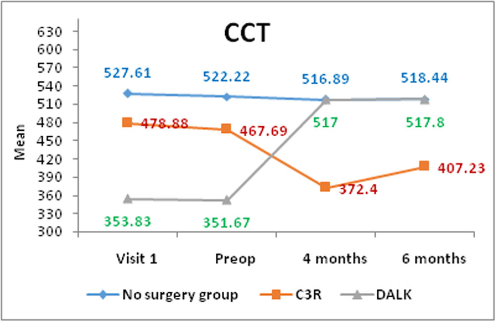
Follow up results at the end of 6 months in the no surgery group (18 eyes) revealed improvement of BCVA, slight increase in MRSE, SimK & Kmax, but the CCT values were reduced.
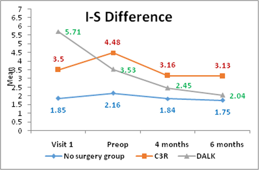
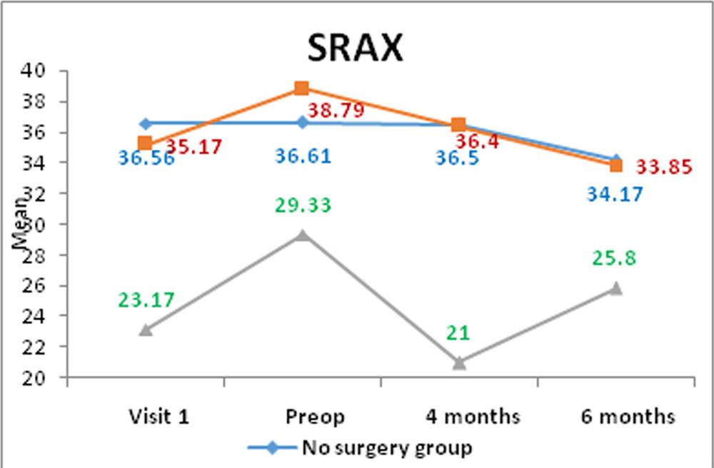
CXL group had 42 eyes. Post operatively these showed stabilization of UCVA, BCVA, MRSE, SimK, Kmax, 3 mm zone, 5 mm Zone, SRAX & I-S difference values with reduction in CCT post operatively at the end of 6 month followup. The 2 eyes which did not under go CXL showed decrease in UCVA, BCVA, CCT & increase in MRSE, SimK, Kmax ,3mm zone, 5 mm Zone, SRAX & I-S diff, at 4th & 6th month of followup when compared to earlier visits. This signifies progression
DALK group had 6 eyes. Post operatively these showed improvement in UCVA & BCVA at end of 6th month followup. The Kmax & 3mm zone values was statistically reduced & the CCT improved postoperatively at the end of 6th month followup. The singe eye which did not undergo DALK showed decrease in UCVA, BCVA, CCT & increase in MRSE, SimK, Kmax ,3mm zone, 5 mm Zone, SRAX & I-S diff, at 4th & 6th month of followup when compared to earlier visits. This signifies progression
The single eye operated for PKP, showed improvement in UCVA & BCVA, with reduction in MRSE values at 6 months post op.
Discussions
Keratoconus in children can be missed in its early stages and progression could be rapid and relentless. This could lead to failure of visual maturation and failure to reach developmental and social milestones. Surgical intervention with potential for a prolonged recovery time can have a profound effect on the developing child. Early detection of KC in children is therefore vital, and more tools are becoming available to allow for this.
70 eyes of 38 patients were included in this study. The incidence in the pediatric age was 0.26% and in relation to number of patients with keratoconus of all ages was 21.93%.
The mean age in our study was found to be 13.26 +/- 2.2 years, which was significantly less as compared to 17+/-3 years as documented by Ertan et al1. Female cases were more in our study (55.3%), which was a similar finding by Elias et al2 (53.3%) & Shetty et al3 (53%). Bilateral KC was in 50% cases in our study, which was less as compared to the study by Shetty et al3 (97%).
Common presenting complaints were poor vision (n=51; 72.9%) & eye rubbing (n=32;45.7%) El-Khoury et al4 reported poor vision (62.5%) & Sharma et al5 reported eye rubbing(55.8%) to be the most common complaints
Clinical findings such as scissoring reflex, vogt straie, fleischer’s ring were seen in 71.1 %, 38% & 58 % of eyes respectively Similar results have been found by Sharma et al5 (vogt straie 47.5 % & fleischer’s ring 50.8%). This highlights that keratoconus detection should not only be relied on external and slit lamp findings, but a topographical analysis should be obtained as topographical changes precede clinical diagnosis.
History of VKC was present in 35 eyes, commonest being palpebral type (n=25). Eyes with a history of VKC had a higher I-S difference as compared to eyes without a history of VKC. The Kmax, posterior BFS & posterior elevation was comparable in eyes with & without history of VKC
Eyes with mixed type of VKC (n=8) had higher values of Kmax, I-S difference, posterior elevation, posterior BFS as compared to palpebral type, all of which were statistically significant (p<0.05). This highlights the severity of the mixed type of VKC and its association with a higher risk of progression of disease. A similar study by Taneja et al6 reported significant differences only in the 3mm zone values comparing mixed & palpebral type of VKC. Cone morphology of the nipple type was found to be the most common type, where as Sharma et al5 documented oval type of cone to be the most common
Based on Rabinowitz classification, 72.9% eyes were diagnosed as KC at first visit, which increased to 80% at second visit (2 eyes from the early KC group, 1 eye from FFKC group & 2 eyes from the normal group). Li et al7 in their study reported similar results (highest rate of progression from the early KC group to clinically detectable KC). This highlights the effect of progression and the necessity for frequent evaluations in all early KC, FFKC & KC suspect eyes.
At diagnosis, based on Krumeich staging, stage 1 KC was the most common presentation. These results were different as compared to a study by Léoni-Mesplié S et al8, which documented stage 2 to be the commonest at diagnosis.
Based on clinical & topographical analysis, 34 eyes were advised surgical intervention at first visit, which increased to 52 eyes at second visit. 49 eyes underwent one of the three surgical procedures (CXL, DALK, PKP). Eyes which did not show progression at second visit were followed up at 4th & 6th months. Spectacle correction was prescribed in these children.
Six month followup in the no surgery group (18 eyes), revealed stable UCVA, BCVA & MRSE values. On topographical analysis, slight elevation in SimK, Kmax & 5 mm Zone values were noted, which were not statistically significant. There was a gradual reduction of CCT through the course of study & it was statistically significant at 4th & 6th months of followup. Hence these cases were advised spectacle correction and were advised frequent followups. This emphasizes the need of frequent clinical & topographical analysis in such cases in the pediatric age group. Léoni-Mesplié S et al8 in their study on 98 eyes of KC not undergoing surgery, documented reduction in UCVA, elevation in Kmax, SimK, & posterior BFS at the end of 2 years.
In the CXL group, 42 out of 44 KC eyes underwent the procedure. There was stabilization of UCVA, BCVA, MRSE, SimK, Kmax, 3 mm zone, 5 mm Zone, SRAX & I-S difference values at the end of 6th month followup as compared to first visit data. There was reduction in CCT noted as compared to first visit, which was statistically significant at 2nd visit, 4th month & 6th month followup. Supporting these conclusions is a study by El Khoury et al4 who noted improvement in BCVA, stabilization of MRSE & reduction in Kmax over a 6 month followup. Many landmark study have evaluated the efficacy of CXL in mild to moderate pediatric KC and have come up with promising results over a longer duration of followup ranging from one to two years9-12. The 2 eyes in the observational group which denied CXL showed reduction in visual acuity & worsening of topographical parameters at 4th & 6th month of followup, when compared to earlier visits. This signified progression of the disease in these eyes.
In the DALK group, 6 out of 7 eyes underwent the procedure. There was improvement in UCVA & BCVA, whereas Kmax reduced at end of 6th month followup. The 3 mm zone values were reduced statistically at the end of 6th month followup. The CCT improved postoperatively at 6th month followup. There was reduction in MRSE, SimK, 5mm zone values, SRAX, I-S difference at end of 6th month followup which was not statistically significant. Similar results were noted in a study by Shetty et al3 over a 2 year followup. The single eye in the observational group which denied DALK showed reduction in visual acuity & worsening of topographical parameters at 4th & 6th month of followup, when compared to earlier visits. This signified progression of the disease in this eye.
In the single eye operated for PKP, improvement in UCVA & BCVA was noted at the end of 6th month of followup as compared pre operatively. There was reduction in MRSE values too at 6th month when compared to preoperatively. Similar outcomes have been observed in various studies comparing PKP versus DALK for the treatment for KC13-16
References
- Ertan A, Muftuoglu O. Keratoconus clinical findings according to different age and gender groups. Cornea. 2008 Dec;27(10):1109-13
- Elias RM, Lipener C, Uras R, Pavês L. Keratoconus: prognosis factors. Arq Bras Oftalmol. 2005 Jul-Aug;68(4):491-4
- Shetty R, Kaweri L, Pahuja N, Nagaraja H, Wadia K, Jayadev C, Nuijts R, Arora V. Current review and a simplified “five-point management algorithm” for keratoconus. Indian J Ophthalmol. 2015 Jan;63(1):46-53
- El-Khoury S, Abdelmassih Y, Hamade A, Slim E, Cherfan CG, Chelala E, Bleik J, Jarade EF. Pediatric Keratoconus in a Tertiary Referral Center: Incidence, Presentation, Risk Factors, and Treatment. J Refract Surg. 2016;32(8):534-41
- Sharma R, Titiyal JS, Prakash G, Sharma N, Tandon R, Vajpayee RB. Clinical profile and risk factors for keratoplasty and development of hydrops in north Indian patients with keratoconus. Cornea. 2009 May;28(4):367-70
- Taneja M, Ashar JN, Mathur A, Vaddavalli PK, Rathi V, Sangwan V, Murthy S. Measure of keratoconus progression in patients with vernal keratoconjunctivitis using scanning slit topography. Cont Lens Anterior Eye. 2013 Feb;36(1):41-4
- Li X, Yang H, Rabinowitz YS. Keratoconus: classification scheme based on videokeratography and clinical signs. J Cataract Refract Surg. 2009 Sep;35(9):1597-603
- Léoni-Mesplié S, Mortemousque B, Touboul D, et al. Scalability and severity of keratoconus in children. Am J Ophthalmol. 2012;154:56-62
- Arora R, Gupta D, Goyal JL, Jain P. Results of corneal collagen cross-linking in pediatric patients. J Refract Surg. 2012;28(11):759–62
- Chatzis N, Hafezi Progression of keratoconus and efficacy of pediatric corneal collagen cross-linking in children and adolescents. J Refract Surg. 2012;28(11):753–8
- Vinciguerra P, Albé E, Frueh BE, Trazza S, Epstein D. Two-year corneal cross-linking results in patients younger than 18 years with documented progressive keratoconus. Am J Ophthalmol. 2012;154:520-526
- Kumar Kodavoor S, Arsiwala AZ, Ramamurthy D. One-year clinical study on efficacy of corneal cross-linking in Indian children with progressive keratoconus. Cornea 2014;33(9):919-22
- Richard JM, Paton D, Gasset A comparison of penetrating keratoplasty and lamellar keratoplasty in the surgical management of keratoconus. Am J Ophthalmol. 1978;86:807-811
- Borderie VM, Sandali O, Bullet J, Gaujoux T, Touzeau O, Laroche L Longterm Results of Deep Anterior Lamellar versus Penetrating Keratoplasty. Ophthalmology 2011 Nov 4
- Watson SL, Ramsay A, Dart JKG, et al. Comparison of deep lamellar keratoplasty and penetrating keratoplasty in patients with keratoconus. Ophthalmol. 2004;111(9):1676-1682
- Zhang YM, Wu SQ, Yao YF. Long-term comparison of full-bed deep anterior lamellar keratoplasty and penetrating keratoplasty in treating keratoconus. J Zhejiang Univ Sci B 2013;14(5):438-450
CHART – 1
| Variables | Yes | No | Total |
| Family History | 1(2.6) | 37(97.4) | 38(100) |
| Eye Rubbing | 32(45.7) | 38(54.3) | 70(100) |
| History of VKC | 35(50.0) | 35(50.0) | 70(100) |
| Systemic findings | 7(18.4) | 31(81.6) | 38(100) |
| Scissoring Reflex | 54(71.1) | 16(22.9) | 70(100) |
| Munson’s Sign | 37(48.7) | 33(47.1) | 70(100) |
| Rizutti Sign | 35(50.0) | 35(50.0) | 70(100) |
| Vogt straie | 27(38.6) | 43(61.4) | 70(100) |
| Fleischer Ring | 41(58.6) | 29(41.4) | 70(100) |
| Prominent Nerves | 37(52.9) | 33(47.1) | 70(100) |
| Apical scarring | 7(10.0) | 63(90.0) | 70(100) |
| Systemic findings | N | % |
| Diabetes Mellitus | 1 | 2.6 |
| Allergic rhinitis | 1 | 2.6 |
| Asthma | 3 | 7.9 |
| Atopy | 1 | 2.6 |
| Microcephaly | 1 | 2.6 |
| Hypothyroidism | 1 | 2.6 |
| Type of VKC | N | % |
| Palpebral | 25 | 71.4 |
| Limbal | 2 | 5.8 |
| Mixed | 8 | 22.8 |
| Total | 35 | 100 |
CHART – 2
| Variables in pre op | History of VKC | P – value M | |||
| Yes (n=35) | No(n=35) | ||||
| Mean(SD) | Range | Mean(SD) | Range | ||
| K max (D) | 53.51(7.6) | 42.6 to 69.9 | 53.01(6.3) | 44.2 to 67.4 | 0.819 |
| IS difference (D) | 4.62(4.4) | 0.2 to 18.1 | 3.08(2.4) | 0 to 9.3 | 0.225 |
| Posterior BFS (D) | 55.82(5.4) | 49.5 to 73.8 | 55.55(2.3) | 50.7 to 60.8 | 0.196 |
| Posterior elevation (mm) | 0.09(0.04) | 0.02 to 0.156 | 0.08(0.04) | 0.015 to 0.208 | 0.733 |
| Variables in pre op | Type of VKC | P – value M | |||
| Palpebral (n=25) | Both (n=8) | ||||
| Mean(SD) | Range | Mean(SD) | Range | ||
| K max (D) | 51.28(5.3) | 42.6 to 61.4 | 62.5(7.4) | 49 to 69.9 | 0.001 |
| IS difference (D) | 3.59(3.0) | 0.2 to 11.7 | 8.44(6.4) | 1.2 to 18.1 | 0.032 |
| Posterior BFS (D) | 53.79(2.1) | 49.5 to 59 | 63.19(6.6) | 54.9 to 73.8 | 0.0001 |
| Posterior elevation (mm) | 0.08(0.04) | 0.02 to 0.15 | 0.12(0.03) | 0.06 to 0.15 | 0.013 |
CHART – 3
Pre operative Topographical Parameters in Surgical Vs Non Surgical eyes
| Variables | Surgical (n=52) | Non-surgical (n=18) | P – value C |
| N(%) | N(%) | ||
| K max
≥48 D <48 D |
44(89.8) 5(10.2) |
9(42.9) 12(57.1) |
<0.001 |
| SRAX
≥22 O <22 O |
34(69.4) 15(30.6) |
13(61.9) 8(38.1) |
0.541 |
| Sim K
≥6 D <6 D |
29(59.2) 20(40.8) |
4(19.1) 17(80.9) |
0.002 |
| IS difference
≥1.5 D <1.5 D |
43(87.8) 6(12.2) |
11(52.4) 10(47.6) |
0.001 |
| CCT
≥ 470 µ <470 µ |
21(42.9) 28(57.1) |
14(66.7) 7(33.3) |
0.068 |
| Posterior BFS
≥55 D <55 D |
26(53.1) 23(46.9) |
5(23.8) 16(76.2) |
0.024 |
| Posterior elevation
≥0.05 mm <0.05 mm |
47(95.9) 2(4.1) |
8(38.1) 13(61.9) |
<0.001 |
CHART – 4
Comparison Graphs of variables between Non Surgical Group, CXL Group & DALK Group visit 1, Visit 2, 4th & 6th Month follow up
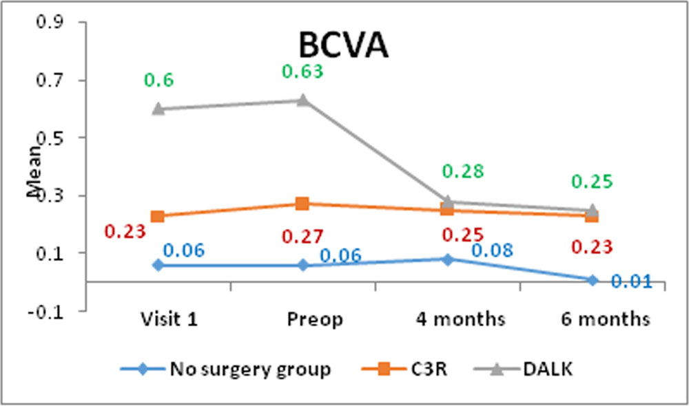
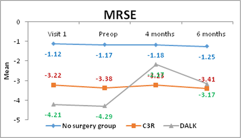
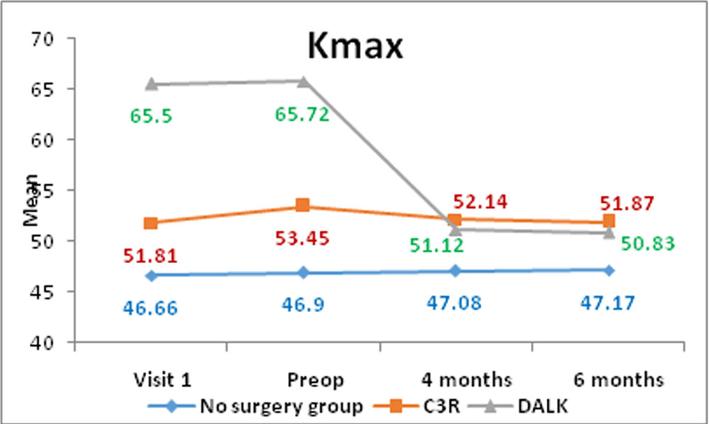
CHART – 5
Comparison Graphs of variables between Non Surgical Group, CXL Group & DALK Group visit 1,Visit 2, 4th & 6th Month follow up
Leave a Comment