

Dr.Vanaja Vaithianathan,Dr.Susil Kumar Pani
Abstract
Aim:
To analyse the most important points to remember during the learning of DMEK.
Method:
DMEK has to be mastered at 2 levels :- The first level is obtaining the donor DM scroll with endothelium without damage. The second is inserting it into the recipient eye & then unfolding the scroll, in the easiest possible way without touching or damaging the tissue.
Result:
For the first step, practising on atleast 10 waste corneal tissues to obtain DM scroll without tearing or damaging is essential. For the second step, again practising in simulated anterior chamber would help. But, in real patient, undistorted anterior chamber is the most important pre-requisite and of course any unscrolling technique can take between less than 5 minutes to upto even 1 hour in the initial stage. This presentation will highlight mistakes and lessons learnt during DMEK practice.
Conclusion:
Though, the learning curve for DMEK is steep, with perseverance it can be learnt & mastered with the greatest benefits to patients.
Key words: DMEK – Wetlab practice – Scroll – Unfolding – Simple Bubble techique.
Introduction:
Corneal endothelial problems, which used to be managed with full thickness Penetrating Keratoplasty (PKP) only, just a few years ago, are now being elegantly managed by removal & replacement of the diseased portion only, i.e Endothelium with the Descemet’s Membrane (DM).
The evolution of Descemet’s Membrance Endothelial Keratoplasty (DMEK) started as EndoKeratoplasty, Posterior Lamellar Keratoplasty (PLEK) ,Deep Lamellar Endothelial Keratoplasty (DLEK), Manual Descemet’s Stripping Endothelial Keratoplasty (DSEK) and Automated DSAEK 1,2,3,4,5, where there is mismatch between the amount of tissue removed & the amount replaced. DMEK is the only Endothelial procedure where the diseased Endothelium with DM is removed & replaced exactly by the same layers with no extra stromal tissue (Fig 1). Also, one cornea can be used for two patients (Fig 2). Hence, it gives the fastest visual rehabilitation & the best quality of vision, compared to any other Endothelial procedure. But, along with the above mentioned advantages, it is also tagged with the disadvantage of procedural difficulty.
Method:
Any Corneal Surgeon, wanting to covert from DSEK or DSAEK to DMEK should do quite a bit of wet lab practice (Fig 3).
The DMEK procedure has the following 4 steps
- Procuring the Donor Scroll, Roll or Sacor (Fig 4)
- Recipient preparation & DM stripping
- Loading & injecting the Sacor into the Anterior Chamber (AC) of the recipient.
- Unscrolling & adhering the scroll to recipient stroma.
Mastering DMEK consists of persevering practice with poor quality non-usable corneas in wet lab.
Step 1: Practice is done with atleast 5 to 10 corneas for obtaining DM scroll, without tearing or damaging.
Step 2: Recipient eye can be simulated by mounting a waste cornea on an artificial Anterior chamber, where the base is covered with a thin opaque plastic paper, so that on-going fluid flow is not needed. DM stripping, side ports & main port are done & the eye is ready for DM stripping & tissue injection (Fig 5).
Step 3: The scroll is loaded & injected into the artificial Anterior Chamber, created for the purpose (Fig 6a, 6b).
Step 4: The unscrolling steps are practised in the Artificial Anterior Chamber which will more or less simulate the recipient eye (Fig 7).
Thus, by practising in wet lab with atleast 5-10 waste corneas the steps of DMEK, an interested corneal surgeon can confidently go on to doing DMEK on patients, without damaging the tissue & also without facing major problems. The first & the last steps are the most difficult parts of DMEK, but can easily be mastered with meticulous practice in wet labs & then moving on to performing on patients.
Nuances in surgical procedure:
Step 1:
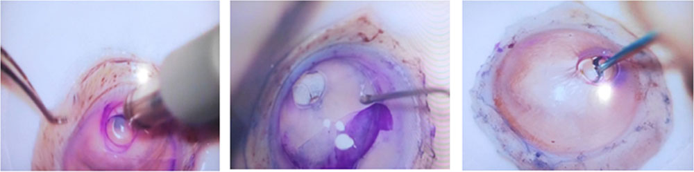
- a) The Donor tissue is placed endothelial side up on the Teflon block and the 10mm diameter trephine with 100 micron depth is used to cut the tissue partial thickness. The point to note, is that, the peripheral tissue outside this 10mm diameter is completely removed all round (Fig 8).
- b) The DM with endothelium submerged in BSS is carefully lifted off the underlying stroma, all round, meticulously, so that, all 360° the DM is free & ready for stripping.
- c) With a curved forceps, the DM is carefully stripped upto 2/3 rd area, folded on, then the underlying stroma is cut full-thickness with a 3mm trephine paracentrally, the folded DM is put back, using BSS flush the whole tissue flipped, S-mark made through the 3mm opening in the stroma on the stromal side of the DM & then Trypan blue double staining of the tissue is done (Fig 9).
- d) The 8mm Trephine is used to cut within the 10mm area & holding just at one edge with an atraumatic microtip forceps, the 8 mm diameter DM with endothelium is stripped off completely & the scroll is placed in a glass bowl, with BSS ready for loading & injecting (Fig 10a, 10b).
Step 2 :
The recipient preparation consists of the following steps:
- Marking the recipient cornea with Blue ink painted 8mm trephine, just epithelium deep. The epithelium can be removed completely if edematous & obscuring the view.
- 2 side ports exactly 180° apart are made & a 3mm keratome main port entry is made in between (Fig 11).
- Preservative free-xylocaine & Trypan Blue are loaded in same syringe & (1ml each) is injected into AC through side port. This stains the DM & also anaesthetises the eye. Then either air or Cohesive Visco elastic is used, which is easy to remove completely, after DM stripping.
- An inverse Sinskey hook is used to score the diseased DM exactly at the 8mm circle mark, all round.
- Then, the DM stripping is slowly carried out all round with the same inverse Sinskey, making sure no islands of DM are left behind, & removed with microforceps.
- Peripheral Iridotomy is a must.
Step 3 :
- The scroll in the glass bowl with endothelium side out is gently drawn into the cartridge by pulling the plunger of the syringe slowly, until the scroll comes into the cartridge body.
- The injection system consists of preparation done with the foldable IOL cartridge, (of Alcon-C Catridge) attached to a small silicon tube cut out from the IV set & attached to a 5cc glass syringe loaded with BSS.
- Then, the sacor is slowly injected into the AC of the recipient, which is only flaccidly filled & not very tightly loaded with BSS.
Once the scroll is in the AC, the wound is securely sutured & then through side port gently BSS is injected, until the scroll gets oriented with double folds, upwards. The endothelium is on the outside.
Step 4 :
Moutsouri’s sign7 used to check the orientation of the scroll (Fig 12a, 12b).
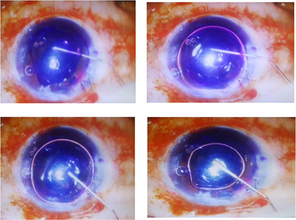
My preferred technique & the easiest one is the use of a single large bubble with a 30G cannula & 2cc syringe, injected gently between the double scroll right at the centre, slowly until the large bubble gently opens up the scroll, which lies on the iris flat (Fig 13).
Then, through the side port at right angle to the scroll direction, the air is slowly withdrawn by the syringe, without allowing the DM to roll back. The AC is now in a collapsed state due to the negative pressure created by the removal of air. This helps in not allowing the DM to roll back.
Then the cannula is inserted below the DM, right at the centre point & injection of air is done slowly until the DM sticks on to the stroma of recipient in a well-centred manner.
The patient is not allowed to get up for atleast 1 hour so that the graft adheres well, after which the eye is seen on Slitlamp for proper adherance & also for raised IOP, which needs to be medically controlled.
Discussion:
Some Do’s & Dont’s provided by Netherlands Institute for Innovative Ocular Surgery(NIIOS)6 also is similar to the method described by us.
The common points are :
- They also recommend superior peripheral iridotomy and not inferior.
- They describe the “rotating corneal rim” technique similar to how we have described.
- They also prefer to keep the DMEK roll continuously submerged in BSS
- The “Moutsouri sign” for identifying the orientation of the scroll using a rod or cannula is common.
- They also insist on keeping the eye soft.
- Air fill for one hour at the end of the surgery is common.
But, few point that are contradictory to our practice in NIIOS, are as follows:
- They recommend Descemetorhexis only under air, while we also do with cohesive.
- They use glass injection system designed by themselves, which is very expensive. They do not recommend plastic injectors.
- They have described various indirect methods for unfolding 6,8.
- Standard tapping
- Air bubble between the rolls
- Bubble rolling “Dapena ” Manoeuvre
- Bubble bumping technique
- 2 parallel cannulas,”Dirisamer” technique
- Single sliding cannula manoeuvre
- Combined techniques
But, we prefer the large single bubble technique, mostly.
- They are not in favour of ‘S’ marks.
- They are recommending only manipulations outside eye for unrolling.
Also, the following practices have to be adhered to, for complete success of DMEK.
- a) The tissue used for DMEK should be of donor age between 50-60 years, preferably, in the beginning, with endothelial cell count of >3000/cubic mm
- b) The choice of DMEK eye is very important. The AC should not be distorted, in any form. Only pseudophakia should be chosen. There should not be any vitreous in AC or decentred IOL, allowing air to escape behind.
- c) If wound is not securely closed or sutured, scroll can get thrown out and lost. Even if DMEK fails, repeat DMEK or DSEK is possible.
Conclusion:
Any Bullous Keratopathies & Endothelial diseases with good anterior segment anatomy, DMEK should be the first choice of surgery, provided the preferred choice of tissue is available.
Mastering DMEK, needs a lot of practice & perseverance but definitely, the result & quality of vision obtained post-DMEK is worth the efforts.
References:
- DMEK – Introduced in 2005 by Gerrit R.J. Melles
- Melles GRJ et al Cornea 1998;34:1102, Ophthalmology 1999; 127:1850-1857, Cornea 2001; 20: 239-243, Cornea 2002, 21:325-327, Cornea 2004;23:286-288, Am I Ophthal 2008;145:222-227
- Busin M et al Ophthalmology 2000; 107:2077-2082
- Terry MA et al Cornea 2001;27: 239-243, Cornea 2005; 24: 59-65, Ophthalmology 2005; 112: 1541-1549, Cornea 2004; 23: 143-153, Ophthalmology.2009; 116: 248-256
- Azar DT et al J Cat.Refract Surg 2001;20:239-243
- niios.com – Netherland Institute for Innovative Ocular Surgery
- Described in detail – Archives of O’logy 2011 Jan; 129:88-94
- Additional unfolding techniques – Ophthalmology 2012
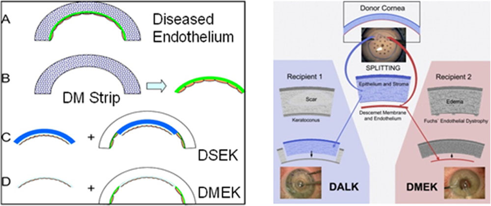
Fig 1 Fig 2
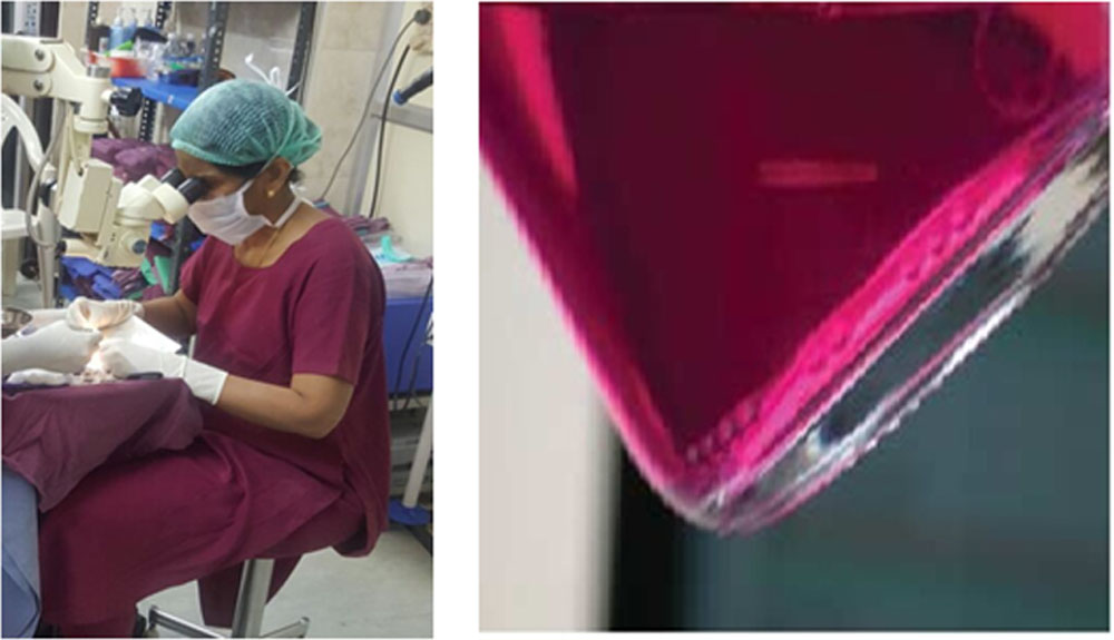
Fig 3 Fig 4
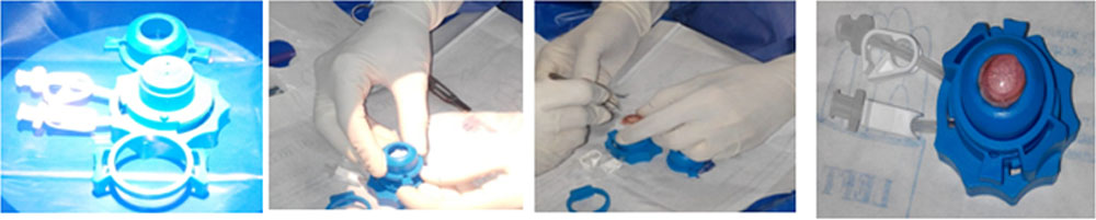
Fig 5
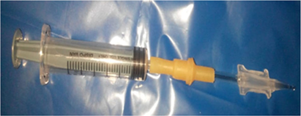
Fig 6a
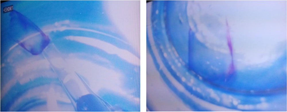
Fig 6b
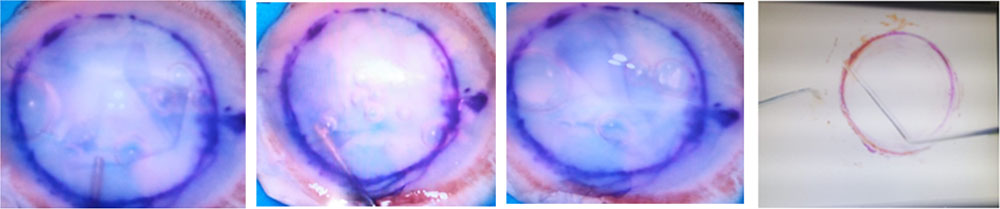
Fig 7 Fig 8
3mm paracentral punching ‘S’ mark on the stromal side through the 3mm punched
Fig 9
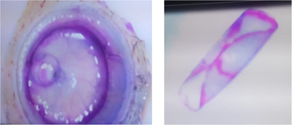
Fig 10a Fig 10b
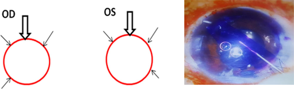
Fig 11 Fig 12 a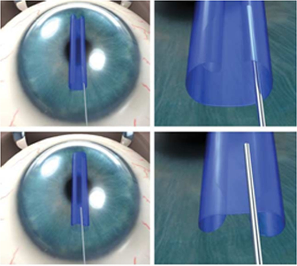
Ref: www.archophthal.com / vol 129 (No1) Jan 2011
Fig 12b
Leave a Comment