

Dr. ANITHA VENUGOPAL, Dr. Sneha Sourabha,Dr.Shalini Singh
ABSTRACT:
AIM:
To describe the clinical features, culture correlation with management and outcomes of pythium keratitis.
METHODS:
Case records of 11 eyes of patients with culture proven pythium keratitis were retrospectively analysed at tertiary eye care centre. History, vision, slit lamp examination was done. Corneal scrapping was performed as a routine to all and Therapeutic Penetrating Keratoplasy done on non-responding cases. 0.2% fortified topical linezolid was started for patients who revealed pythium in culture from corneal scrapping.
RESULTS:
Mean age was 47 years with 7 males and 4 females. All the ulcers involved the visual axis except one. Six patients had infiltrates less than 3×3 and rest of the cases had size more than 3×3. 45.45% (n=5) cases showed Pythium on culture from the scrapping material. A total of 72.72% (n=8) cases underwent TPK. Corneal button of all the cases revealed Pythium on culture. 27.27% (n=3) cases resolved completely with medical management. 27.27% (n=3) cases resolved after TPK. 36.36 %(n=4) cases progressed to untoward complications and 0.9% (n=1) case was lost to follow up.
CONCLUSION:
This cases series emphasizes the similarity of clinical features of pythium keratitis with fungal keratitis leading to delay in diagnosis and poor prognostic outcome. Early diagnosis with morphological features of culture growth, adding topical linezolid and early TPK in non-responding patients may show better outcomes. Studies with larger no of patients are recommended.
BODY
Introduction
Pythium species are aquatic fungi affecting crops and mammals.1 Human infection, known as Pythiosis manifests as Subcutaneous granulomatous lesion, systemic arteritis or Keratitis.1 Pythium insidiosum known to be the commonest organism affecting the humans is classified in Kingdom Protoctista, Phylum Oomycota.1 Risk factors found to be are contact-lens wear, farming exposure, contaminated water exposure and trauma4,8-10Pythium Keratitis (PK) share clinical and morphological features with fungal keratitis leading to delayed diagnosis and untoward complications with vision loss.5 Newer diagnostic techniques are being developed for faster and easier identification with changing trend of the treatment from sole surgical to medical therapy.
Methods
Case records of 11 patients diagnosed with PK between July 2017 and May 2018 were analyzed retrospectively. Detailed history and visual acuity were taken to all the patients. Slit Lamp Examination was done to all the patients noting the ulcer features like size, shape, extent and depth of involvement of the ulcer, presence of hypopyon, corneal integrity and intra ocular findings wherever possible. Corneal scrapping was performed as a routine on all the ulcers and stained with Gram Stain and 10 % Potassium hydroxide (KOH)with . The infective scrapping materials and the corneal buttons after Therapeutic Penetrating Keratoplasty (TPK) were cultured on Blood agar and Potato dextrose agar.All the patients who showed pythium in cultures of scrapping material were started on with fortified 0.2% topical linezolid. No responsive and worsening cases underwent surgical intervention like TPK and evisceration.
Results
Pythium Keratitis was diagnosed in 11 eyes using culture for identification over a period of 9 months (July 2017 and May 2018). The demographic and clinical details are tabulated in TABLE 1. All cases evaluated belonged to age group of 24-70 with mean age 47years with 63.63% (n=7) and 36.36%(n=4) females. No patient had any systemic co morbid disorders like diabetes mellitus or hypertension. Patients presented with subepithelial and stromal infiltrates radiating in a reticular pattern towards limbus. All the ulcers involved the visual axis except one. Six patients had infiltrates less than 3×3 and rest of the cases had size more than 3×3. Four patients presented with hypopyon. The microbiological details are enlisted in TABLE 2. Cases were clinically presumed to be fungal and started on antifungal agents after obtaining scrapping material for light microscopy and culture.
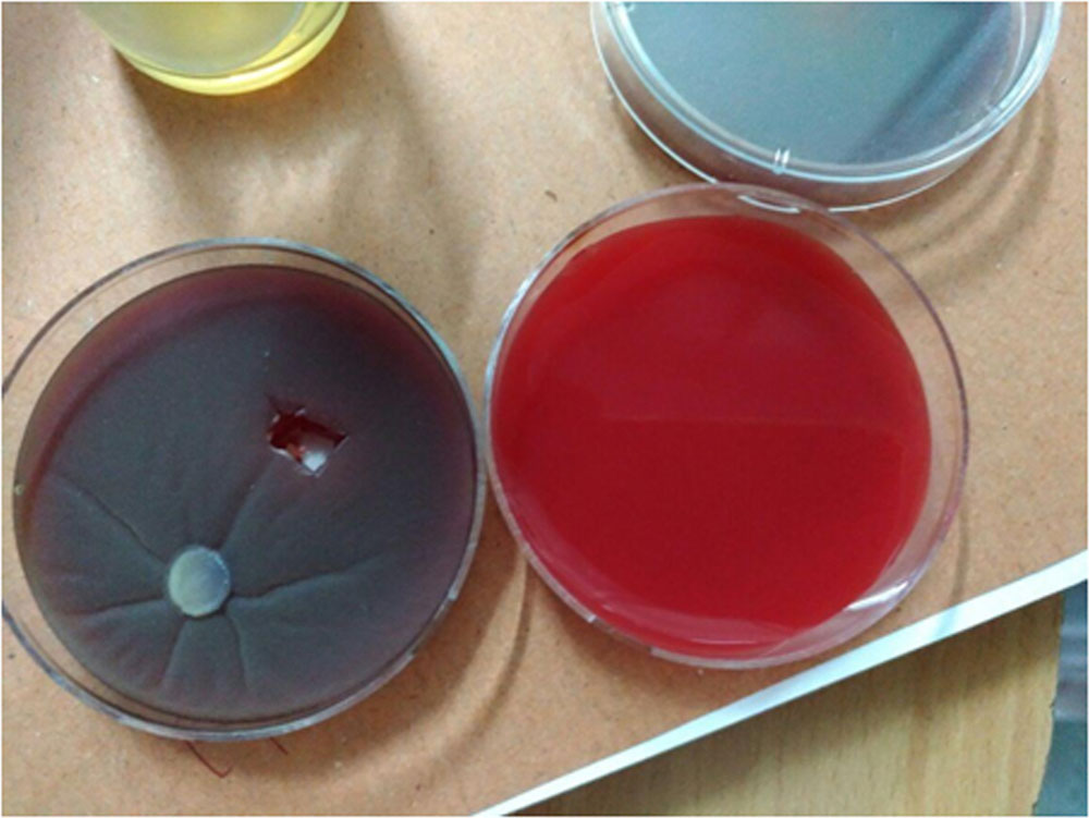
In all cases, fungal likebroad aseptatehyaline filaments with ribbon like folds were noted with 10% Potassium hydroxide (KOH)staining but identification of the organism was not possible. Added to this, one sample showed gram negative organism. The sample materials were inoculated into Blood agarand Potato dextrose agar. The colonies were flat, colourless to creamy white with fine radiations on the culture medium. 45.45% (n=5) cases showed Pythium on culture from the scrapping material. A total of 72.72% (n=8) cases underwent TPK of which 37.5% (n=3) underwent repeat TPK. Corneal button of all the cases revealed Pythium on culture. The final outcomes are shown in TABLE 3. 27.27% (n=3) cases resolved completely with medical management. 27.27% (n=3) cases resolved after TPK. 36.36 %(n=4) cases progressed to untoward complications and 0.9% (n=1) case was lost to follow up.
Discussions
Pythium Keratitis is an emerging morbid infectious condition of the eye being reported from various countries in recent years.It is either being delayed in diagnosis or mis-diagnosed as fungal keratitis thus leading to untoward complications with repeated surgeries and vision loss mainly due to less familiarity with the clinicians and the microbiologists.
Our study noted that PK occurs in middle age group with no sex predilection similar to other studies.6Consistent with other studies, our study also showed incidence of PK to be high among farmers.4Studies have shown majority of patients to be healthy individuals.4Thalassaemia patients have been shown to be a risk factor mainly for systemic pythiosis with relatively less for ocular keratitis and mainly in Thailand. None of our patients were known thalassaemic or had any other systemic co morbid illness.7Studies have shown that contact-lens wear, farming exposure, contaminated water exposure, and trauma as risk factors to develop PK.4,8-10 Our study showed foreign body exposure to be the most common predisposing factor with two cases giving history of native medicine application.
Clinically ulcers showed varying sizes at presentation with or without involving visual axis with underlying dense stromal infiltrates surrounded by subepithelial and superficial stromal opacity in a reticular pattern radiating towards limbus, some with hypopyon. These features were similar in our patients as well.2,7,11
PK is mis or late diagnosed also because of lack of faster, economical and specific diagnostic technique.Studies showed that direct microscopy of corneal scrapings of PK revealed broad, aseptate, hyaline filaments with ribbon-like folds in 10 % KOH confusing with fungal hyphae. KOH staining of all the scrapping materials of our cases revealed similar findings.2,12In our study diagnosis on cultures are made on morphology of the colonies unlike few studies which used zoospore formation in the water based medium thus leading to positive diagnosis early as 2 to 3 days over weeks for the zoospore formation or even negative in some cases where they had to take the support of DNA sequencing and other expensive methods for confirmation.11,12,14Colonies on blood agar and potato dextrose agar colonies were flat, colourless to creamy white with fine radiations on the culture medium and similar features were found in our cultures of scrapping materials and corneal buttons after TPK.2,2,14,Many newer, simpler, economical and faster diagnostic techniques by various staining methods have been proposed like Gomorimethenamine silver (GMS), Potassium iodide – sulphuric acid (IKI-H2SO4). which were not used in our study.15
Cases did not respond or worsened with topical antifungals alone as pythium do not contain ergosterol on which most of the antifungals act.3,13Initially the sole treatment of PK was surgical namely TPK, evisceration or enucleation to save the globe or to eliminate the infected tissue for both ocular as well as systemic infection.4,7,11 In our study as well majority underwent surgical intervention.Recently trend has been slowly changing to medical management with use of drugs like linezolid azithromycin with possible favorable response.6,16 In our study we used fortified linezolid which showed possible better results in patients who were started with topical linezolid after culture revealed for pythium.
Highlights of our study are
- Clinical findings and light microscopy from scrapping though has few distinctive features still misleads us towards fungal keratitis.
- Use of morphological characteristics of colonies in culture for in early diagnosis.
- Possible favorable response of medical treatment with fortified topical linezolid.
- Early surgical intervention with TPK also possibly shows better response especially with combined with topical linezolid.
References
- Imwidthaya P. Human pythiosis in Thailand. Postgrad Med J. 1994;70:558–60.
- Thanathanee O, Enkvetchakul O, Rangsin R, Waraasawapati S, Samerpitak K, Suwan-apichon O. Outbreak of Pythium keratitis during rainy season: a case series. Cornea. 2013;32:199–204.
- Tanhehco TY, Stacy RC, Mendoza L, Durand ML, Jakobiec FA, Colby KA. Pythium insidiosum keratitis in Israel. Eye Contact Lens. 2011;37:96–8.
- Krajaejun T, Pracharktam R, Wongwaisayawan S, Rochanawutinon M, Kunakorn M, Kunavisarut S. Ocular pythiosis: is it under-diagnosed? Am J Ophthalmol. 2004;137:370–2.
- Schlosser E, Gottlieb D. Sterols and the sensitivity of Pythium species to filipin. J Bacteriol. 1966;91:1080–4.
- Bagga B, Sharma S, MadhuriGuda SJ, Nagpal R, Joseph J, Manjulatha K, et al. Leap forward in the treatment of Pythium insidiosum keratitis. Br J Ophthalmol. 2018 Mar 15; [Epub ahead of print]
- Agarwal S, Iyer G, Srinivasan B, Agarwal M, PanchalamSampath Kumar S, Therese LK. Clinical profile of pythium keratitis: perioperative measures to reduce risk of recurrence. Br J Ophthalmol. 2018;102:153–7.
- Krajaejun T, Sathapatayavongs B, Pracharktam R, Nitiyanant P, Leelachaikul P, Wanachiwanawin W, et al. Clinical and epidemiological analyses of human pythiosis in Thailand. Clin Infect Dis. 2006;43:569–76.
- Badenoch PR, Coster DJ, Wetherall BL, Brettig HT, Rozenbilds MA, Drenth A, et al. Pythium insidiosum keratitis confirmed by DNA sequence analysis. Br J Ophthalmol. 2001;85:502–3.
- Lekhanont K, Chuckpaiwong V, Chongtrakool P, Aroonroch R, Vongthongsri A. Pythium insidiosum keratitis in contact lens wear: a case report. Cornea. 2009;28:1173–7.
- Sharma S, Balne PK, Motukupally SR, Das S, Garg P, Sahu SK, et al. Pythium insidiosum keratitis: clinical profile and role of DNA sequencing and zoospore formation in diagnosis. Cornea. 2015 Apr;34(4):438–42.
- Vanittanakom N, Supabandhu J, Khamwan C, Praparattanapan J, Thirach S, Prasertwitayakij N, et al. Identification of emerging human-pathogenic Pythium insidiosum by serological and molecular assay-based methods. J ClinMicrobiol. 2004;42:3970–4.
- Neufeld A, Seamone C, Maleki B, Heathcote JG. Pythium insidiosum keratitis: a pictorial essay of natural history. Can J Ophthalmol. 2018;53:e48–50.
- Kosrirukvongs P, Chaiprasert A, Uiprasertkul M, Chongcharoen M, Banyong R, Krajaejun T, et al. Evaluation of nested pcr technique for detection of Pythium insidiosum in pathological specimens from patients with suspected fungal keratitis. Southeast Asian J Trop Med Public Health. 2014;45:167–73.
- Mittal R, Jena SK, Desai A, Agarwal S. Pythium Insidiosum Keratitis: Histopathology and Rapid Novel Diagnostic Staining Technique. Cornea. 2017;36:1124–32.
- Ramappa M, Nagpal R, Sharma S, Chaurasia S. Successful Medical Management of Presumptive Pythium insidiosum Keratitis. Cornea. 2017;36:511–4.
|
TABLE 1 : Demographic details of Pythium Keratitis patients |
||||
| SL NO / Age / Sex / Occupation | Mode of injury | Time of Presentation | Vision at presentation | Ulcer size |
| 1 / 37 / M / Farmer | Vegetative matter | 3 days | 6/24 | 4×2 |
| 2 / 70 / F / Housewife | NIL | 20 days | HM | 4×3 |
| 3 / 28 / M / Farmer | Vegetative matter | 1 day | 6/6 | 1×1 |
| 4 / 24 / M / Coolie | insect fall | 3 days | 5/60 | 3×2 |
| 5 / 60 / F/ Housewife | Breast milk | 1 month | FCF | 2×2 |
| 6 / 36 / M / Painter | Dust | 2 days | 2/60 | 5×4 |
| 7 / 57 / F / Coolie | Paint | 2 months | HM | 3×2 |
| 8 / 50 / M / Coolie | Vegetative matter | 2 days | 1/60 | 2×2 |
| 9 / 55 / F / Farmer | NIL | 3 days | 6/36 | 5×2 |
| 10 / 49 / M / Coolie | dust, breast milk | 1 week | HM | 4×6 |
| 11 / 60 / M / Farmer | NIL | 4 days | HM | 3×3 |
| TABLE 2: Microbiological details of Pythium Keratitis patients | ||||
| Scrapping culture positive in no of days | Initiation of topical Linezolid | TPK | Corneal button culture positive in no of days | Initiation of topical Linezolid |
| No growth | NO | Yes | No growth | NO |
| No growth | NO | Yes | Pythium / 2 | NO |
| Pythium / 2 | YES | NO | continued | |
| Pythium / 2 | YES | NO | continued | |
| No growth | NO | Yes | Pythium / 3 | NO |
| Pythium / 2 | YES | Yes | Pythium / 2 | YES |
| Pythium / 2 | YES | Yes | Pythium / 2 | YES |
| Pythium / 2 | YES | NO | continued | |
| No growth | NO | Yes | Pythium / 2 | YES |
| No growth | NO | Yes | Pythium / 2 | YES |
| No growth | NO | Yes | No growth | NO |
| TABLE 3 : Final outcome of Pythium Keratitis patients | ||||
| SL NO | Repeat TPK | Corneal button culture | Final Outcome | Vision |
| 1 | Yes | Pythium | Eviceration | |
| 2 | NO | Resolved +graft opaque | FCF | |
| 3 | NO | Resolved | 6/6 | |
| 4 | NO | Resolved | 6/9p | |
| 5 | NO | Scleral melt & evisceration | ||
| 6 | NO | Graft clear | 6/36 | |
| 7 | Yes | Pythium | Anterior Staphyloma | |
| 8 | NO | Resolved | 6/18 | |
| 9 | NO | Clear graft | 6/24 | |
| 10 | Lost to follow up | |||
| 11 | Yes | Pythium & pseudomonas aeruginosa | Anterior staphyloma | |
Chart 1: Slit Lamp Photo of Pythium Keratitis
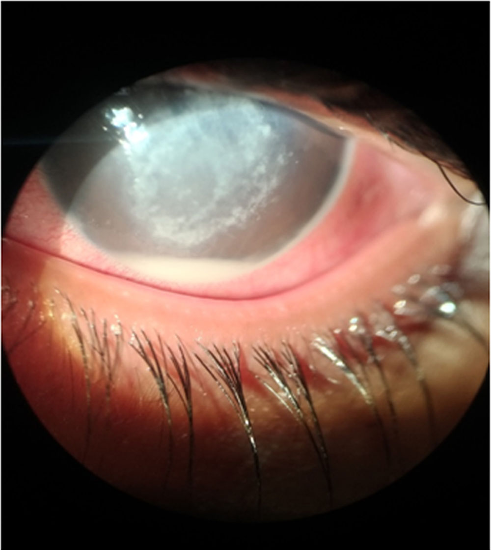
Chart 2: Growth of Pythium in Blood Agar
Leave a Comment