

Dr.GIRDHARI GUPTA,Dr.Shukla A K,Dr.Smita Singh,Dr.M AZHAR MOHD. YUSUF SHEIKH
Purpose :
To assess the need and complianceof treatment in patients with diabetic retinopathy.
Study design :
A prospective observational study. To assess need and compliance to treatment for Diabetic retinopathy.
Results :
7884 known diabetic patients registered at NCD clinics were screened at PHC/ CHC level by Non-mydriatic Fundus Camera. DR in atleast one eye was noted in 608 patients (7.7%) who were referred to base hospital for investigation (OCT, FFA and USG B-Scan) and treatment (PRP/Grid LASER and/or Intravitreal Anti- VEGF).Visual acuity of 12.7% eyes was <6/60. Sight Threatening Diabetic Retinopathy was seen in 50.19% eyes. DME was noted in 18.5 % eyes, 18.1% eyes had Severe NPDR, 11.8% eyes had PDR. In spite of free treatment with provision of transport out of 608 referred patients only 251 (41.3%) patients attended.Out of 151(29.9%) eyes requiring PRP/Grid Laser treatment could be completed in 57 (38.1%) eyes. Out of 152(30.3%) eyes requiring Intravitreal Anti-VEGF only 91 (18.2%) eyes could be treated.
Conclusion :
Lack of awareness, socio-economic constraints and accessibility to eye care reduces compliance in spite of need of treatment to prevent visual deterioration due to Diabetic Retinopathy.
INTRODUCTION
BURDEN OF DIABETIC RETINOPATHY
Zheng et al. in 2012 ,Globally the number of people with DR will grow from 126.6 million in 2010 to 191.0 million by 2030, and it is estimated that the number with vision-threatening diabetic retinopathy (VTDR) will increase from 37.3 million to 56.3 million, if prompt action is not taken. (1)
Study on epidemiology of Diabetic Retinopathy published in 2015 by Ryan Lee et al showed that out of the estimated 285 million people with diabetes mellitus globally, approximately one third have Diabetic Retinopathy out of which, a further one third of DR is vision-threatening DR, which also includes diabetic macular edema (DME)(2)
According to the study by All India Ophthalmology Society (AIOS) on prevalence of diabetic retinopathy in India among 5130 known diabetic patients screened across India in 194 centres in 2014 the prevalence was 21.27 % with about 12.27 % in Central India and 34.06 % in North India (3)
NEED OF TREATMENT FOR DIABETIC RETINOPATHY
Study by Narendran et al showed the prevalence of near normal vision in about 70.2 %, Visual impairment in 11.9 % and severe visual impairment in 1.5 % of known diabetic patients. (4)
Study by Olivarius showed that in patients diagnosed with clinical, often symptomatic type 2 diabetes, 6.3% were visually impaired. Among those patients with reduced sight, 76% had cataract and 58% retinopathy (5)
Study by Shreshta et al showed that diabetes accounts for Legal blindness in 83% of visual impairment among persons with youth-onset diabetes, and 33% among persons with older age onset diabetes . Around two-thirds of diabetics have an increased possibility of visual impairment after 35 years of suffering the condition, and are 25 times more likely to go blind, compared with other conditions. Visual impairment increased with increase in grade and severity of diabetic retinopathy (6).
Diabetic retinopathy is the leading cause of blindness among the working age adult population aged 20–74 years. Diabetic retinopathy can be treated with modalities that have been proven to decrease the risk of severe vision loss by > 90% if early detection is made (7)
Diabetes alone can increase the risk of blindness 25 times (8).
COMPLIANCE OF DIABETIC RETINOPATHY SCREENING AND TREATMENT
Clausette E. Hall et al assessed the need of people living with diabetes and diabetic retinopathy in 5 rural district hospitals in Kilimanjaro region, Tanzania with a population of 1,376,702. Mixed methods approach (Qualitative and Quantitative) was used, concluded the need of comprehensive, accessible diabetic education to prevent diabetic retinopathy and observed that low level of self-efficacy,high level of anxiety and depression have a negative impact on compliance for screening of diabetic retinopathy.(9)
Salah Memon et al in a observational case study, assessed the acceptance of retinal screening, laser treatment uptake and subsequent follow-up in diabetics in urban diabetes care centre, Karachi. In this study, 100% compliance was observed for retinal screening, 70% for laser treatment uptake and 21.25% of the pateints who availed laser treatment attended follow-up. (10)
MATERIAL AND METHODS
A rural diabetic population based screening was done to assess the need and compliance to treatment.
Type of study :- Prospective Cross-sectional observational study
Place of study : – Government health facilities (CHCs) and Eye OPD, Tertiary care hospital.
Duration of study : – 18 months
StudySubjects:- Known diabetic population in rural area
Inclusion Criteria:
- All known diabetic patients at government health centres CHCs and attending
eyeopd.
Exclusion criteria:
- Patients not giving consent for study.
- Patients who are uncooperative.
- Hazy media
- Patients with any posterior segment pathology other than diabetic retinopathy
METHODOLOGY:-
7884 Known diabetic patients registered at NCD clinics were screened at PHC/ CHC level by Non-mydriatic Fundus Camera. DR in at least eye was noted in 608 patients who were referred to base hospital for investigation and treatment. Free transport facility was provided to referred patients from their residence to base hospital. Out of 608 referred patients,252 patients came to base hospital for further investigation and treatment.
After taking informed consent from the patient, data was collected on a pre-designed proforma and a separate file was made for each patient to collect the data of follow-up visits.
1.Demographic details of the patient- Unique id, Age, sex, address, contact number was noted.
2.Known duration of diabetes, Type of DM, Nature of treatment, control of DM, Random blood sugar, blood pressure was noted.
3.Best corrected Visual acuity was tested using a ETDRS vision chart, intraocular pressure was noted.
4.Anterior segment examination by torch light, slit-lamp was done to rule out cause of hazy media and lens status was noted by distant direct ophthalmoscopy.
5.Examination of posterior segment was done with the direct ophthalmoscopy, Slit- lamp(90D) and if required with indirect ophthalmoscopy and grading of diabetic retinopathy noted down.
6.B-scan ultrasonography was undertaken in hazy media for posterior segment assessment. 7. Fundus Photograph, Optical Coherence Tomography was done for each patient irrespective of grade of diabetic retinopathy, Fundus Fluorescein Angiography was performed wherever required.
7.According to above investigations, need for treatment was assessed among Laser photocoagulation and Intravitreal anti-vegf.
VISUAL ACUITY
The presenting visual acuity of the patients were measured separately for each eye using ETDRS chart cut out with “E” optotypes at a distance of 4 meters and improvement with pinhole was noted. The LogMAR 0 correspond to 6/6 equivalent of Snellen‘s chart, LogMAR 0.2 corresponds to 6/9 , LogMAR 0.3 correspond to 6/12, LogMAR 0.5 to 6/18, LogMAR 0.6 correspond to 6/24, LogMAR 0.8 corresponds to 6/36, LogMAR 1 .0 correspond to 6/60. Visual acuity measurement were performed in good illumination. Patients who read the largest letter confirms VA 6/60 , were then shown small size letter E the corresponding visual acuity was recorded. Patient who were unable to read the largest letter at 4 meter were retested at 2 m and VA was recorded as 3/60 . Testing included the ability to count fingers ,handmovements or to perceive light , when necessary.
VISUAL ACUITY: ETDRS LOGMAR NOTATION AND ITS CORRESPONDING SNELLEN’S NOTATION(11)
| ETDRS LogMAR Notation(Visual Acuity from 4 meters) | Corresponding Snellen‘s vision chart Notation(Visual acuity from 6 meters) |
| LogMAR 0 | 6/6 |
| LogMAR 0.20 | 6/9 |
| LogMAR 0.30 | 6/12 |
| LogMAR 0.50 (~0.48) | 6/18 |
| LogMAR 0.60 | 6/24 |
| LogMAR 0.80 (~0.78) | 6/36 |
| LogMAR 1.00 | 6/60 |
According to WHO criteria of Blindness Visual Status was divided into Six categories :
| Category | Best Corrected Visual Acuity (BCVA) of better eye | Indian definition |
| 1 | 6/6 to 6/18 | Near Normal |
| 2 | < 6/18 to 6/60 | Visual impairment / Low Vision |
| 3 | <6/60 to 3/60 | Severe visual impairment |
| 4 | <3/60 to 1/60 | Blind |
| 5 | <1/60 to PL+ | Blind |
| 6 | No PL | Total Blindness |
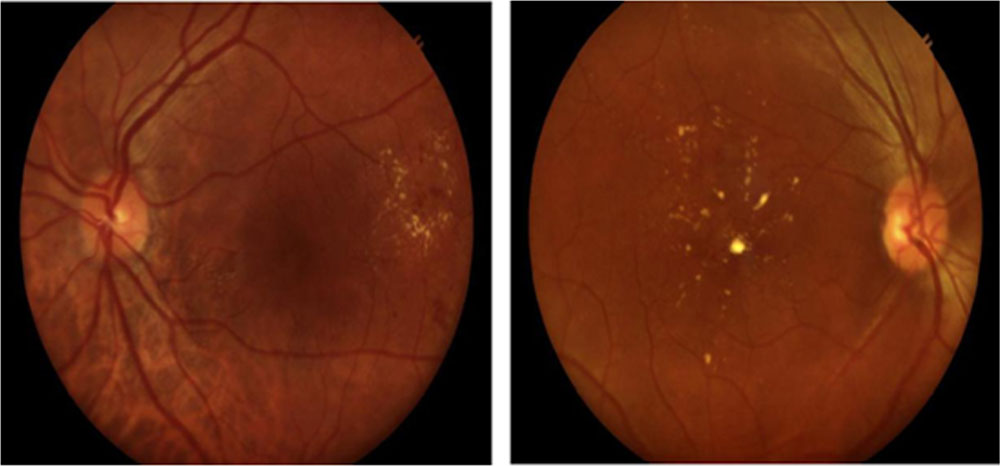
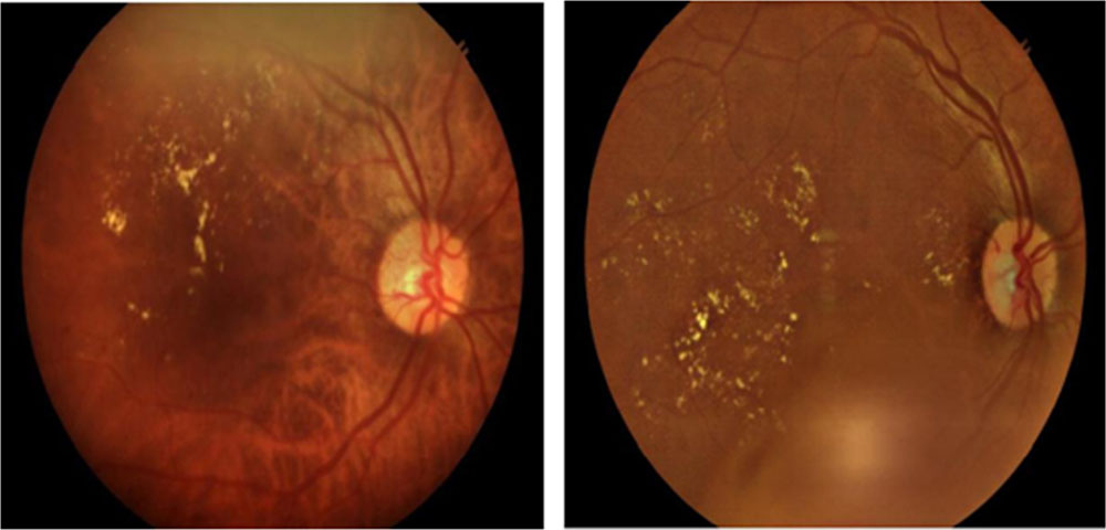
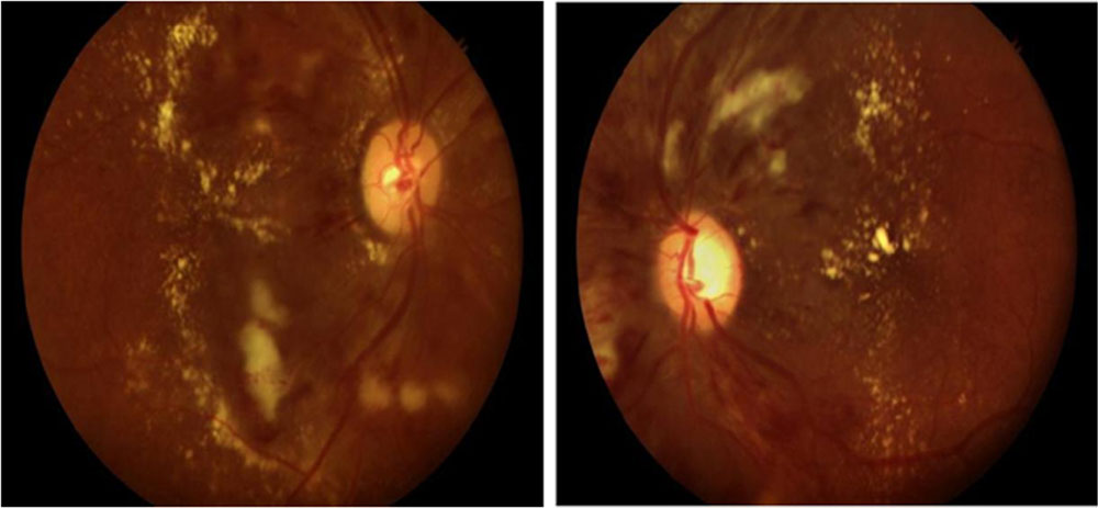
Grading of diabetic retinopathy was done accoerding to ETDRS Revised modified Airlie House diabetic retinopathy classification.(12)
| A. Nonproliferative Diabetic Retinopathy (NPDR) |
| 1. Mild NPDR: |
| a. At least one microaneurysm |
| 2. Moderate NPDR: |
| a. Hemorrhage/microaneurysm OR
b. Soft exudates (cotton wool spots), venous beading, and intraretinal c. microvascular abnormalities definitely present d. Criteria not met for severe NPDR, very severe NPDR, or PDR |
| 3. Severe NPDR:
a. Hemorrhage/microaneurysm in all 4 quadrants OR b. Venous beading in at least two quadrants OR c. Intraretinalmicrovascular abnormalities in at least one quadrant |
| 4.Very Severe NPDR:
a. Any two or more of criteria for severe NPDR b. Criteria not met for PDR |
| B. Proliferative Diabetic Retinopathy (PDR) |
| 1. Early PDR:
a. New vessels b.Criteria not met for high-risk PDR |
| 2. High-risk PDR:
a. Neovascularization of the disk (NVD) ≥1/3 to 1/2 disk area OR b. Neovascularization of the disk and vitreous or preretinalhemorrhage OR c. Neovascularization elsewhere (NVE) ≥1/2 disk area AND vitreous or pretretinalhemorrhage |
| 3. Severe PDR:
a. Posterior fundus obscured by preretinal or vitreous hemorrhage OR b. Center of macula detached |
| Clinically Significant Macular Edema (CSME) |
| 1. Thickening of the retina ≤500 µm from the center of the macula
OR 2. Hard exudates and adjacent retinal thickening ≤500 µm from macular center OR 3. Zone of retinal thickening at least 1 disc area in size located ≤1 disc diameter from the center of the macula |
SIGHT THREATENING DIABETIC RETINOPATHY:
Severe NPDR, PDR,PDR WITH HRC, Advanced PDR and presence of DME irrespective of grade of DR were considered as sight- threatening diabetic retinopathy and patients with STDR were considered for treatment ( Laser/ Intra-vitreal injection).
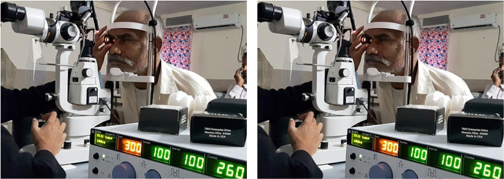
Pan retinal photocoagulation was completed in 3 sittings for each eye with average number of 1500 spots. Macular Grid was done in those patients having leakages surrounding macula on Fundus Fluorescein Angiography. Intravitreal injection was given to those patients having macular edema on OCT.
Fundus Photograph with diabetic retinopathy changes
MILD NPDR MILD NPDR WITH CSME
MODERATE NPDR MODERATE NPDR WITH CSME
SEVERE NPDR WITH CSME PDR WITH CSME
PDR WITH HRCADVANCED PDR
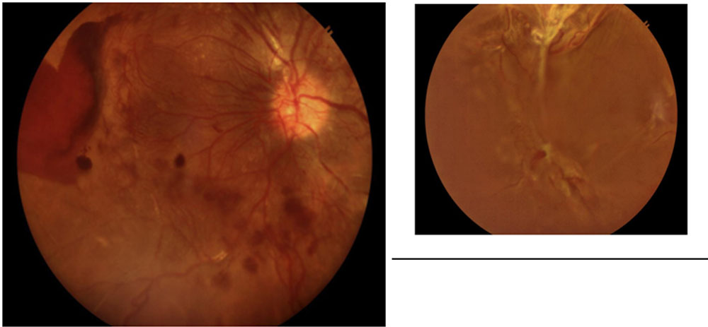
LASER PHOTOCOAGULATION INTRA VITREAL ANTI-VEGF
DATA MANAGEMENT & ANALYSIS –
Data was collected in Diabetic Retinopathy Screening Clinical proforma and in Diabetic retinopathy Android application in a tablet. Collected data was in coded form and it was entered and managed in Epi Info 7 Database. Statistical analysis was done by using descriptive and inferential statistics using chisquare test and software used in the analysis were SPSS 22.0 version and GraphPad Prism 6.0 version and p<0.05 is considered as level of significance.
ETHICAL CONSIDERATION –
Ethical approval was sought from the Ethical committee of the Institute and Department of Ophthalmology, MGIMS , Sewagram. The participants were informed about the study and were requested for participation.
RESULTS:
During the study period a total of 7884 known diabetic patients registered at NCD clinics were screened at PHC/ CHC level by Non-mydriatic Fundus Camera. DR in at least one eye was noted in 608 patients (7.7%) who were referred to base hospital for investigation (OCT, FFA and USG B-Scan) and treatment (PRP/Grid LASER and/or Intravitreal Anti- VEGF).
COMPLIANCE TO TREATMENT:
In spite of free treatment with provision of transport, out of 608 referred patients only 252 patients attended with compliance of 41.4%.(Table 1)
Table 1 : compliance for treatment
| known diabetic patients | Referred patients | DR patients attended the hospital | Compliance | |
| Male | 4420 (56.06%) | 334 (54.93%) | 164 (65.07%) | (49.10%) |
| Female | 3464 (43.97%) | 274 (45.07%) | 88 (34.93%) | (33.33%) |
| Rural | 5613 (71.2%) | 450 (74.01%) | 151(59.92%) | (33.35%) |
| Urban | 2270 (28.8%) | 158 (25.99%) | 101 (40.08%) | (63.92%) |
Poor compliance was observed in Rural population and females, while good compliance was observed in urban population and males comparatively.
NEED FOR TREATMENT:
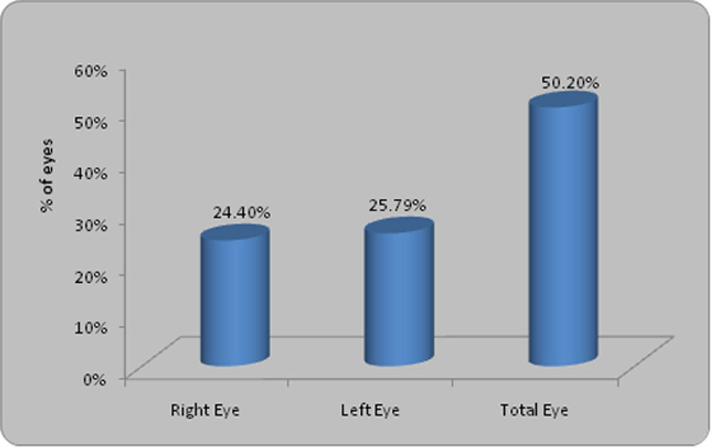
Out of 252 DR patients, 122 (48.41%) patients and out of 504 eyes,253 eyes (50.19%) were having sight threatening diabetic retinopathy and needed for treatment.
Majority of patients 324 (64.29%) have near normal vision(6/6-6/18), 23.02% have visual impairment (<6/18-6/60), 4.37% have severe visual impairment(3/60-6/60), 8.15% have blindness (<3/60-PL+) and only 1 eye has absolute blindness.
18.25% eyes have severe non proliferative diabetic retinopathy, 11.9% eyes have proliferative diabetic retinopathy,18.5% eyes have diabetic macular edema.
Mean age of male was 58.84±8.88 in years and of female was 60.56±9.67. Patients with Type 2 DM were 121(99.20%) and only 1 patient (0.80%) with Type 1 DM. 113 patients (92.6%) were taking oral hypoglycaemic drugs, 8 patients(6.6%) were on insulin and 1 patient (0.8%) was on ayurvedicmedicine.
Association of known duration of DM with patients needed for treatment
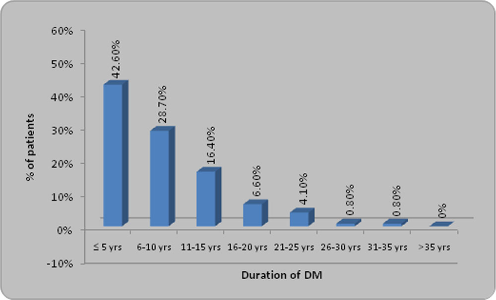
It was observed that majority of patients(42.6%) have < 5 years of known duration of DM. Majority of patients (73%) were associated with uncontrolled DM, only 27% have contolled blood sugar.
It was observed that 65 patients (53.27%) have history of hypertension and 57 patients (46.73%) were not hypertensive.
Out of 151(29.9%) eyes requiring PRP/Grid Laser treatment could be completed in 57 (38.1%) eyes. Out of 152(30.3%) eyes requiring Intravitreal Anti-VEGF only 91 (18.2%) eyes could be treated.
DISCUSSION
Study on epidemiology of Diabetic Retinopathy published in 2015 by Ryan Lee et al showed that out of the estimated 285 million people with diabetes mellitus globally, approximately one third have Diabetic Retinopathy out of which, a further one third of DR is vision-threatening DR, which also includes diabetic macular edema (DME).(2)
In our study, 608 patients were found to have diabetic retinopathy (DR)out of 7884 diabetics (7.7%), out of which 252 patients with compliance of 41.4% attended, among which 122 patients (48.41%) have sight threatening diabetic retinopathy. Low prevalance of DR was due to screening of undilated pupil and due to hazy media.
Study by Narendran et al showed the prevalence of near normal vision in about 70.2 %, Visual impairment in 11.9 % and severe visual impairment in 1.5 % of known diabetic patients.(4)
Study by Shreshta et al showed that diabetes accounts for Legal blindness in 83% of visual impairment among persons with youth-onset diabetes, and 33% among persons with older age onset diabetes . Around two-thirds of diabetics have an increased possibility of visual impairment after 35 years of suffering the condition, and are 25 times more likely to go blind, compared with other conditions. Visual impairment increased with increase in grade and severity of diabetic retinopathy.(6)
In our study, Majority of patients 324 (64.29%) have near normal vision(6/6-6/18), 23.02% have visual impairment (<6/18-6/60), 4.37% have severe visual impairment (4.37%).
Salah Memon et al in a observational case study, assessed the acceptance of retinal screening, laser treatment uptake and subsequent follow-up in diabetics in urban diabetes care centre, Karachi. In this study, 100% compliance was observed for retinal screening, 70% for laser treatment uptake and 21.25% of the pateints who availed laser treatment attended follow-up.(10)
In our study, compliance for treatment in urban population was (63.92%).so results are similar to previous study.
Sheth NR et al in 2017 assessed the knowledge and attitude of the general population regarding diabetes mellitus and diabetic retinopathy with an aim to understand the shortcomings in awareness programs. The authors found 50 patients were randomly selected.42%- females and 58% – males. Mean age – 58.5 years. Age range 40 to 71 years. Patients with good sugar control -26% and poor control – 74%. 94% of the patients were taking treatment for DM and 6% were not on any treatment. 48 % of the patients were illiterate, 10% were graduate and 42% had education below 12th standard. 50% were aware about DM affecting the eye .38% had taken eye treatment and 62% had not. 26% were aware of DM affecting eye in spite of good control and 26 % aware of the need of check-up in poor control. 40% aware of the complications related to DM. The authors concluded that better literacy rates is contributory to public awareness, however trend for poor practice needs to be radically changed with aggressive public motivation emphasizing the necessity of screening and follow ups.(13)
Srinivasan NK et al in 2017 in a hospital-based, cross-sectional study documented the Knowledge, Attitude and Practice (KAP) patterns of diabetic patients regarding diabetes and diabetic retinopathy, to determine association between them, and to identify barriers to compliance with follow up and treatment regimes. The authors found of the 288 patients in the study, 42% had good knowledge about diabetes, but only 4.5% had good knowledge about retinopathy. Good knowledge about diabetes was significantly associated with positive attitude towards diabetes and good practice patterns regarding retinopathy; awareness of retinopathy was also significantly associated with good practice. A total of 61.1% of patients did not have periodic eye examination; most common barrier identified was lack of awareness about the necessity for this (38.5%). The authors concluded that Good knowledge about the disease was significantly associated with positive attitude and good practice patterns. Knowledge about diabetic retinopathy was poor among the patients in our study. Lack of awareness concerning the need for screening for retinopathy was a major barrier to regular screening. There is an urgent need to educate diabetic patients about this potentially blinding complication of diabetes.(14)
CONCLUSION:
Lack of awareness, socio-economic constraints and accessibility to eye care reduces compliance in spite of need of treatment to prevent visual deterioration due to Diabetic Retinopathy.
References:
(1) Zheng Y, He M, Congdon N. The worldwide epidemic of diabetic retinopathy. Indian Journal of Ophthalmology. 2012;60(5):428-431. doi:10.4103/0301-4738.100542.
(2) Epidemiology of diabetic retinopathy, diabetic macular edema and related vision loss .Ryan Lee, Tien Y. Wong, and CharumathiSabanayagam.
(3) Prevalence of diabetic retinopathy in India: The All India Ophthalmological Society Diabetic Retinopathy Eye Screening Study 2014. Salil S Gadkari, Quresh B Maskati, Barun Kumar Nayak . Indian Journal Of Ophthalmology
(4) .Narendran V, John RK, Raghuram A, Ravindran RD, Nirmalan PK, Thulasiraj RD. Diabetic retinopathy among self reported diabetics in southern India: a population based assessment. The British Journal of Ophthalmology. 2002;86(9):1014-1018.
(5) Niels de Fine Olivarius, VolkertSiersma, GitteJuulAlmind, NielsVestiNielsen. Prevalence and progression of visual impairment in patients newly diagnosed with clinical type 2 diabetes: a 6-year follow up study. BMC Public Health, 2011, Volume 11, Number 1, Page 1.
(6) .Shrestha GS, Kaiti R. Visual functions and disability in diabetic retinopathy patients. Journal of Optometry. 2014;7(1):37-43. doi:10.1016/j.optom. 2013.03.003.
(7) Role of Early Screening for Diabetic Retinopathy in Patients with Diabetes Mellitus: An Overview. Praveen Vashist, Sameeksha Singh, NoopurGupta, and RohitSaxena. Indian J Community Med. 2011 Oct-Dec; 36(4): 247–252.
(8) . Alexander L.J. Appleton and Lange; Connecticut: 1989. Primary care of the posterior segment.p. 193.
(9). Claudette E. ,Anthony B. Hall,,Gerjo Kok, Joyse Mallya and Paul Courtright.A needs assessment of people living with diabetes and diabetic retinopathy.BMC Research Notes20169:56 .https://doi.org/10.1186/s13104-016-1870-4
(10). Memon S, Ahsan S, Alvi R, Fawwad A, Basit A, Shera S, Sheikh SA, Fahim MF Retinal Screening Acceptance, Laser Treatment Uptake and Follow-up Response in Diabetics Requiring Laser Therapy in an Urban Diabetes Care Centre.JColl Physicians Surg Pak. 2015 Oct;25(10):743-6. doi: 10.2015/JCPSP.743746
(11) Prospective Evaluation Of Visual Acuity Assessment: A Comparison Of Snellen Versus Etdrs Charts In Clinical Practice. Peter K. Kaiser, MD. Trans Am Ophthalmol Soc. 2009 Dec; 107: 311–324.PMCID: PMC2814576
(12) . Grading diabetic retinopathy from stereoscopic color fundus photographs-an extension of the modified Airlie House classification. ETDRS report number 10. Early Treatment Diabetic Retinopathy Study Research Group.Ophthalmology. 1991 May;98(5 Suppl):786-806.
Leave a Comment