


Dr. Anubhav Goyal, Dr. Giridhar Anantharaman, Dr. Mahesh G
Abstract
Purpose:
To design a low cost, non-contact smartphone based screening system in retinopathy of prematurity and to illustrate its potential clinical application as a teleophthalmology system.
Method:
Neonatal intensive care unit based bed-side ROP documentation donebetween January 2018 and May 2018. Images captured by using a smartphone and non-contact +40D, +28D or +20D indirect condensing lenses. Coaxial light source of the phone was used to acquire digital image of the fundus. With our usual smartphone based camera we extracted highquality, still images from the video clip.
Results:
Total of 220 eyes of 110infants were screened for ROP. 50 out of 220 eyes were diagnosed as ROP and only 20 eyes were diagnosed to Type 1 ROP showing an incidence of 9.09%.We used smartphone imaging in 22 out of total 50 eyes diagnosed to have ROP. It is a light weight, user-friendly, high quality smartphone based fundus imaging with field of view varying from 30 degree, 55 degree and 90 degree with +20D, +28D and +40D indirect condensing lenses respectively, which gives excellent images for ROP documentation. It can also be used as telescreening device.
Conclusion:
The currently described system was able to take consistently high quality fundus photographs for bed-side documentation of ROP in neonatal ICUs using readily available instruments that are portable with simple power sources.
Keywords:
Neonatal intensive care unit, retinopathy of prematurity,condensing lens-smartphone-MIIRETCAM (CSM) device assembly.
Introduction
Retinopathy of prematurity (ROP) is a leading cause of childhood blindness worldwide. The incidence of ROP in India is reported between 38 –51.9% in low birth weight infants.1Incidence of ROP in India is increasing because of advancing neonatal care andimproved neonatal survival rate. According to Early Treatment for Retinopathy of Prematurity early detection and prompt treatment can reduce unfavourable outcomes secondary to high-risk prethreshold ROP.2
The current gold standard method for ROP screening requires indirect ophthalmoscope with the condensing lens.3But this method is subjective and correct diagnosis can be missed if Ophthalmologist is inexperienced or when the infant is too sick to allow adequate fundus examination. Nowadays, digital fundus photography is used in ROP screening to facilitate consultation in difficult cases and also contribute to medicolegal issues. However, it is not easy to take a fundus photograph of a newborn without professional equipment such as the RetCam system,4 which is costly and unaffordable in most of the local medical healthcare systems. The ease of use, portability, and cost-effectiveness of smartphone fundoscopy can share the advantages of telemedicine, either to document the fundus status or to consult experienced senior Ophthalmologists and also helps in medicolegal issues.5Besides the light source of smartphones is safe for human eyes. Retinal irradiance from a smartphone (iPhone) is less than that from an indirect ophthalmoscope.8
The smartphone has become the basic necessity in today’s world. Due to advanced technology, cost-effectivity and ease of handling for capturing images, smartphones are commonly used as the clinical imaging device in ophthalmology.3 Modern smartphone camera is already equipped with a high-quality optical system and a coaxial light source which can be used to capture high-quality retinal images. In this study we used condensing lens with smartphone to capture widefield, bedside fundus images of ROP infants in an Indian population.
Methods
It is an observational study. Bedside fundus images of preterm infants with ROP were captured in neonatal intensive care unit (NICU) between January 2018 to May 2018. All infants were awake during fundus examination and smartphone imaging. Pupils were well dilated and only topical anaesthesia was administered for applying a paediatric wire speculum and performing globe rotation. One nurse assisted in holding infant’s head stable.
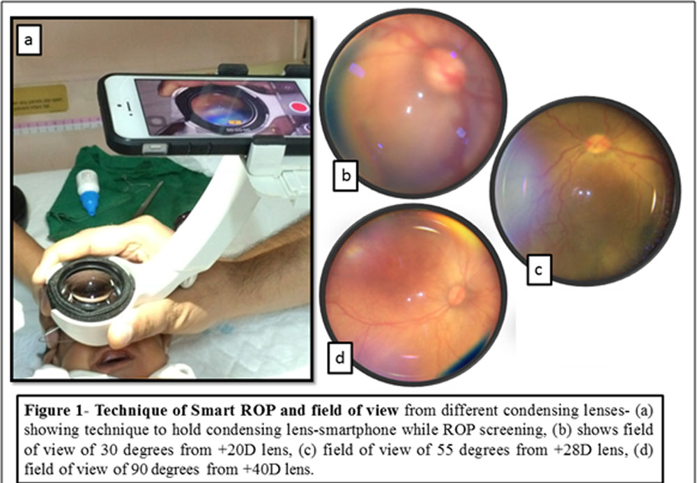
Images were captured with a smartphone, iPhone 5S (Apple Inc., Cupertino, CA, USA) and either 20D, 28D or 40D lens (Volk Optical Inc., Mentor, OH, USA). 20D, 28D, and 40D lenses give a field of view of 30 degrees, 55 degrees and 90 degrees respectively. Condensing lens and a smartphone (iPhone 5S) under original camera settings is used to record a video. The constant coaxial inbuilt light source and capturing snapshots out of the video help us in documenting retinopathy of prematurity. MIIRetCam10 was used to hold lens and smartphone at a fixed working distance. Smartphone was aligned in a straight axis at a particular distance and stabilized with a supporting handle.The infant’s forehead was supported by one hand and the another hand can be used occasionally to rotate the globe for better periphery visualization.
The eyelids were widely opened by the use of pediatric wire speculum and pupil should be fully dilated to minimise glare during fundoscopy with the nurse monitoring the infant during the course of examination. Hazy vitreous and vasculosalentis were other reasons which hampered good quality image acquisition. The recording was done by video mode of approximately one-minute duration and the video was kept on during the whole examination. Video mode automatically focuses to give clear imaging. If the light reflection is too severe, the light intensity can be reduced by covering the flash with a layer of Micropore tape (3M, St.cPaul, MN, USA).7 Once the recording was completed, still images were captured from the video sequence by taking a screenshot (by pressing and holding Home and Sleep/Wake button simultaneously). Images captured were transferred and read by specialists (Ophthalmologist and Neonatologists) elsewhere by internet guided inbuilt softwares in smartphone, customising telemedicine in ROP screening.
Single Ophthalmologist (AG) trained in ROP screening captured all images. Image quality was graded as good and poor based on ability of grader to determine the dilatation and tortuosity of vessels or presence of different stages of ROP in the images. “Good” was defined as an image in which there was a clear view of optic nerve and vessels and the grader could easily discern the dilatation and tortuosity of vessels or presence of various ROP stages with or without glare artefact, and “poor” image in which the grader was unable to clearly determine the characteristics of the vessels.
Telemedicine refers to the use of telecommunication and information technologies in order to provide clinical health care at a distance. It involves capturing and transporting patient data for subsequent interpretation by a remote expert.
Table 1 highlights the comparison of different imaging modalities used for fundoscopy in infants with ROP.
| Imaging modality | Field of view | Setting for use | Staffing requirement | Advantages | Disadvantages |
| Smartphone ROP-
Condensing lens • +20D • +28D • +40D |
30° 55° 90°
|
• Special care baby unit
• OPD • Theatre |
•Ophthalmologist | • Non-contact based
• Portable • Wide field of view • Cost effective • Able to image till oraserrata through sclera depression |
• High-resolution images
• Only colour imaging available
|
| NIDEK
Camera[3] |
30° | • Special care
baby unit •Outpatient department • Theatre |
•Ophthalmologist | • Non-contact based
• Portable |
• Low resolution images
• Narrow field of view • Only colour imaging available • Unable to image till oraserrata
|
| 3Netra Neo widefield camera[13] | 120° | • Special care baby unit
• OPD • Theatre |
•Ophthalmologist
• Nursing staff tomonitor vital signs |
• Portable
• Wide fundal field of view • Fast image acquisition |
• Contact based
• Heavy weight camera • Unable to image till oraserrata • Costly |
| RetCam
Widefield camera[14,15] |
130° | • Special care baby unit
• OPD • Theatre |
•Ophthalmologist
• Nursing staff tomonitor vital signs |
• Portable
• Wide field of view • Fast image acquisition • Colour and FFA imaging available
|
• Contact based
• Heavy weight camera • Sedation essential for high quality angiograms • Unable to image out to oraserrata • Costly |
| Optos
ultrawidefield camera[17] |
200° | • OPD
|
•Ophthalmologist
• Nursing staff to monitor vital signs • Ophthalmic photographer |
• Non-contact
• Fast image acquisition • High resolution • Wide field of view • Colour imaging available. |
• Non-portable
• Unable to image out to oraserrata • Costly
|
Results
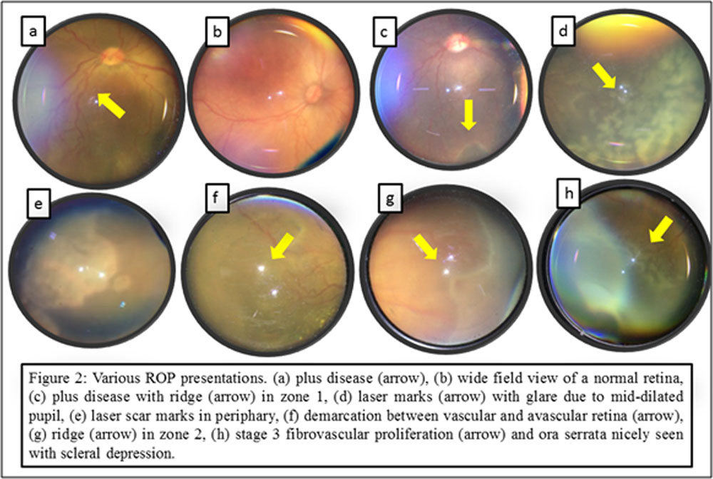
A total of 220 eyes of 110preterms infants were screened for ROP in neonatal intensive care unit (NICUs) of 17 referral hospitals in Kochi, Kerala between January 2018 to May 2018. All infants less than 1700 g birth weight and less than 35 weeks gestational age were screened according to latest Indian screening guidelines.12 ROP was observed in 50eyes with an incidence of 22.72% out of which type 1 high-risk prethreshold ROP was seen in 20 eyes with an incidence of 9.09%. Smartphone based fundoscopy was used to document ROP in 22 out of 50 eyes diagnosed as ROP. Image quality was “good” in 20 out of 22 (90.90%) eyes undergone smartphone imaging. 2 eyes showing poor vessel and retinal differentiation were graded as “bad”. 6 eyes with zone 1 ROP were documented without globe contact or rotation. Glare and artefacts were seen in 16 out of these 22 eyes (72.72%) but did not hamper in image clarity in most of the eyes. Using different condensing lenses varying from +20D, +28D and +40D give us the advantage of wide field visualization with a view of 30 degrees, 55 degrees and 90 degrees respectively. Peripheral retinal fundoscopy till oraserrata was possible in all eyes. Figure 1 showing the technique of Smart ROP image capture and comparison of various images taken from different condensing lenses. Various presentations of ROP are seen in figure 2.
Discussion
Use of smartphone to capture retinal images has several advantages. First, a smartphone has an almost coaxial and constant light source that can directly photograph the retina. Second, the smartphone camera can adjust focus automatically on the retina. Third, fundoscopy with a smartphone is an inexpensive, non-contact and portable way to image the retinal in patients with ROP. Fourth, the smartphone and MIIRETCAM being a lightweight gadget, the
condensing lens-smartphone-MIIRETCAM assembly can be supported with a single hand to capture retina pictures. This is probably the best and the only way for a single Ophthalmologist who has to visit multiple NICU setups on the same day to achieve good documentation. Fifth, easy availability and quick data transfer facility make it useful and effectivetelescreening tool to discuss ROP even in remote places. Use of +28D and +40D in addition to +20D condensing lenses have an advantage of wide field visualization of retina. It can also help in visualization till far periphery with minimal scleral depression. Although the quality of the picture is not equivalent to those taken by professional equipment like RetCam, the cost performance has more value in developing countries like India. In addition,availability of a smartphone in local and remote hospitals or relatively poor medical healthcare environments is easy nowadays. This technique is also helpful in general patient consultation by experienced Ophthalmologists, and the ease of sharing images makes smartphone fundoscopy an extremely useful technique for ROP screening and telemedicine.7
Using only one hand to hold smartphone assembly and other hand for eyeball rotation ease us to capture retinal periphery images equivalent to professional wide field fundus
imaging systems. Lin SJ et al explained manual adjustment oflens and smartphone light but our device assembly facilitates automatically adjusted coaxial image with minimum effort.7On an average, the time taken by an experienced Ophthalmologist for a full examination is approximately 1 minute which is slightly longer than that required by a professional widefield retinal imaging system. Cost-affectivity, ease of handling and telescreening facility makes smartphone imaging more useful than any other imaging modality.
KIDROP composed of 3-4 trained Ophthalmic staff, RETCAM shuttle and a laptop equipped with additional special software to record ROP images which are assessed by
Ophthalmologist elsewhere.13 While Smartphone ROP needs single trained person (mainly Ophthalmologist) and a mobile light weight device to hold smartphone and condensing lens using inbuilt video settings of phone to record ROP. Smartphone based fundus camera have reasonably good field of view of 55 degree with +28D and 90 degree with +40D condensing lens, which is only minimally less than 3 netraneo (120 degree)13, RetCam (130 degree)14 or Optos imaging system (200 degree)3,17. Probe weight of contact imaging devices like 3 netra neo is 340 gms and that of Ret Cam shuttle is still heavier exerting undue pressure overeyeball during examination, a problem that would not be expected of a non-contact system.17Moreover the cost of condensing lens ( easily available in clinic and personal smartphone) is far less than price of 3 netra neo, Ret Cam shuttle or Optos fundus camera, which makes it affordable even for beginners. Smart ROP screening is possible with all the smartphones available in market.
Smartphone based fundus camera was able to obtain clear images of the retina with wide field of view to grade the presence of ROP in premature infants. We found good quality image in 89.28% eyes captured for ROP which is comparable with 90% to 99% good quality image seen in Pictor non-contact camera16, RetCam.14,15, 3 netra neo13 and Optosdual wavelength scanning laser Ophthalmoscope17 .It is anticipated that newer smartphone based cameras with better image acquisition will aid in increasing this percentage even further.
Smartphone fundoscopy has certain limitations. Light emitted by the flash is much strong than the light source of binocular indirect Ophthalmoscope or professional wide field imaging systems. It is used only for documentation of ROP in infants already diagnosed after ROP screening by indirect Ophthalmoscopy. It is an initial experience with this novel technique of mobile smartphone ROP imaging, capturing images with single hand. Presence of single examiner for capturing and accessing ROP images warrants more studies to test image accuracy. Strength of this study is using single hand for stabilising mobile smartphone over the infant’s eye, use of non-contact technique, utilising second hand for visualising retinal periphery and without the use of bulky and costly imaging cameras we were able to capture wide field fundus images for ROP documentation. Our initial experience using portable, high data storage capacity, cost-effective mobile smartphone can suggest that a mobile phone based retinal camera can play an important role in infants presenting with ROP. Smartphone fundoscopy can serves as an effective educational tool for educating NICU staff and parents which may help with compliance. It also provides reliable, defensible medico-legal documentation. Smartphone based fundoscopy can be used to visualise fundus till oraserrata. Digital fundus photographs can be sent immediately and conveniently through Whatsapp or emails. This technique can take advantage of expanding smartphone fundoscopy for telemedicine in developing country like India where cost and availability is a considerable problem.
References
- Sen P., Rao C., Bansal N. Retinopathy of prematurity: an update. Sci J Med Vis Res Foun 2015; XXXIII(2): 93–96.
- Early Treatment for Retinopathy of Prematurity Cooperative Group. Revised indications for treatment of retinopathy of prematurity: results of early treatment of retinopathy of prematurity randomized trial. Arch Ophthalmol 2003; 121: 1684–1696.
- Fung T.H., Smith L.M., Patel C.K. Contemporary imaging of retinopathy of prematurity. Infant 2014;10(5):143-146.
- Ells A.L., Holmes J.M., Astle W.F., Williams G., Leske D.A., Fielden M., et al. Telemedicine approach to screening for severe retinopathy of prematurity: a pilot study. Ophthalmology. 2003;110:2113e2117.
- Bastawrous A. Smartphone fundoscopy. Ophthalmology. 2012;119:432-433.
- Lord R.K., Shah V.A., San Filippo A.N., Krishna R. Novel uses of smartphones in ohthalmology. Ophthalmology. 2010;117:1274.
- Lin S.J., Yang C.M., Yeh P.T., Chang Ho T. Smartphone fundoscopy for retinopathy of prematurity.Taiwan Journal of Ophthalmology 2014;4:82-85.
- Kim D.Y., Delori F., Mukai S. Smartphone photography safety. Ophthalmology. 2012;119:2200e2201.
- Maamari R.N., Keenan J.D., Fletcher D.A., Margolis T.P. A mobile phone-based retinal camera for portable wide field imaging. Br J Ophthalmol. 2014;98:438e441.
- Sharma A., Subramaniam S.D., Ramachandran K.I., Lakshmikanthan C., Krishna S., Sundaramoorthy S.K. Smartphone-based fundus camera device (MII Ret Cam) and technique with ability to image peripheral retina. Eur J Ophthalmol 2016; 26(2): 142–144.
- Haddock L.J., Kim D.Y., Mukai S. Simple, inexpensive technique for high-quality smartphone fundus photography in human and animal eyes. J Ophthalmol.2013;2013:518479.
- Jalali S., Anand R., Kumar H., Dogra M.R., Azad R., GopalL. Programme planning and screening strategy in retinopathy of prematurity. Indian J Ophthalmol 2003; 51(1): 89–99.
- Vinekar A., Gilbert C., Dogra M., Kurian M., Shainesh G., Shetty B. et al. The KIDROP model of combining strategies for providing retinopathy of prematurity screening in underserved areas in India using wide-field imaging, tele-medicine, non-physician graders and smart phone reporting. Indian J Ophthalmol 2014;62:41-9.
- Chiang M.F., Wang L., Busuioc M., Du Y.E., Chan P., Kane S.A., Lee T.C., Weissgold D.J., Berrocal A.M., Coki O., Flynn J.T., Starren J. Telemedical retinopathy of prematurity diagnosis: accuracy, reliability, and image quality. Arch Ophthalmol. 2007; 125:1531–1538. [PubMed: 17998515].
- Thanos A., Yonekawa Y., Todorich B., Moshfeghi D., Trese M.T. Screening and treatments using telemedicine in retinopathy of prematurity. Eye Brain. 2016 Aug 17;8:147-151.
- Sasipan G.P., David K.W., Sharon F.F. Retinal imaging in premature infants using the Pictor noncontact digital camera. J AAPOS. 2014 August ; 18(4): 321–326.
- Patel C.K., Fung T.H.M., Muqit M.M.K., Mordant D.J., Brett J., Smith L., Adams E. Ultrawide field imaging of retinopathy of prematurity using the Optos dual wavelength scanning laser ophthalmoscope. Eye (Lond) 2013;27:589-96.
Leave a Comment