

Dr. Sayantan Bhattacharya, Dr. Sil Asim Kumar
Purpose:
Retrospective analysis of 40 patients who underwent Toric IOL implantation by a single surgeon at a rural eye hospital.
Methods:
40patients with pre-op astigmatism ranging from 1.25 D to 4.5 D underwent Toric IOL implantation in cataracts of varying grades by a single surgeon having surgically induced astigmatism (SIA) 0.25 to 0.5 D, with IOL power ranging from16.5 D to 24.5 D over a period of 2 years. In all cases, both manual& automated Keratometry, Ultrasonic axial measurement and manual marking of axis (pre-op & intra op) was done. IOL power was calculated using online TORIC IOL calculators
Results:
Of these 40 patients 14 patients (35%)had an unaided post operative visual acuity of 6/6 Snellen’s, 10 patients(25%) had post op -0.5/+0.5 D cyl, another 9 pts(22.5%) had 0.75 D cyl,4 (10%)with -1.0 D cyl and in only 3 cases(7.5%) the final refractive outcome was a cylindrical power of -1.25 or more.
Conclusion:
Toric IOLs calculated using only a few basic parameters gave reliable refractive outcomes
Introduction:
Cataract surgery has faced a monumental transformation since the ages of ICCE, where post operatively patients were ridden with multiple complications and a poor quality of vision.
In this day and age of refractive cataract surgery where intraocular lenses have changed the post surgical expectations of cataract patients, Toric IOLs have dealt with the challenge of higher order preoperative astigmatisms and making the patient’s life almost spectacle free.
Toric intraocular lenses (IOLs) were first introduced in 1992 by Shimizu et al. as 3-piece nonfoldable polymethyl methacrylate implants to be inserted through a 5.7 mm incision. Since then, toric IOL implantation has been firmly established as the procedure of choice to correct significant corneal astigmatism in cases undergoing cataract surgery since it offers increased predictability and enhanced safety.
Purpose of study:
In a rural set up, where dealing with cataract related blindness was more of a priority to providing premium ophthalmic care initially, we at our two and a half decade old institute have now gradually gone from treating the most neglected and ignored to delivering premium IOLs, albeit using the most basic and cheap methods, minimally required in calculating IOL power. This has also helped us keep the cost of doing premium IOLs ( Toric ) in this study to the optimum.
Study design:
A retrospective analytical study done to assess the outcomes of TORIC IOL implantation following cataract surgery at a peripheral set up in rural West Bengal after proper preoperative counselling of every patient.
Inclusion and Exclusion criteria:
Cases with irregular astigmatism resulting from corneal scars or ectatic disorders are not ideal candidates for toric IOL implantation, hence were excluded. They are unlikely to achieve complete refractive correction with toric IOLs; however, the amount of astigmatism may be reduced with a decreased dependence on spectacles or contact lenses and such cases may be considered for surgery after adequate counselling.
Zonular instability and posterior capsular dehiscence are contraindications for implanting toric IOLs, as a stable capsular bag-IOL complex is essential for the rotational stability of the IOL.
Poor pupillary dilatation is also a relative contraindication, as it may hamper the visualization of the alignment marks which are located in the periphery of the toric IOL. Patients that have undergone prior vitreoretinal procedures, buckling, and glaucoma drainage surgeries may not achieve the intended results with toric IOLs due to their primary pathology as well as the surgically induced changes in the anatomical configuration.
Of paramount importance is preoperative patient counselling since it is absolutely essential to address unrealistic patient expectations at the stage of planning itself.
Patients who desire good uncorrected near vision may be counselled for toric multifocal IOLs.
Materials and Methods:
40 patients with pre-op astigmatism ranging from 1.25 D to 4.5 D underwent Toric IOL implantation in cataracts of varying grades by a single surgeon having surgically induced astigmatism (SIA) 0.25 to 0.5 D, with IOL power ranging from16.5 D to 24.5 D over a period of 2 years.
Intraocular Lens Selection
Various toric IOLs are available commercially, with different material, design, and range of toricity .The choice of IOL depends on the surgeon comfort, patient expectations, financial considerations and availability. A monofocal or multifocal toric IOL may be selected based on patient’s preference and preoperative assessment.
In all cases both manual & automated keratometry, (Bausch & Lomb manual keratometer and Topcon Auto-refractokeratometer) Ultrasonic axial measurement (TOMEY AL-100 A-scan/Biometer) and manual marking of axis (pre-op & intra op) was done.
IOL power was calculated using online TORIC IOL calculatorsThe AcrySof online toric calculator and the iTRACE calculator employ a fixed ratio to convert power from IOL to corneal plane. The TECNIS calculator incorporates the anterior chamber depth based on the axial length and keratometry values, and the Holladay formula incorporates the ELP in its calculations.
The online calculators have been revised to incorporate corrections for posterior corneal astigmatism. The revised AcrySof toric calculator incorporates the Barrett toric algorithm, and the Tecnis calculator received FDA approval in 2016 to incorporate posterior corneal astigmatism compensation.
Manual techniques
The technique used was the commonly followed three-step technique for toric IOL alignment, which involves the preoperative marking of the reference axis, intraoperative alignment of the reference marks with the degree gauge of the fixation ring and intraoperative marking of the target axis. The reference marks are commonly placed in the 3’o, 6’o, and 9’o clock positions to improve predictability, though some surgeons may prefer to mark only the horizontal 3’o and 9’o clock positions, or only the inferior 6’o clock position. The marking may be performed with a skin-marking pen in a free-hand manner, or with the help of various devices such as a thin slit-beam, weighted thread, pendulum marker or Nuijts-Solomon bubble marker. This is followed by the intraoperative alignment of these reference marks to the degree gauge on a fixation ring, and the target axis is then marked with a corneal meridian marker.
Since a change in patient position from sitting to supine may induce significant cyclotorsion, and up to 28° of cyclotorsion has been observed in previous studies, the patient should be sitting erect with the back resting against a wall and a straight-ahead gaze while marking the reference axis to avoid inadvertent errors. The cornea was kept adequately dry and adequate topical anaesthesia administered to improve patient comfort during marking.
The three-step marking method is fairly accurate, and a mean error of 2.4° ± 0.8° has been observed during axis marking with a bubble marker, with a total error of 4.9° ± 2.1° in toric IOL alignment The manual marking methods have inherent sources of errors, such as smudging of the dye, irregular, and broad marks. Moreover, they are associated with a significant learning curve and intersurgeon variability may be observed in the accuracy of marking. Anterior stromal puncture using a 26-gauge bent needle stained with sterile blue ink has been described for reference axis marking, to obtain precise reference marks with no smudging.
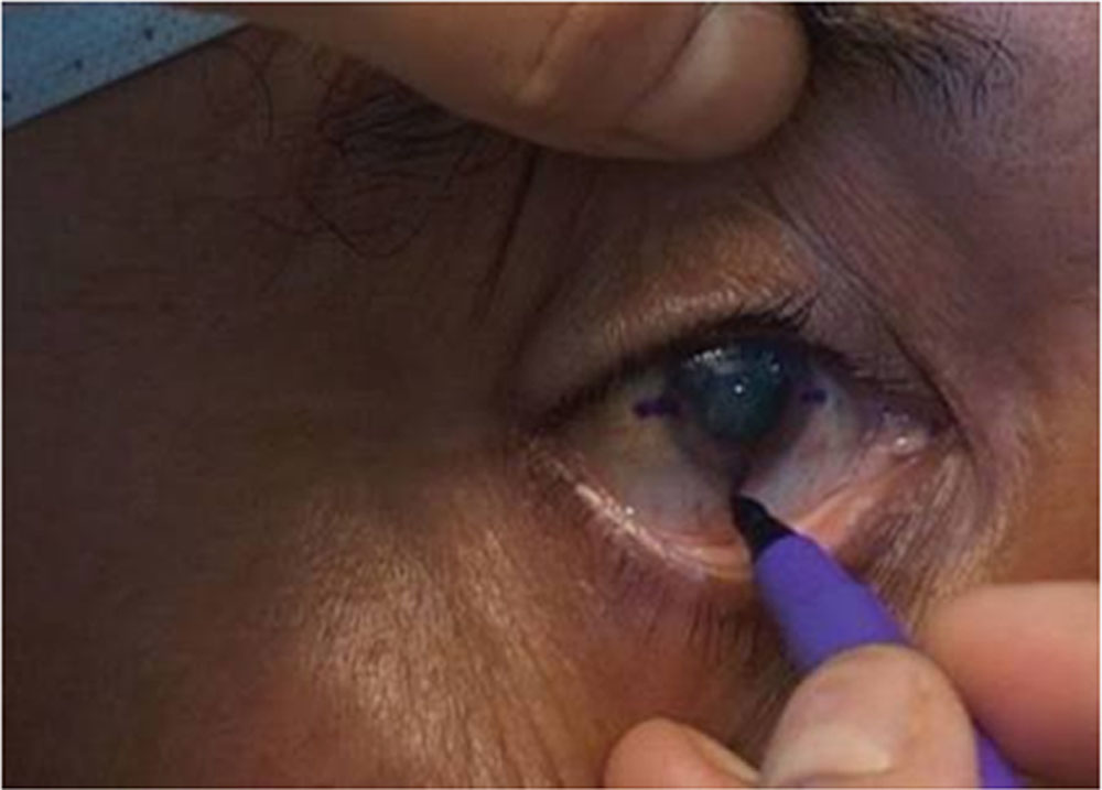
Figure1.Slit lamp marking for Toric IOLs
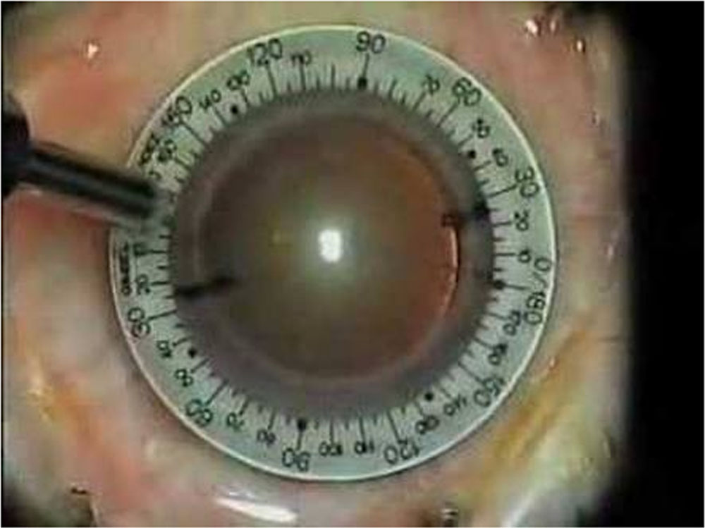
Figure 2: On table marking for Toric IOls
Results:
Of these 40 patients who underwent Toric IOL implantation,14 patients (35%) had an unaided post operative visual acuity of 6/6 Snellen’s with nil post operative astigmatism ,10 patients(25%) had post operative astigmatism in range of -0.5 to +0.5 D cyl, another 9 pts(22.5%) had 0.75 D cylindrical astigmatism post surgery ,4 pts (10%)with -1.0 D cyl and in only 3 cases(7.5%) the final refractive outcome was a cylindrical power of -1.25 or more.
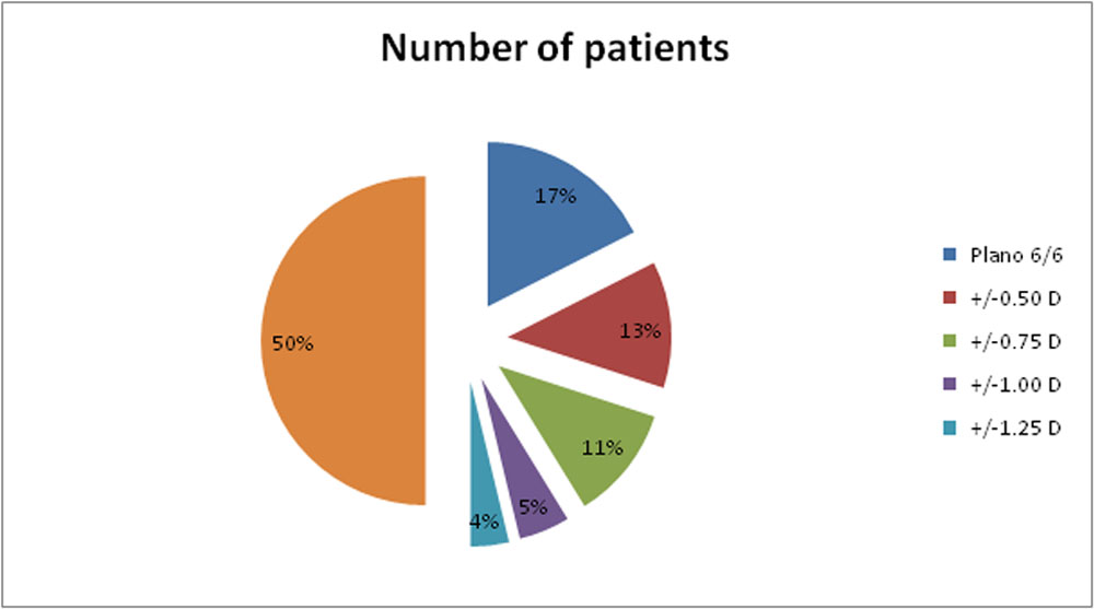
Figure 3: Pie chart showing percentage wise distribution of astigmatism Post toric IOL implantation
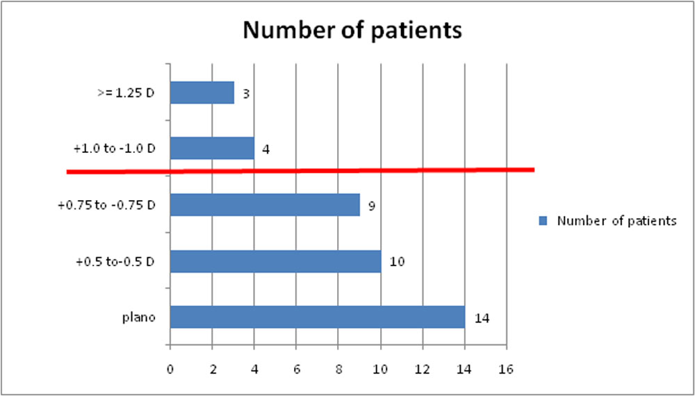
Figure 4: Illustrates 82.5% patients fall below the red line, i.e.; post operative astigmatism less than or equal to +/-0.75D
A significant proportion of patients (82.5%) had post operative astigmatism less than or equal to +/-0.75 D which was termed satisfactory considering the preoperative astigmatism and the basic methods of biometry and keratometry implemented in our study.
Conclusion:
The outcomes after Toric IOL implantation are influenced by numerous factors, right from the preoperative case selection and investigations to accurate intraoperative alignment and postoperative care. However, this study reiterates the fact that basic methods of IOL power calculation with manual keratometry and ultrasound Biometry can yield fairly reliable outcomes, more so in a rural set up where cost-effectivity is of paramount importance.
Limitations of this study: The major limitation of our study is its relatively small sample size since a larger pool of patients would further corroborate our findings.
References:
- Shimizu K, Misawa A, Suzuki Y:
Toric IOLs: correcting astigmatism while controlling axis shift. J Cataract
Refract Surg 1994, 20:523–526.
- Grabow HB. Toric IOL report. Ann Ophthalmol 1997, 29:161–163
- Cronje S, Harris WF:
A comparison of keratometric variation in manual and
automated keratometry. South African Optometrist 1999, 58:31–38.
- Ferreira TB, Berendschot TT, Ribeiro FJ.
Clinical outcomes after cataract surgery with a new transitional
toric intraocular lens. J Refract Surg. 2016;32:452–9.
- Vale C, Menezes C, Firmino-Machado J, Rodrigues P, Lume M, Tenedório
P, et al.
Astigmatism management in cataract surgery with precision (®) toric
intraocular lens: A prospective study. Clin Ophthalmol. 2016;10:151–9.
Leave a Comment