

Dr. Neha sawant, Dr. Gaurav Patil
Abstract :
Context: Iris Claw Lens (ICIOL) vs. Scleral Fixated Intraocular Lens (SFIOL)
Aims:
To compare the effectiveness, safety and complexity of ICIOL and SFIOL implantation.
Settings and Design: Retrospective Observational Study at a tertiary eye care centre.
Methods and Material: 65 patients presenting to our institute for cataract extraction who had an inadequate posterior capsular support during surgery and subsequently underwent SFIOL implantation or ICIOL implantation from December 2017 to September 2018 were selected for this study.
Statistical analysis used: Independent sample t-test and Chi-square test.
Results:
Out of 65 patients, 31 were in ICIOL group and 34 in SFIOL group. Follow-up done for six weeks. Mean best corrected visual acuity at final follow-up was 0.48±0.27 in ICIOL group and 0.53±0.25 in SFIOL group. There was no statistically significant difference between two groups (p value 0.418). Mean intraocular pressure on the first post-operative day in ICIOL group was significantly higher than SFIOL group (p value 0.043). The ICIOL group showed higher rates of hyphema and anterior chamber reaction in the early post-operative period which resolved by final follow-up. Cystoid macular oedema, suture erosion and vitreous haemorrhage were more common in SFIOL group. Mean surgical time of ICIOL implantation (18.11±3.11 min) was significantly shorter than SFIOL (63.05±6.32 min) (p value < 0.0001)
Conclusions:
Both ICIOL and SFIOL have similar levels of efficacy and safety in cases of complicated cataract surgeries with inadequate capsular support but ICIOL implantation is a much simpler and speedy procedure.
Key-words: SFIOL, retro-pupillary iris claw, Posterior capsular rupture
Key Message:
The iris claw lens implantation is a much simpler and faster procedure which can be done at the time of the primary surgery and can be considered a procedure of choice in cases of complicated cataract surgery leading to posterior capsular rupture.
Introduction:
The most commonly performed procedure in the field of ophthalmology is cataract surgery and in spite of various technical advancements posterior capsular rent (PCR) remains its commonest intra-operative complication.1,2,3 In this situation of inadequate capsular support when intraocular lens (IOL) implantation is being contemplated the surgeon has mainly three options: implanting an anterior chamber lens (ACIOL), a scleral fixated lens (SFIOL) or an iris claw lens (ICIOL). Over the years it has been observed that ACIOL implantation is often associated with a higher rate of complications4,5,6,7 and has therefore fallen out of use.
Various studies done on SFIOLs have found favourable results8,9,10,11 but the surgical technique of scleral fixation is a technically demanding procedure with a steep learning curve.11,12,13 Furthermore complications like cystoid macular oedema (CME), IOL tilting, decentration and conjunctival erosion due to trans-scleral sutures can occur.14
The Iris Claw lens was designed by Prof. Jan G.F Worst in 1978.15 The technique of its retro-pupillary iris fixation was developed by Andreas Mohr in 2002.15,16
The current study was performed to determine whether ICIOL can be safely used in place of SFIOL in cases of inadequate posterior capsular support during cataract surgery.
Subjects and Methods:
This was a retrospective observational study comprising 65 patients carried out at a tertiary eye care centre. ICIOL Group included 31 patients who had retro-pupillary ICIOL implantation (polymethyl methacrylate (PMMA) IOL with an 8.0-mm length and 5.50-mm optical zone, A-constant of 117) and SFIOL Group included 34 patients who had SFIOL implantation (PMMA IOL 6 mm optic, with 12.75 mm overall diameter, A-constant of 118.2) following lack of posterior capsular support during cataract surgery between the time period of December 2017 to September 2018. The patients were followed up to six weeks post-operatively (post-op).
Approval of the Ethics Committee was obtained before proceeding with the study. A written informed consent was obtained from all patients before the surgery.
Inclusion criterion:
Patients undergoing cataract surgery who had a PCR or zonular dialysis large enough to preclude in-the-bag IOL implantation were included in this study.
Exclusion criterion:
Patients with pre-existing chronic uveitis or pathologies of macula, cornea, retina or patients who were known cases of glaucoma as well as those presenting with traumatic cataract were excluded from the study.
Patients were selected based on these inclusion and exclusion criterion out of those who underwent cataract surgery between December 2017 and September 2018. Data from patient evaluation done pre-operatively and then followed up post-operatively on day one, week one and six weeks was collected.
Pre-operative data:
Personal information
History obtained from patient regarding name, age, sex and address.
Best corrected visual acuity (BCVA)
Recorded using Snellen’s Chart and later converted to logarithm of minimum angle of resolution (logMAR) for statistical analysis.
Slit lamp examination
In view of cataract and to rule out candidates based on exclusion criterion.
Fundus
To exclude pre-existing retinal/ optic nerve pathology.
Intraocular pressure (IOP)
Recorded using Non-contact Tonometry (NCT).
A-scan
For lens power calculation. SRK-T formula was used to calculate power.
Intra-operative data: Surgical time.
Post-operative data (Day one, week one and six weeks post-op):
BCVA
Recorded using Snellen’s Chart and later converted to logMAR.
Anterior chamber (AC) cells
Documented according to standardization of uveitis nomenclature (SUN) classification (Table 1)
| Grade | Cells in field* |
| 0 | <1 |
| 0.5+ | 1-5 |
| 1+ | 6-15 |
| 2+ | 6-25 |
| 3+ | 26-50 |
| 4+ | >50 |
* per high power field 1mm x 1mm
Table 1 – AC cells
AC flare
Documented according to SUN classification (Table 2)
| Grade | Description |
| 0 | None |
| 1 | Faint |
| 2 | Moderate(iris and lens details clear) |
| 3 | Marked (iris and lens details hazy) |
| 4 | Intense(fibrin) |
Table 2 – AC flare
IOL dislocation
IOL decentrations or luxations causing visual disturbance were documented as present or absent. Minor decentrations while remaining in visual axis and not hampering vision were taken as absent.
IOP
Recorded by NCT
Cystoid Macular Edema (CME)
Fundus evaluation of each patient was done post-op on to look for CME. It was documented as either present or absent. If present such patients were further evaluated by optical coherence tomography (OCT)
Hyphema
Documented as present or absent
Suture related complication
These were looked for in SFIOL patients. Documented as either present or absent.
Pupillary distortion
Documented as either present or absent.
Method of implantation:
Iris claw IOL (Figure 1):
In case of vitreous prolapse in the anterior chamber, anterior vitrectomy was done. Two side ports were made diagonally opposite, i.e. at 9’ o′ clock and 3’ o′ clock positions. Iris claw IOL was introduced into the anterior chamber such that haptics were in line with the side ports. Holding the optic of the lens with an iris claw holding forceps, one haptic was pushed under the iris with gentle manipulation. Simultaneously, enclaving was done by a sinsky hook which was passed through the paracentesis on the same side and pressed on the iris below. End point was noting the dimple at the site of enclavation. Similarly, haptic encalvation in the other side was done.
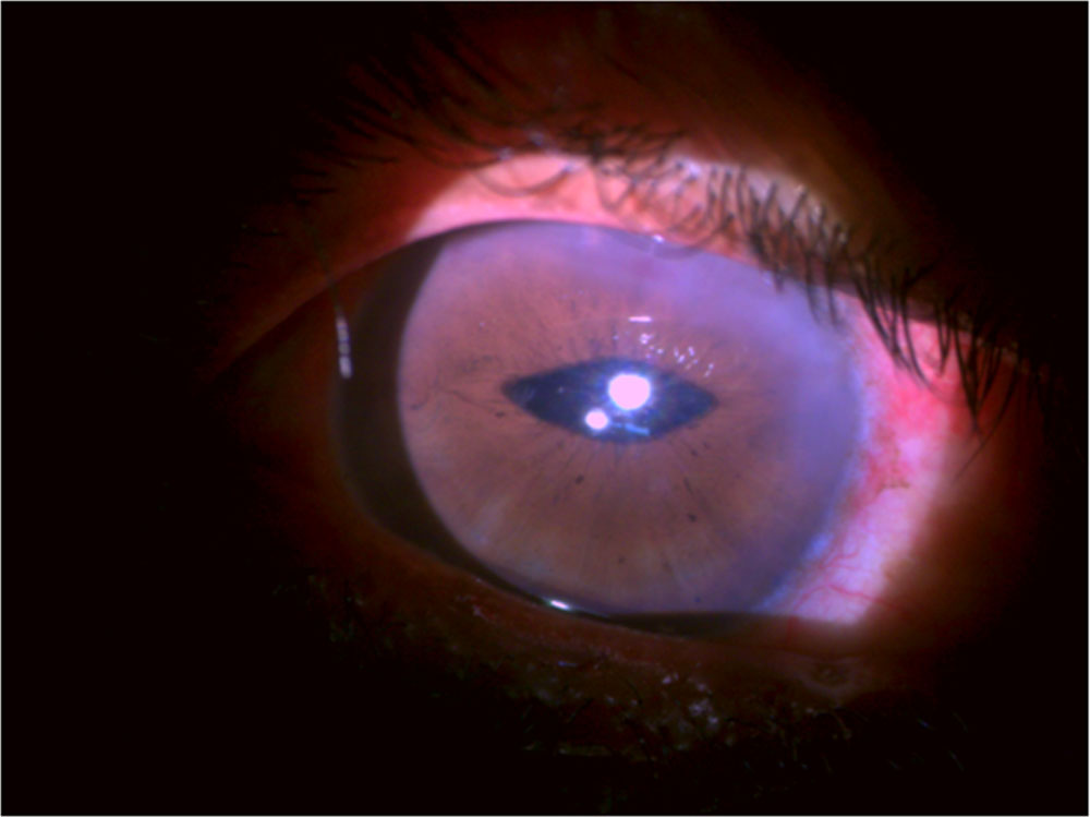
SFIOL (Figure 2):Figure 1 – Iris Claw lens (site of enclavation)
Peritomy done. Two partial thickness scleral flaps 2 mm posterior to the limbus created at the 3 0’clock and 9 0’ clock positions. A doubled arm 10-0 prolene suture with straight needle was used and the needles were rail-roaded out of the eye through the bed of the opposite scleral flap using a bent 25g needle introduced through the scleral bed. A limbal section was
fashioned and the sutures were drawn out of the eye, and cut into two halves. Each half of the sutures were passed through the fixation eyelet on the superior and inferior haptic of the IOL at the point of maximum haptic spread. The IOL was introduced into the posterior chamber, and the sutures were tightened and tied & the suture knots were buried in the scleral bed and the scleral flap sutured. The viscoelastic was cleared from the AC . The sclerocorneal and conjunctival peritomies were closed with 10-0 nylon sutures.
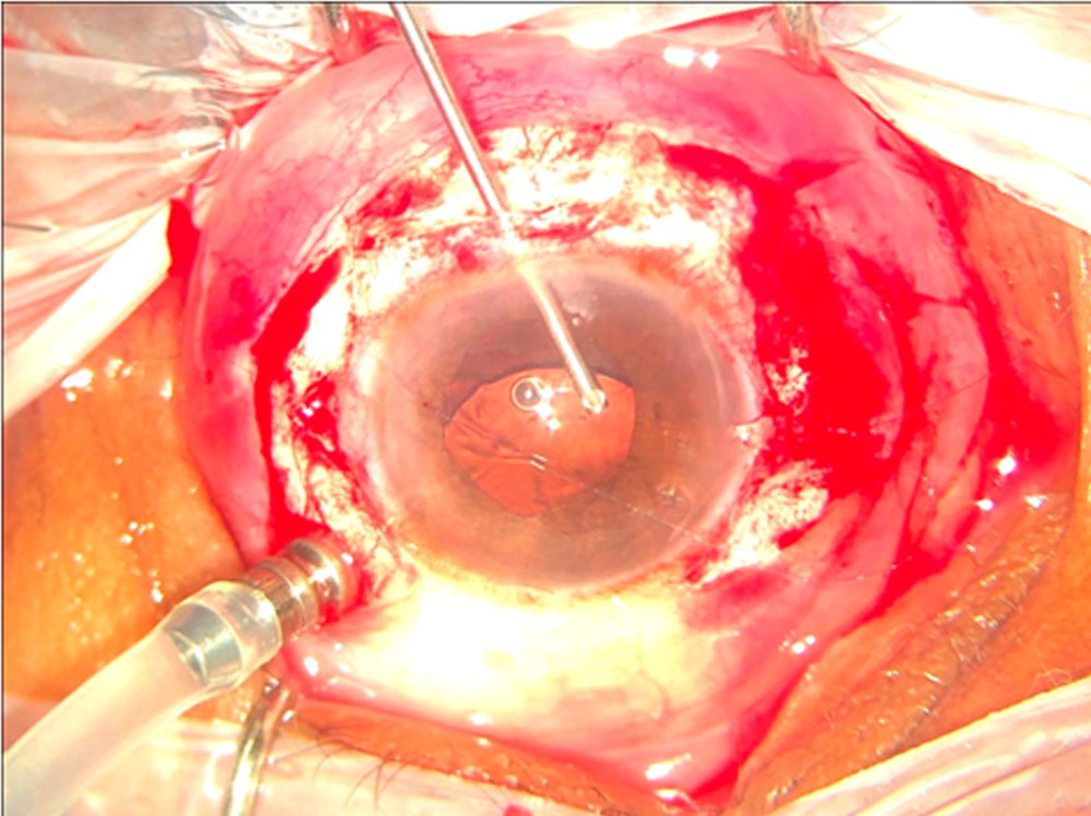
Figure 2 – SFIOL implantation
At the end of both the procedures subconjunctival Inj. Gentamycin+ Dexamethasone was given.
The procedure was said to be primary if cataract removal and IOL implantation was done in the same sitting and secondary if IOL implantation was done on a later date.
Data was evaluated using the independent sample t and Chi square tests. Level of significance was set at 0.05
Originally the study was planned to include 100 patients. But due to loss of follow-up only 65 patients could be included. Patients who did not complete six weeks of post-op follow-up were removed from the study.
Results:
Out of the 65 eyes which could be included in the study, 31 underwent iris claw lens implantation (ICIOL group) while 34 underwent scleral fixated IOL implantation (SFIOL group).
The two groups were matched in terms of the involved eye (P=0.543), gender (P=0.493) and age
ICIOL group:
This group included 12 female and 19 male patients. Out of these 28 patients had undergone small incision cataract surgery while three had undergone phacoemulsification. The mean age was 62.06±8.02 years. Out of these 31 patients, 29 had ICIOL implantation done as a primary procedure in the same sitting while in two patients it was done as a secondary procedure: In one patient due to excessive vitreous thrust ICIOL implantation was abandoned and later done one week after the primary surgery and in another patient iris claw lens of the required power was unavailable therefore the ICIOL implantation was done after two days.
SFIOL group:
This group comprised 13 female and 21 male patients. Out of these 26 patients had undergone small incision cataract surgery while eight had undergone Phacoemulsification. The mean age was 61.85±7.39 years. All the SFIOL implantations were done as secondary procedures four weeks to two months after the primary surgery.
Best Corrected Visual Acuity (Figure 3):
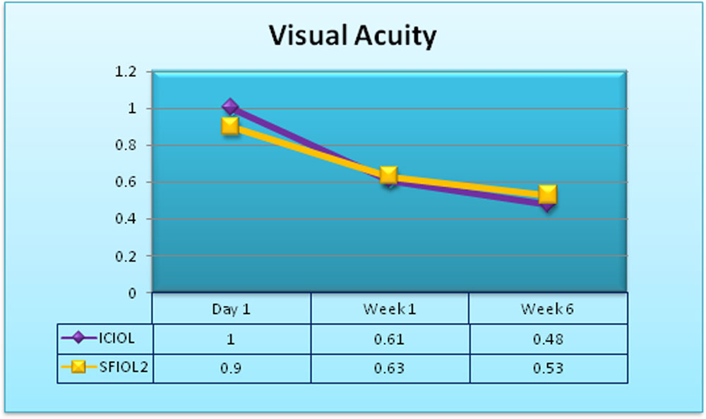
Figure 3 – Visual acuity (LogMAR)
The BCVA in terms of logMAR on day one was slightly better in the SFIOL group (0.90 ±0.53) as compared to the ICIOL group (1.00 ±0.53). This difference was statistically insignificant with a p value of 0.45
BCVA was similar at week one (ICIOL mean BCVA 0.61±0.42 and SFIOL mean BCVA 0.63±0.23)
The ICIOL group (0.48±0.27) fared slightly better than that of SFIOL (0.53±0.25) at the final follow-up though this difference was not statistically significant.
IOP Changes (Table 3):
| Cases | Mean | SD | p-value | ||
| IOP
Pre-op |
SFIOL | 34 | 12.50 | 3.81 | 0.208 |
| Iris Claw | 31 | 11.39 | 3.24 | ||
| IOP
Day 1 |
SFIOL | 34 | 13.18 | 7.55 | 0.043 |
| Iris Claw | 31 | 16.71 | 6.25 | ||
| IOP
Day 7 |
SFIOL | 34 | 13.59 | 4.42 | 0.448 |
| Iris Claw | 31 | 12.84 | 3.47 | ||
| IOP
Week 6 |
SFIOL | 34 | 12.71 | 3.65 | 0.224 |
| Iris Claw | 31 | 11.61 | 3.52 | ||
Table 3 – IOP changes
Pre operative IOPs were similar in both groups of patients.
On the first postoperative day, the IOP in ICIOL group ranged from 6 mmHg to 36mmHg, with a mean of 16.71 ± 6.25 mmHg, and in SFIOL group it ranged from 4 mmHg to 30 mmHg, with a mean of 13.18 ± 6.94 mmHg. There was a statistically significant difference between the groups (P = 0.043).
At the first postoperative week, the IOP in ICIOL group ranged from 11 mmHg to 20 mmHg, with a mean of 14.28 ± 2.47 mmHg, and in SFIOL group it ranged from 10 mmHg to 21 mmHg, with a mean of 13.86 ± 3.42 mmHg, with no statistically significant difference between the groups (P = 0.441)
At six weeks post operatively there was no statistically significant difference in the IOP of the two groups (p value 0.224) with ICIOL group IOPs ranging from 8 mmHg to 19 mmHg, mean 11.61±3.52 mmHg and SFIOL group IOP ranging from 6 mmHg to 19 mmHg, mean 12.71±3.65 mmHg.
Early Post-Op Complication:
The early post-op complication were seen within the first month of the surgery
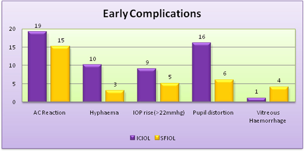
Figure 4: Early post-op complication
AC reaction:
In the ICIOL group AC cells and flare was seen in 19(61.29%) cases on the first post-op day which resolved by one week in 15 of the cases while the remaining four resolved by one month post-op. Fifteen (44.11%) cases of SFIOL patients had AC reaction out of which ten resolved by first post-op week while five resolved by first post-op month. There was no AC reaction in final follow-up in any patient. There was no significant difference in ICIOL and SFIOL groups in this regard.
IOP raised (above 22mmHg):
Nine (29.03%) cases in ICIOL group and five (14.28%) cases in SFIOL group had IOP raised above 22mmHg. This difference was significant with a p value of < 0.05. Oral and topical hypotensives were given.
Hyphema:
Ten (32.25%) cases in ICIOL group on day one had hyphema in the anterior chamber out of which eight (25.08%) resolved by the first week while two took three weeks to resolve.
Only three (8.82%) cases in the SFIOL group had hyphema and all of these resolved by the first week post-op. This difference was significant with a p value of 0.031
Pupillary distortion:
This was by far more commonly seen in the ICIOL group of patients with 16 (51.61%) patients having pupil ovalisation while pupillary distortion was seen only in six (17.64%) cases in the SFIOL group.
Vitreous haemorrhage:
Vitreous haemorrhage was seen in only one (3.22%) case in the ICIOL group while it was seen four (11.76%) cases in SFIOL group
Late Complications:
These complications were seen from one month up to 6 weeks post-op.
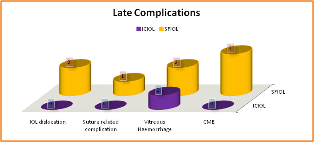
Figure 5 – Late post-op complications
IOL dislocation:
IOL dislocation or dis-enclavation was not seen in any of the patients in the ICIOL group. Two (5.88%) patients in the SFIOL group had significant IOL dislocations and had to be taken up for re-surgery.
Suture related complications:
These were not present in the ICIOL group. One (2.94%) patients had conjunctival erosion due to sutures in SFIOL group which was treated by trimming the suture
Vitreous haemorrhage:
Persistent vitreous haemorrhage was seen in one patient in ICIOL group which had not resolved by the time of last follow-up at the end of six weeks. The patient was followed up every week as the haemorrhage had not resolved but at two months post-op the patient was lost to follow-up. Two patients in the SFIOL group had vitreous haemorrhage at the end of six weeks. Both the patients were followed up weekly and in both the cases the haemorrhage had resolved by the end of two months.
CME:
None of the patients had CME at the end of six weeks in the ICIOL group. Three (8.82%) cases had fundus changes suggestive of CME in SFIOL group. These cases were evaluated by OCT and referred to vitreoretinal surgeon for further treatment.
Surgical Duration:
Average duration of procedure after aphakia in iris claw implantation was 18.11±3.11 min while in SFIOL group it was 63.05±6.32 min. This difference was significant with a p value of <0.0001
Discussion:
The ideal conclusion of all cataract surgeries is in-the-bag implantation of the IOL, but this becomes a challenge when there is an absence of adequate posterior capsular support. At present ACIOLs, SFIOLs and iris-claw IOLs are the various options available for the correction of aphakia in such a situation.17,18,19
Logic dictates that the easiest method which entails the lowest complication rate but also has the best visual result should be chosen.
Angle supported anterior chamber IOLs have been implanted in the past as these have a advantage of being technically easy and minimally invasive. However, complications like progressive injury to the endothelial cells of cornea, decompensation, anterior chamber inflammation, secondary glaucoma and CME have been reported6,20,21
In a study published by Evereklioglu et al., SFIOLs were found to have a better outcome than ACIOLs23 but these are not just technically difficult but are also associated with higher chances of retinal detachment (4%) and suture breakage (24%) as seen in a study done by Asadi and colleagues.23
In a study done by Güell and colleagues it was found that implantation of an iris-claw lens in the anterior chamber may cause damage to the corneal endothelial cells.24
The implantation of an iris-claw lens behind the iris plane combines the advantages of being a posterior chamber lens with that of a short operation time as well as an easy operation technique.
We have been using SFIOLs and ACIOLs to correct aphakia at our centre since the past decade but being a high volume set up the need for a faster, safe and less technically demanding procedure which could be satisfactorily performed by most surgeons was felt. This made us turn to posteriorly fixated Iris Claw lenses. The rationale behind posterior fixation instead of anterior is the reduced chance of endothelial decompensation along with the lens being nearer the nodal point of the eye which is a more physiological position for an intraocular lens.14
To the best of our knowledge four studies25,26,27,28 have been conducted in the past comparing these two methods but none of them have been conducted in India. Our study also differs from them in having a larger sample size.
All our patients were followed up for six weeks. At the end of this period the mean BCVA in the ICIOL group ranged from 2.1 to 0.18 with a mean of 0.48±0.27 which was similar to that of SFIOL group where the BCVA ranged from 1.85 to 0.18 with a mean of 0.53±0.25. This finding is in agreement with previous studies.25,26,27,28 In our study the BCVA at the first post operative day was worse in the ICIOL group which may have been due to the fact that majority of cases in this group were done as primary procedures and therefore had more inflammation. The mean improvement in BCVA in the ICIOL group was more than that in the SFIOL group (-1.32±0.71 versus -1.07±0.67) but the difference was not statistically significant (p value 0.149). Overall, this result agrees with the results from other studies25,27,28
Hsing and Lee had reported a final BCVA of 6/12 or better in 58% of eyes after retro-pupillary iris claw implantation.29 In our study at final follow-up 14 out of 31 eyes (45.16%) had a visual acuity equal to or better than 6/12 in ICIOL group while 10 out of 34 eyes (29.41%) in the SFIOL group had a visually acuity equal to or better than 6/12.
In our study IOP elevation was a significant complication in the early post-operative period in the ICIOL group with 29.03% of patients having a IOP higher than 22mmHg. In this respect our study differs from that of Rashad et al25 and Hara et al26 where initial IOP rise was more in SFIOL cases. This may be explained by the fact that increased inflammation in ICIOL group was causing an initial trabecular meshwork blockage which later subsided once inflammation reduced. The final IOP was similar in both the groups. We did not perform a peripheral iridectomy in any of the cases. The need for performing peripheral iridectomy in retro-pupillary implantation of the iris claw lens is controversial. Previous studies like those conducted by Sathyan et al and Helvaci et al have found that there were no incidences of pupillary block in retro-pupillary ICIOL implantation even when a peripheral iridectomy was not performed.30,31 This may be explained by the fact that due to the vaulted design of the lens there is adequate space between the iris and the lens optic for aqueous drainage. There were no incidences of pupillary block in our study as well. It was felt that the mechanism for rise in IOP in some cases could be inadequate anterior vitrectomy. We recommend a thorough anterior vitrectomy before implantation of the iris claw lens.
Hazar et al27 had reported AC reaction rates of 20.8% in ICIOL cases and 9.6% in SFIOL cases. In our study AC inflammation in the form of cells and flare was slightly more in the ICIOL(61.29%) group as compared to the SFIOL group(44.11%) on the first post-op day. It reduced to 12.9% and 10.2% respectively by the first post-op week. By the time of the final follow-up AC reaction had resolved in all the patients in both the groups. AC inflammation in the ICIOL group can be explained by the iris handling and primary setting while that in the SFIOL group can be explained by the prolonged duration of the procedure.
Similar to a previous study32, in our study there were no incidences of IOL dis-enclavation in the ICIOL group but there were two cases of IOL dislocation in the SFIOL group which had to be taken up for re-suturing.
Gonnerman et al had reported an incidence of CME in 8.7% cases of retro-pupillary iris claw implantation.35 There was no incidence of CME in the ICIOL group in our study similar to the study by Forlini et al.33 It may be prudent to note that the absence of detection of CME in our study may be because follow-up done only till 6 weeks could be inadequate for CME detection. CME was seen in three cases in SFIOL group(8.82%) which was lesser than the findings of Kumar and colleagues(15.62%).35 Prolonged surgical time and intraocular manipulation have been described as two risk factors for CME.36
Vitreous haemorrhage was only seen in one patient in the ICIOL group which was suspected to be due to an unrecognised globe perforation at the time of peribulbar block. This vitreous haemorrhage persisted up to the 6th week follow-up with the final BCVA of this patient remaining HM+. Follow-up was maintained up till two months at which time the vitreous haemorrhage was still present. Vitrectomy was planned if the vitreous haemorrhage remained unresolved by the next month but the patient was lost to follow-up.
In the SFIOL group vitreous haemorrhage was seen in 11.76% of patients similar to other studies.27,31 It resolved in 5.88% by final follow-up while in the rest it resolved by the end of two months.
SFIOLs have been known to have suture related complications ranging from conjunctival erosion to suture breakage24. Various studies27,31 have found these to be to the tune of 4.76% to 3.2%. Similarly our study had a suture related complication rate of 2.94%
By far the commonest complication of ICIOL seen in our study was pupillary ovalization which was seen in 51.61% cases. Only 17.64% of cases in the SFIOL group had pupillary distortion. This was a cosmetic complication and did not require any intervention.
Other complications like hyphema were more in the ICIOL group in the early postoperative period but resolved by the final follow-up.
We can see through this study that the early post-op complications are more in the patients implanted with ICIOLs as compared to those implanted with SFIOLs. This may be because SFIOL implantation was done as a secondary procedure after a duration of four weeks to two months during which time inflammation from the primary procedure would have a chance to subside whereas ICIOL implantation was done mainly as primary procedure. In spite of this at the end of six weeks both groups of patients had comparable visual outcomes. There was no re-surgeries done in the ICIOL group whereas two cases(5.88%) from the SFIOL group had to be taken up for re-surgery.
The most significant difference in the two groups was in the surgical time taken for performing the procedure. This finding has been confirmed by other studies as well.27,29 ICIOL required 18.11±3.11 min post aphakia for the completion of the procedure which was less than a third of the time required for SFIOL(63.05±6.32 min). This may be due to surgical challenges like construction of scleral flaps, finding the correct fixation positions for the haptics, extensive vitrectomy to avoid retinal detachment as well as the suturing of sclerocorneal flaps and conjunctival peritomies. Surgical time is an indicator for the complexity of the procedure and therefore it can be interpreted that ICIOL is a significantly simpler procedure as compared to SFIOL. Also the fact that ICIOLs can be easily combined with the primary procedure makes them more convenient for the patient as well as the institute especially in a high volume community set up where loss to follow-up post surgery is not unusual and the cost of a secondary procedure has to be borne by the institute.
There are two key limitations of our study: the relatively short follow-up period and the retrospective study design. The present report compiles the findings at the end of six weeks. The study, however, will continue to follow-up the cohort for a longer duration and also enroll more patients into this study in a prospective manner.
References:
- Chan E, Mahroo OA, Spalton D. Complications of cataract surgery. Clin Exp Optom 2010; 93: 6: 379–389
- Reddy MK. Complications of cataract surgery. Indian J Ophthalmol 1995;43:201-9
- Hashemi H, Alipour F, Rezvan F. Intraoperative Complications of Cataract Surgeries in Iran: 2000-2005 Iranian Cataract Surgery Survey. Iranian Journal of Ophthalmology 2011;23(1):3-10
- Wagoner MD, Cox TA, Ariyasu RG, Jacobs DS, Karp CL. American Academy of Ophthalmology. Intraocular lens implantation in the absence of capsular support: A report by the American Academy of Ophthalmology. Ophthalmology 2003;110:840–59
- Apple DJ, Mamalis N, Loftfield K, Googe JM, Novak LC, Kavka-Van Norman D, et al. Complications of intraocular lenses. A historical and histopathological review. Surv Ophthalmol. 1984;29:1–54
- Smith PW, Wong SK, Stark WJ, Gottsch JD, Terry AC, Bonham RD. Complications of semiflexible, closed-loop anterior chamber intraocular lenses. Arch Ophthalmol. 1987;105:52–7
- Apple DJ, Brems RN, Park RB, Norman DK, Hansen SO, Tetz MR, et al. Anterior chamber lenses. Part I: Complications and pathology and a review of designs. J Cataract Refract Surg. 1987;13:157–74.
- McAllister AS, Hirst LW. Visual outcomes and complications of scleral-fixated posterior chamber intraocular lenses. J Cataract Refract Surg. 2011;37:1263–9
- Krause L, Bechrakis NE, Heimann H, Salditt S, Foerster MH. Implantation of scleral fixated sutured posterior chamber lenses: A retrospective analysis of 119 cases. Int Ophthalmol. 2009;29:207–12
- Bading G, Hillenkamp J, Sachs HG, Gabel VP, Framme C. Long-term safety and functional outcome of combined pars plana vitrectomy and scleral-fixated sutured posterior chamber lens implantation. Am J Ophthalmol. 2007;144:371–7
- Kjeka O, Bohnstedt J, Meberg K, Seland JH. Implantation of scleral-fixated posterior chamber intraocular lenses in adults. Acta Ophthalmol. 2008;86:537–42
- Uthoff D, Teichmann KD. Secondary implantation of scleral-fixated intraocular lenses. J Cataract Refract Surg. 1998;24:945–50
- Yang YF, Bunce C, Dart JK, Johnston RL, Charteris DG. Scleral-fixated posterior chamber intraocular lenses in non-vitrectomized eyes. Eye (Lond) 2006;20:64–70
- Kwong YY, Yuen HK, Lam RF, Lee VY, Rao SK, Lam DS. Comparison of outcomes of primary scleral-fixated versus primary anterior chamber intraocular lens implantation in complicated cataract surgeries. Ophthalmology. 2007;114:80–85
- Thall EH, Miller KM, Rosenthal P. Clinical Refraction: Optics, Refraction and Contact Lenses. SanFrancisco USA: American academy of Ophthalmology 1999-2000; 159
- Ozcan AA, Ceran BB. Scleral fixation – Where are we. Int J Ophthalmol Eye Rec 2014;2:1-2.
- Rao R, Sasidharan A. Iris claw intraocular lens: A viable option in monocular surgical aphakia. Indian J Ophthalmol 2013;61:74-5
- Gicquel JJ, Langman ME, Dua HS. Iris claw lenses in aphakia. Br J Ophthalmol 2009;93:1273-5.
- Sawada T, Kimura W, Kimura T, et al. Long-term follow-up of primary anterior chamber intraocular lens implantation. J Cataract Refract Surg. 1998;24:1515-1520
- Snellingen T, Shrestha JK, Huq F, Husain R, Koirala S, Rao GN, et al. The South Asian cataract management study: Complications, vision outcomes, and corneal endothelial cell loss in a randomized multicenter clinical trial comparing intracapsular cataract extraction with and without anterior chamber intraocular lens implantation. Ophthalmology 2000;107:231-40
- Marques FF, Marques DM, Osher RH, Freitas LL. Longitudinal study of intraocular lens exchange. J Cataract Refract Surg 2007;33:254-7.
- Evereklioglu C, Er H, Bekir NA, Borazan M, Zorlu F. Comparison of secondary implantation of flexible open-loop anterior chamber and scleral-fixated posterior chamber intraocular lenses. J Cataract Refract Surg 2003;29:301-8.
- Asadi R, Kheirkhah A. Long-term results of scleral fixation of posterior chamber intraocular lenses in children. Ophthalmology 2008;115:67-72
Güell JL, Velasco F, Malecaze F, Vázquez M, Gris O, Manero F-J.Secondary Artisan-Verysise aphakic lens implantation. Cataract RefractSurg 2005; 31:2266–2271 - Rashada DM, Omar MM, et al., Retro-pupillary fixation of iris-claw intraocular lens versus trans-scleral suturing fixation for aphakic eyes without capsular support. Journal of Egyptian Ophthalmological Society 2015, 108:157–166
- Hara S, Borkenstein AFM, Ehmer A, Auffarth GU. Retro-pupillary fixation of iris-claw intraocular lens versus transscleral suturing fixation for aphakic eyes without capsular support. J Refract Surg. 2011; 27: 729–735
- Hazar L, Kara N, Bozkurt E, Ozgurhan EB, Demirok A. Intraocular lens implantation procedures in aphakic eyes with insufficient capsular support associated with previous cataract surgery. J Refract Surg. 2013; 29: 685–691.
- Saleh M, Heitz A, Bourcier T, et al. Sutureless intrascleral intraocular lens implantation after ocular trauma. J Cataract Refract Surg. 2013; 39: 81–86
- Hsing YE, Lee GA. Retropupillary iris claw intraocular lens for aphakia. Clin Experiment Ophthalmol. 2012;40:849-854
- Sathyan S, Kurian R. Comparison of retropupillary fixated iris-claw lenses and sulcus placed intraocular lenses for the treatment of aphakia. Kerala J Ophthalmol 2017;29:207-12
- Helvaci S, Demirduzen S, Oksuz H. Iris-claw intraocular lens implantation: Anterior chamber versus retropupillary implantation. Indian J Ophthalmol 2016;64:45-9
- Upadhyay AM, Nataraj A. Visual outcomes and complications of posterior iris-claw intraocular lens implantation in aphakic postvitrectomized eyes. Kerala J Ophthalmol 2017;29:184-8
- Forlini M, Soliman W, Bratu A, Rossini P, Cavallini GM, Forlini C. Long-term follow-up of retro-pupillary iris-claw intraocular lens implantation: A retrospective analysis. BMC Ophthalmol 2015;15:143.
- Gonnermann J, Klamann MK, Maier AK, Rjasanow J, Joussen AM, Bertelmann E, et al. Visual outcome and complications after posterior iris-claw aphakic intraocular lens implantation. J Cataract Refract Surg 2012;38:2139-43
- Kumar, Santosh et al. “Visual Outcome and Complications of Various Techniques of Secondary Intraocular Lens.” Oman Journal of Ophthalmology 2017;10.3:198–204.
- Malinowski SM, Mieler WF, Koenig SB, Han DP, Pulido JS. Combined pars plana vitrectomy–lensectomy and openloop anterior chamber lens implantation. Ophthalmology. 1995;102:211-216
Acknowledgement:
Dr. Amrita Kapoor
Dr. Nitant Shah
Dr. Aparna Paranjpe
Dr. Tushar Waghule
Dr. Dhanashree Ratnaparkhi
Dr. Sonalika Dubey
Dr. Arindam Bhattacharyya
Dr. Raghavendra Borgaonkar
Ms. Renu Wadhawa
Leave a Comment