

Dr. Puspa Kumari,Dr. DEEPAK MARIANUS LAKRA,Dr. Rajiv Kumar Gupta
INTRODUCTION:
Cataract is a major cause of impaired vision and blindness. Cataract is complete or partial opacification in human lens or in the capsule(1). More than 80% of all cataracts are age related and pathophysiology is complex (1, 2).
Dyslipidemia affects many organs of the body. Dyslipidemia is associated with wide range of eye diseases including glaucoma, hypertensive and diabetic retinopathy, age related macular degeneration, retinal vein occlusion (3, 4). Lipoproteins are complexes of lipids and proteins that are needed for transport of cholesterol, triglycerides and fat soluble vitamins (5). Based on relative density, plasma lipoproteins are divided into 5 classes- chylomicrons, very low density lipoprotein (VLDL), intermediate density lipoprotein (IDL), low density lipoprotein (LDL) and high density lipoprotein (HDL). Each lipoprotein class comprises a family of particles that vary slightly in density, size, and migration during electrophoresis and protein composition (5-7).
The current study aimed to assess plasma lipid levels including cholesterol, triglycerides (TG), LDL, VLDL and HDL in patients with cataract.
AIM:
To find any association between dyslipidemia and cataract in tribal population of Jharkhand.
MATERIALS AND METHODS:
This cross sectional hospital based study was performed in a tertiary care centre of Jharkhand between June 2017 to March 2018.
A total of 223 patients of senile cataract were included in this study. Cataract diagnosis was made by using slit-lamp bio microscopy and retro illumination. Detailed history was obtained from each patient. Complete ophthalmological and systemic examination was done. Patient’s height (in cm) and weight (in kg) were recorded and body mass index (BMI) was calculated according to the equation:
BMI=Weight (in kg)/ {Height (in cm)} 2
The fasting blood sample were collected and sent for evaluation of fasting blood glucose and lipid profile to the central pathological and biochemical laboratory department. Analysis of fresh blood samples were conducted within four hours of collection. All these data were recorded on a questionnaire form and the form was used as a source for data entry and analysis.
Dyslipidemia was defined as any of the following:
- Hypercholesterolemia (total cholesterol concentration ≥ 220 mg/dl or
- Hypertriglyceridemia (triglyceride concentration ≥ 150 mg/dl or
- The patients on treatment of hyperlipidemia.
Treatment of dyslipidemia was defined as the use of pharmacological treatment to lower blood lipids during the previous 2 weeks.
Patients aged less than 45 years old, those with traumatic and congenital cataract were excluded from the study.
Study results were compared under the following headings:
- Age: Patients older than 45 years old were included.
- Gender: To have an idea if there is any sex difference concerning dyslipidemia and cataract.
- BMI (Body mass index): Important to be included in his study because of it’s direct and indirect relationship with dyslipidemia. Analysis of data was performed using Chi square and T-Tests.
P values less than 0.05 has been taken to be statistically significant. Statistical analysis was performed by using statistical package for social sciences (SPSS; Version 18.0) software.
RESULTS:
The study included 223 patients with senile cataract (145 men and 78 women).
The age range of the patients was between 45-88 years (Mean 58±7).
The age and sex distribution for the total sample included in the study is illustrated in figure
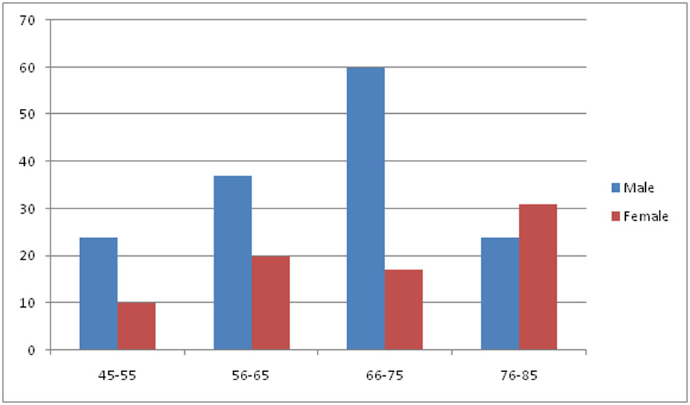
FIGURE 1: Age and sex distribution
| Male | Female | ||
| Age Group | 45-55 | 24 | 10 |
| 56-65 | 37 | 20 | |
| 66-75 | 60 | 17 | |
| 76-85 | 24 | 31 | |
| Total | 145 | 78 | |
Table 1: Age and sex distribution for total sample included in this study.
According to the lipid status it was seen that 156 (70%) patients had dyslipidemia and 67(30%) had no dyslipidemia.
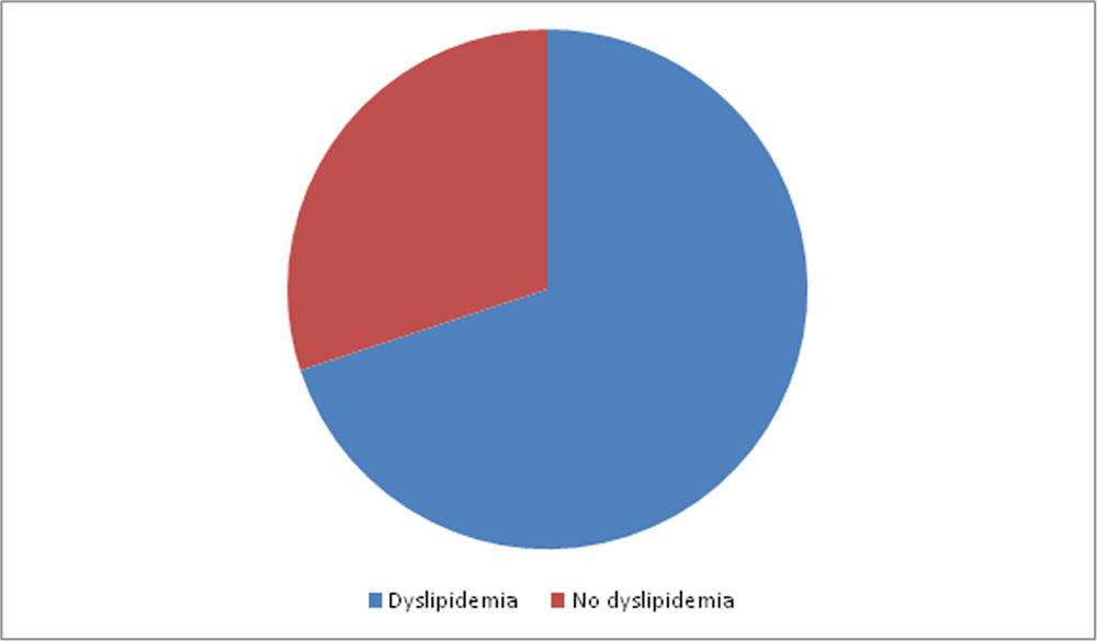
The sex and age distribution according to the patients lipid status involved in this study are illustrated in figure 2 and table 2.
| Parameter | No Dyslipidemia | Dyslipidemia | Total | ||||
| N | % | N | % | ||||
| Sex | Males | 30 | 13.45 | 104 | 46.63 | 134 | 223 |
| Females | 36 | 16.14 | 53 | 23.76 | 89 | ||
| Age(years) | 45-55 | 12 | 5.38 | 22 | 9.86 | 34 |
223 |
| 56-65 | 21 | 9.41 | 36 | 16.14 | 57 | ||
| 66-75 | 24 | 10.76 | 52 | 23.31 | 76 | ||
| 76-85 | 10 | 4.48 | 46 | 20.62 | 56 | ||
| Total | 67 | 156 | |||||
| 223 | |||||||
Table 2: Sex and age distribution of patients according to their lipid status
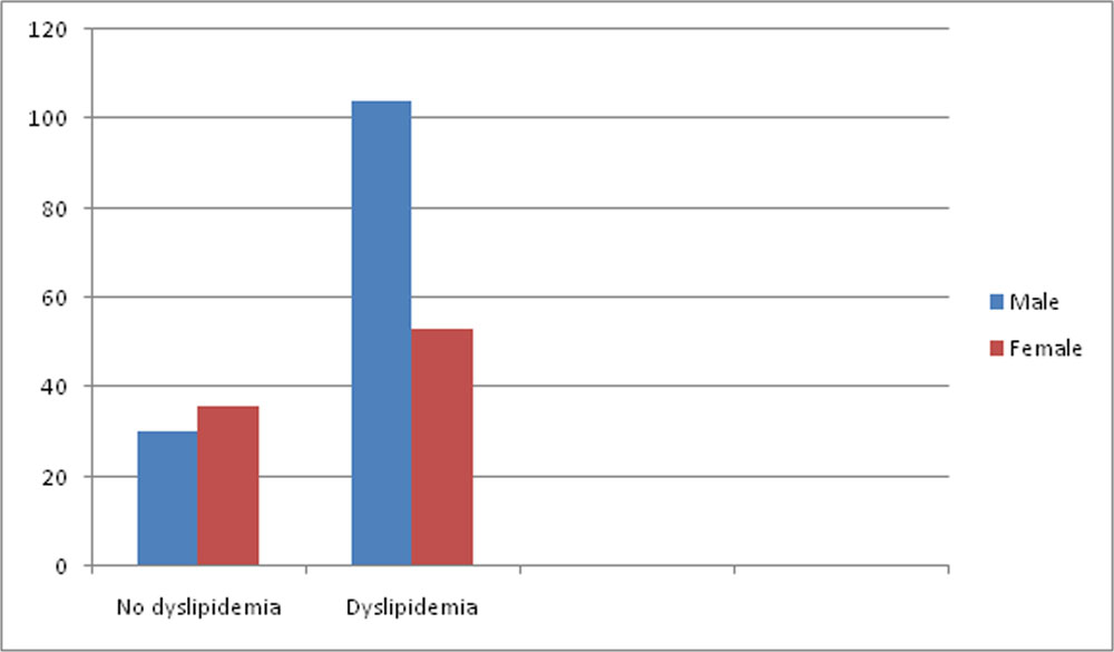
Figure 2:Sex distribution of patients according to lipid status.
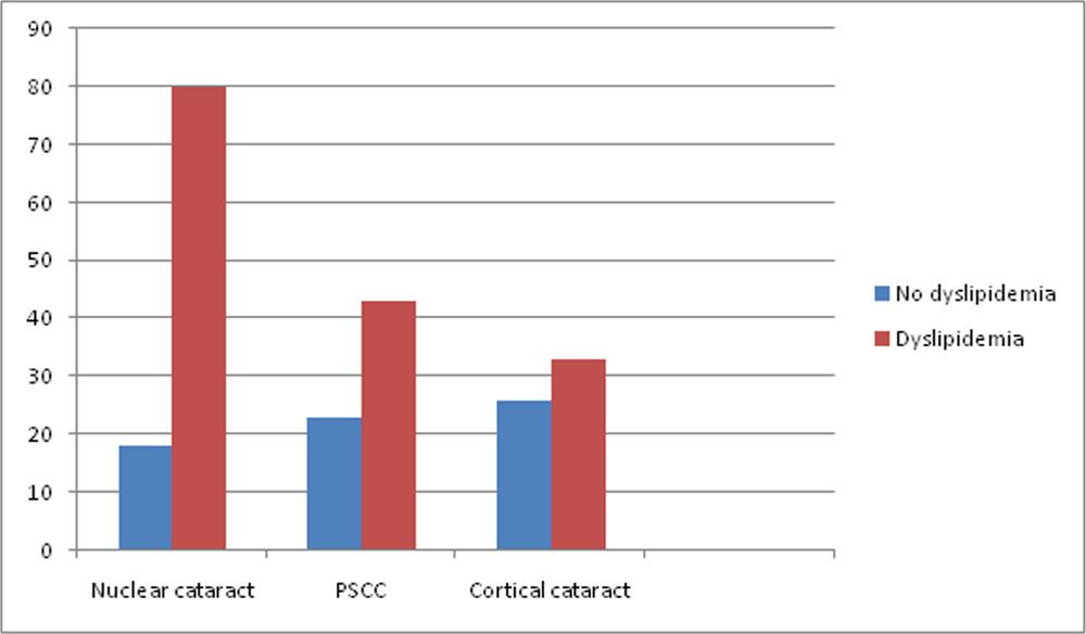
Of the total 35.87% of patients with dyslipidemia had nuclear cataract, 14.79% with those having dyslipidemia had cortical cataract and 19.28% had posterior subcapsular cataract. ( Table 3 and Figure 3).
| Parameter | No dyslipidemia(n=67) | Dyslipidemia(n=156) | P=value | ||
| N | % | N | % | ||
| Nuclear | 18 | 8.07 | 80 | 35.87 | <0.05 |
| PSCC | 23 | 10.31 | 43 | 19.28 | >0.05 |
| Cortical | 26 | 11.65 | 33 | 14.79 | <0.05 |
Table 3: Relationship between cataract types and lipid status.
High BMI is related with cortical cataract.
| Parameter | Nuclear cataract(n=98) | PSCC(n=66) | Cortical cataract(n=26) | ||||||
| Body mass index(BMI) | Mean | ±SD | P- value | Mean | ±SD | P-value | Mean | ±SD | P- value |
| 26.2 | 0.43 | >0.05 | 24.6 | 1.32 | >0.05 | 29.3 | 0.32 | <0.05 | |
Table 4: Comparison between types of cataract in terms of BMI.
DISCUSSION:
More males than females were found to have dyslipidemia this is because of the larger sample of male patients. Age factor is directly related to abnormal lipid profile, elderly have dyslipidemia in comparison with younger people (6-8). In this study, large number of cataract patients included three type of cataracts-nuclear, posterior subcapsular and cortical were found to have dyslipidemia. Nuclear cataract occupied highest percent compared to other types of cataract.
In a study carried in China in 2006(Beijing study) it was seen that by comparing the types of cataract in terms of lipid status there is a significant association with cortical cataract (9). This differs from our study concerning nuclear cataract. Dyslipidemia was significantly associated with cortical cataract and this may be explained by the effect of diabetes or may be due to dyslipidemia as a direct or indirect cause (10). This study report shows prevalence of dyslipidemia was insignificant with posterior subcapsular cataract which is against the finding of Hiller et al (11), which stated that a high level of triglyceride was associated with an increased risk of posterior subcapsular cataract.
There was significant association between cortical cataract and high BMI, it was previously proven that patient with high BMI had abnormally high lipid profile especially dyslipidemia (5-7).
According to study performed by Ning Cheung and Tien Y Wong, et al, it was seen that there is important relationship between cataract and obesity and provided valuable results for use of weight loss strategies to reduce the burden of cataract in individuals with obesity(12).
Another study done by GhaemMaralani H, et al, to see if the effect of metabolic syndrome and it’s components on incidence of different types of cataract change with time, it was seen that low HDL and high glucose were associated with an increased 10 year incidence of cortical and posterior subcapsular cataract respectively(4).
The results of this study, regarding cortical and post subcapsular cataract are consistent with the results of our study.
CONCLUSION:
According to this study, dyslipidemia has a direct or indirect relationship with nuclear and cortical cataract and should be considered as an etiology for cataract.
REFERENCES:
- Andrikopoulos GK, Alexopoulos DK, Gartaganis SP. Pseudoexfoliation syndrome and cardiovascular diseases. World J Cardiol. 2014;6(8):847-54. doi: 10.4330/wjc.v6.i8.847. [PubMed: 25228963].
- Glacet-Bernard A, Coscas G, Chabanel A, Zourdani A, Lelong F, Samama MM. Prognostic Factors for Retinal Vein Occlusion. Ophthalmology. 1996;103(4):551-60. doi:10.1016/s0161-6420(96)30653-2.
- Quiroga L, Lansingh VC, Samudio M, Pena FY, Carter MJ. Characteristics of the corneal endothelium and pseudoexfoliation syndrome in patients with senile cataract. ClinExpOphthalmol. 2010;38(5):449-55. doi:10.1111/j.1442-9071.2010.02313.x. [PubMed: 20456430].
- Sabanayagam C, Wang JJ, Mitchell P, Tan AG, Tai ES, Aung T, et al. Metabolic syndrome components and age related cataract: the Singapore Malay eye study. Invest Ophthalmol Vis Sci. 2011;52(5):2397-404. doi: 10.1167/iovs.10-6373. [PubMed: 21228391].
- Michael A Pesce. “Disorder of lipoproteins metabolism”. In: Anthony S. Fauci, Dennis L Kasper, Dan L Longo, Eugene Braunwald, Stephen L Hauser, J Larry Jameson, Joseph Loscalzo; Harrison’s Principles of Internal Medicine. 17th, USA, The McGraw-Hill Companies(2008):1656.
- Eunice S Wang and Nancy Berliner.”Lipid Metabolism”. In: Thomas E Androli, Charles CJ Carpenter, Robert C Griggs, Ivor J Benjamin; Cecil Essential of Medicine, 7th edition; Canada, Saunders Elsevier; (2007):474.
- Virgil F Fairnanks.”Introduction to lipid disorders”. In: Ernest Butler, Barry S Coller, Marshal A Lichtman; Beutler-Williams hematolgy, 6th edition, New York, McGraw-Hill Companies, (2000): 1654.
- Green M.”Bright Futures: National guidelines for health supervision of infants, children and adolescents”. National Centre for Education in Maternal and Child Health, Arlington, VA (1994).
- Shuang Wang., et al.” Dyslipidemia and eye diseases in the Adult Chinese Population: The Beijing Eye Study”. PLos One 7.3(2012):e26871.
- Jack J Kanski and Brad Bowling. Lens: Acquired cataract; Clinical Ophthalmology: Systematic approach; 7th edition 9:273-280.
- Hiller R, Sperduto RD, Reed GF, D’Agostino RB, Wilson PW. Serum lipids and age-related lens opacities: a longitudinal investigation: the Framingham Studies. Ophthalmology 2003;110:578-83.
- Ning Cheung and Tien Y Wong. “Obesity and eye diseases”. Survey of Ophthalmology 52.2 (2007):180-195.
Leave a Comment