

Dr.Zain Khatib,Dr.Vijay Shetty,Dr.Suhas Haldipurkar
Abstract:
Purpose:To compare the accuracy of isolated manual marking and smartphone assisted manual marking with the gold standard Verion Image guided system for toric IOL implantation.
Setting: Laxmi Eye Institute, Panvel.
Design:Non-randomized prospective observational study.
Methods:42 eyes of 36 patients planned for phacoemulsification with toric IOL implantation with corneal astigmatism >1D as measured on Lenstar LS 900 optical biometer were included. Patients were preoperatively registered and photographed on the Verion Image guided system. In the operating room, the patient’s eye was manually marked at the 6 o clock limbus (Manual axis-90 degree) on slit lamp in sitting position. Next, using the smartphone android app- iToric Patwardhan, the exact mark axis was confirmed (smartphone axis). Following this the patient was taken on the operation table where the mark axis was confirmed with Verion digital overlay on an external screen (Verion axis).The absolute angular deviation of manual axis from Verion was compared with the absolute angular deviation of the smartphone axis from Verion as the primary outcome measure.
Results: The mean angular deviation between the smartphone axis and the Verion axis was 2.62 degrees, which was significantly lower (p <0.05) than that between manual axis and Verion axis (4.60 degrees). Moreover, the Intraclass Correlation Coefficient (ICC) between the smartphone axis and Verion axis was 0.88 indicating a strong agreement between the two.
Conclusions:Smartphone assisted manual marking significantly improves the accuracy of manual marking alone when comparing with the VerionDigital Marker system for toric IOL implantation.
Full Text:
Introduction:
The prevalence of corneal astigmatism greater than 1.5 dioptres in patients undergoing cataract surgery has been estimated to be between 15% to 29% from various studies1-4.Over the years there have been many methods described to correct astigmatism in cataract surgery including arcuate keratotomy, limbal relaxing incisions, clear corneal incisions etc. However, toric IOL implantation with phacoemulsification cataract surgery has been proven to be the most effective way to correct pre-op corneal astigmatism. The accuracy of toric IOLs depends on the following factors: acccuracy of keratometry measuring devices, accuracy of the calculator being used (accounting for posterior corneal astigmatism), accuracy of preoperative reference markings 5,6, and finally accuracy of IOL placement in the capsular bag. The preoperative reference markings are done just prior to surgery with the patient in the sitting position and head erect. These markings are performed either free hand or with the help of various marking systems including bubble marker, pendulum marker, gravity marker, digital marker etc7-9.
The Verion Digital Marker (Alcon Laboratories, Inc.) is a highly sophisticated instrument where toric IOLs can be aligned without the need for preoperative manual markings. The Verion Digital Marker (VDM) not only avoids the need for pre-op markings, but has also been shown to significantly reduce to IOL misalignment errors caused by manual marking methods10.However due to its high cost it may not be economically viable for a majority of ophthalmologists.
With constantly evolving technology, modern smartphones have inbuilt gyroscopes that are capable of measuring rotational acceleration and axis orientation. These gyroscopes along with high resolution smartphone cameras have been used to develop “apps” which can determine the exact axis of the corneal limbal marks as a reference to find the correct alignment for a toric IOL during surgery. Such apps include the “iToric Patwardhan” on the android platform (Dr S.Patwardhan) and the “toriCAM” on the iOS platform (Dr G. Barrett – iOS), both of which are available free of cost.
In this study, we intend to determine whether by using the “iToric Patwardhan” android app to assist manual marking, the accuracy of manual marking can be improved or not with respect to the gold standard Verion Digital Marker.
Aim of the study:
To compare the accuracy of isolated manual marking and smartphone assisted manual marking with the gold standard Verion Image guided system for toric IOL implantation.
Need for the study:
The VDM system has been proven to reduce the inaccuracies caused by preoperative manual marking methods for toric IOL implantation, however it may not be economically viable. Using the smartphone assist for manual marking can be an easy and cost effective approach if it can improve the accuracy of isolated manual marking. To our knowledge, there have been no previous published reports that compare the use of smartphone assisted manual marking with the VDM system.
Methodology:
Study design: Prospective observational study
Study site: Laxmi Eye Institute, Panvel
Sample size: The sample size was calculated by using pilot data of 15 eyes from the current study. Using the paired samples t-test for the primary outcome measure, the mean difference was found to be 2.18, and the standard deviation of differences was found to be 4.07. Based on this data, keeping the significance level as 0.05 with a power of 90%, the minimum required eyes were 39. Thus, a total of 42 eyes of 36 patient undergoing phacoemulsification cataract surgery with toric IOL implantation were included in the study.
Inclusion criteria:
- Patients planned for phacoemulsification cataract surgery with toric IOL implantation, with corneal astigmatism >1D as measured on Lenstar LS 900 optical biometer.
- Patients who gave writteninformed consentforthe st
Exclusion criteria:
- The eyes which would fail to register automatically on the Verion system intraoperatively were excluded from the study.
Study procedure:
Patientsadvisedcataractextractionwith phacoemulsification and toricIOLimplantation were enrolledafteraninformedconsent.The incision location, the axis of implantation and the toric IOL power were decided pre-operatively using online calculators. Initially, the Verion Reference Unit was used to capture a high resolution reference image of the patient’s eye in sitting position. Just before the surgery, these patients were reference marked on slit lamp in sitting position. Once the patient’s face was comfortably positioned on the chin rest, a single free hand mark was made at the 6 o clock limbus as accurately as possible at 90 degrees (Fig 1). This mark at 90 degrees would be considered as the manual axis mark (MA).
Next, using the android app “iToric Patwardhan”, an image of the eye was captured in 2x zoom with the smartphone flashlight on. Once the image was captured, the outer white circular ring was aligned manually with the limbus (Fig 2: white arrow). The red protractor line was then aligned such that one end of it passes through the centre of the circle and the other end through the 6 o clock limbal mark (Fig 2: red arrow). The exact axis of the mark was noted on the top right corner of the screen (Fig 2: blue arrow). In Fig 2, the axis can be seen to be at 89 degrees. This would be considered as the smartphone axis of the mark (SA).
After this patient was shifted on the operating table and registration was done on the Verion system by the operating surgeon. The cases which would fail to register with the VDM were excluded from the study. Once the Verion overlay was turned on, the orientation and axis of the mark was noted by the operating surgeon in the operating microscope, and by an observer on the VERION™monitor (Fig 3: red arrow). In Fig 2, the axis can be seen to be at 97 degrees.This would be considered as the Verion axis of the mark (VA).
The angular deviations of the manual axis (MA) and smartphone axis (SA) were compared with the Verion axis (VA) and calculated as follows:
Angular deviation in degrees of the manual axis mark from the Verion axis mark (MAD) was calculated by the formula: MAD= MA-VA.
Similarly, angular deviation of smartphone axis mark from the Verion axis mark (SAD) was calculated as AD = SA-VA.
In the examples shown in Figures 1, 2 and 3: MA=90 , SA=89, VA=97,
so MAD=90-97=-7 degrees, and SAD=89-97=-8 degrees.
A “-” sign indicated an anticlockwise deviation, and a “+” sign indicated a clockwise deviation from Verion axis. Finally, the absolute angular deviation for both manual and smartphone axes were calculated by removing the “sign” (AMAD-Absolute manual angular deviation, ASAD-Absolute smartphone angular deviation).
Primary outcome measure: To compare the absolute AD of manual marking from VDM (AMAD) vs absolute AD of smartphone assisted marking from VDM (ASAD).
Secondary outcome measures:
- To study the agreement level between smartphone assisted marking and Verion digital marking.
- To study the clockwise and anticlockwise angular deviation of manual and smartphone assisted marking vs VDM
Statistical Methods:
Data were entered inMsExcel (© Microsoft, USA) andanalysed using Stata Version 15 (© StataCorp, College Station, Texas, USA) and MedCalc Statistical Software version 18.6 (MedCalc Software bvba, Ostend, Belgium; https://www.medcalc.org; 2018). For the primary outcome variables, we calculated the means and standard deviations, or median and inter-quartile range (IQR). The Shapiro Wilk test was used to detect normality of variables and decide whether to use parametric or non-parametric tests. Themeans were comparedusingthe paired t-test and themedianswerecomparedusing theWilcoxon rank sum test. Agreement testing was done using the intraclass correlation coefficient (ICC) and Bland Altman plots.
Results:
The demographic data of the study is summarized in table 1.
The primary outcome measure statistics are summarized in table 2 and figure 4. The mean ASAD (2.62 degrees) was found to be significantly lower than the mean AMAD (4.60 degrees) with a p value of <0.05.
The box and whisker plot in figure 4 also shows a much narrower distribution of ASAD as compared to AMAD, indicating increased accuracy of smartphone assisted marking as compared to isolated manual marking.
The agreement testing between the Verion axis and smartphone axis demonstrated an intraclass correlation coefficient 0.88 with the 95% Confidence interval of0.78 to 0.94. This indicated a strong agreement between Verion and smartphone axis marking.
The Bland Altman plot (Figure 5) also confirmed the agreement between Verion axis and smartphone axis by demonstrating the mean difference line between the two being close to zero and most of the points lying between two standard deviations of the difference.
Figure 6 demonstrates the box plots for the actual angular deviations (without removing the sign) of manual axis and smartphone axis from Verion axis to study the clockwise and anticlockwise errors of each. The % of eyes with clockwise, anticlockwise and no deviation for manual and smartphone axis are summarized in table 3.
Discussion:
Our results demonstrated that the mean absolute angular deviation between smartphone assisted marking and VDM (2.62) was significantly lower than that between manual marking and VDM (4.6), proving that smartphone assisted marking improves accuracy.
Both manual and smartphone assisted axes showed a greater clockwise error than anticlockwise error as compared to VDM (Table 3), though this was not clinically significant. While in almost 20% of eyes smartphone assisted manual marking showed no deviation from VDM, only 2.38% of isolated manual markings matched with VDM. In both manual and smartphone axes, more than 75% of eyes showed similar orientation error from VDM (either clockwise or anticlockwise), indicating that even though smartphone axis has a lower mean error than manual axis, in most cases the orientation of error from VDM remains the same.
For toric IOLs, since a deviation of 3˚ from intended axis roughly corresponds to a 10% loss of toricity, marking inaccuracy of >3˚ should be considered as a significant source of error. Hence if VDM is considered the gold standard as of today, as per our study performing manual marking alone can potentially translate into significant post-operative refractive errors, although this was not evaluated in our study. However, with the above results, we can conclude that if smartphone assistance is used along with manual marking, it can reduce the risk of potential post-op refractive errors.
By using intraclass correlation coefficient and Bland Altman plots, we could study the agreement between the smartphone axis and Verion axis. The results demonstrated strong agreement between the two (0.88), reinforcing the fact that smartphone assistance with manual marking can be as accurate as using a Verion system.
One of the drawbacks in our study is of the assumption that the Verion Image Guided System is the gold standard for accurate alignment of the toric IOLs. Both manual and smartphone axis comparisons have been made with the Verion axis. Even though Verion has been shown to have superiority over manual marking methods, there could be some machine errors that may reduce its accuracy.
Conclusion:
Smartphone assisted manual marking significantly improves the accuracy of manual marking alone when comparing with the VERION™ Digital Marker system for toric IOL implantation.
References:
- Lyall D, Srinivasan S, Jia Ng, et al. Changes in corneal astigmatismamong patients with visually significant cataract. Can J Ophthalmol.2014;49:297–303.
- Yuan X, Song H, Peng G, et al. Prevalence of corneal astigmatismin patients before cataract surgery in Northern China. J Ophthalmol.2014.
- Ninn-Pedersen K, Stenevi U, Ehinger B. Cataract patients in adefined Swedish population 1986–1990. II. Preoperative observations.Acta Ophthalmol (Copenh). 1994;72:10–15.
- Hoffer KJ. Biometry of 7500 cataractous eyes.Am J Ophthalmol.1980;90:360–368.
- Visser N, Berendschot TT, Bauer NJ, et al. Accuracy of toricintraocular lens implantation in cataract and refractive surgery. JCataract Refract Surg. 2011;37:1394–1402.
- Farooqui JH, Sharma M, Koul A, et al. Evaluation of a newelectronic pre-operative reference marker for toriciol implantationby two different methods of analysis: adobe photoshop versusiTrace. Adv Ophthalmol Vis Syst. 2015;2:57.
- Ma, J.J.K. and Tseng, S.S. Simple method for accurate alignment in toric phakic and aphakic intraocular lens implantation. J Cataract Refract Surg. 2008; 34: 1631–1636.
- Carey, P.J., Leccisotti, A., McGilligan, V.E., Goodall, E.A., and Moore, C.B.T. Assessment of toric intraocular lens alignment by a refractive power/corneal analyzer system and slitlamp observation. J Cataract Refract Surg. 2010; 36: 222–229.
- Cha, D., Kang, S.Y., Kim, S.-H., Song, J.-S., and Kim, H.-M. New axis-marking method for a toric intraocular lens: mapping method. J Refract Surg. 2011; 27: 375–379
- Webers V.S.C., Bauer N.J.C., Visser N., Berendschot T.T.J.M., van den Biggelaar F.J.H.M., Nuijts R.M.M.A. Image-guided system versus manual marking for toric intraocular lens alignment in cataract surgery. Journal of Cataract and Refractive Surgery, 43 (6) , pp. 781-788.
Legends for figures:
Figure 1: Manual marking at 6 o clock limbus (90 degrees)
Figure 2: Finding the exact axis of the mark with itoric app, white arrow- align the outer ring with the limbus, red arrow – align the protractor line with the 6 o clock mark, blue arrow – mark axis
Figure 3: Finding the exact axis of the mark with Verion overlay
Figure 4: Box and whisker plot- AMAD vs AMAD
Figure 5: Bland Altman plot for agreement between smartphone and VDM axis
Figure 6: Box and whisker plot- actual angular deviation
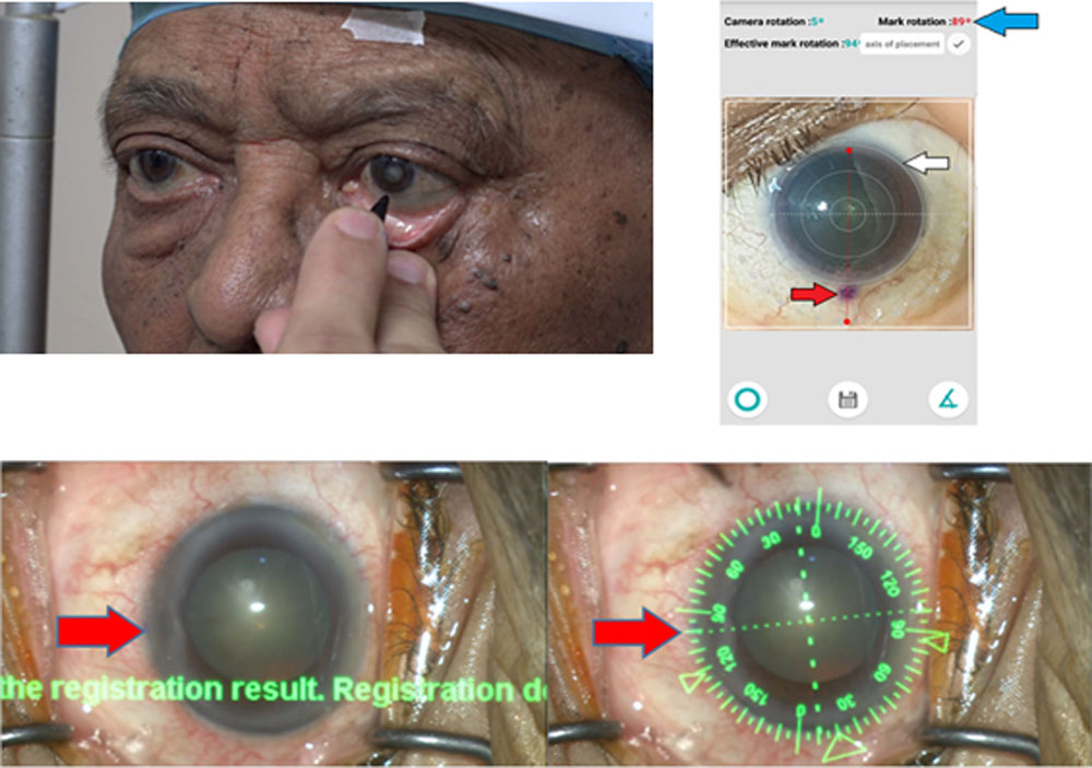
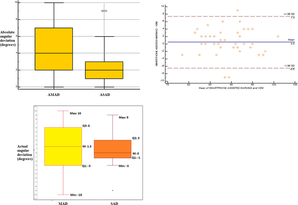
Table 1: Demographic data
| Number of patients | 36 |
| Number of eyes | 42 |
| Mean (SD) Age | 62 years (10 years) |
| Males | 23 (63.9%) |
| Females | 13 (36.1%) |
| Right eye | 23 (54.8%) |
| Left eye | 19 (45.2%) |
Table 2
| AMAD | ASAD | Significance (p value) | |
| Mean ± SD (degrees) | 4.60 ± 2.96 | 2.62 ± 2.40 | 0.0023 |
| Median (degrees) | 4.00 | 2.00 |
Table 3
| MA-VA | SA-VA | |
| % of eyes with clockwise deviation | 52.38 | 45.24 |
| % of eyes with anticlockwise deviation | 45.24 | 35.71 |
| % of eyes with no deviation | 2.38 | 19.05 |
| % of eyes with same orientation in both groups | 76.19 | |
| % of eyes with opposite orientation in both groups | 23.81 | |
Leave a Comment