

Dr.Ajay Aurora, A03243
Purpose: To evaluate the efficacy and safety of two Novel devices for injecting Drugs/ tamponade material into Suprachoroidal space (SCS) manually or using mechanised foot controlled Injection System.
Methods: SCS Injection (SCSI) was performed using two novel devices (Suprachoroidal Injection Device (SCID) 1and 2) in Goats eyes and one of the devices chosen for subsequent experimentation based on ease and safety of injection.
- A) Following experiments were performed on Goats eyes:
1) SCSI of Triamcinolone in Goats eye to assess the ease of injection and distribution of Triamcinolone with sequential Ultrasound Biomicroscopy (UBM) Scanning
2) SCSIof Hyaluronic acid in Goats eyes to assess the ease and safety of injection of Hyaluronic acid and assess with UBM. The hyaluronic acid was injected manually / with foot controlled injection device specially made for the experiment
- B) Ten human Eyes with recalcitrant macular edema, with or without associated glaucoma were injected 0.1- 0.2ml preservative free triamcinolone in the SCS and evaluated with sequential SDOCT, Fundus photography and Indirect Ophthalmoscopy.
- C) Scanning Electron Microscopy (SEM) of the available Triamcinolone preparations (Zuprata , Aurocort, Corteye and Tricort)
- D) Suprachoroidal Injection of Ozurdex Implant
Results: After Injection the drug spread uniformly in the Suprachoroidal space (SCS).
Triamcinolone was well tolerated and reduced macular edema in7/10 eyes without increasing the intraocular pressure even in known glaucoma patients. No eye developed any retinal or Choroidal injury. Scanning Electron Microscopy revealed the particle size varying from 1-12 μm. Tricort had all the particles in range below 5μm while other three had particles from 1-7μm (Zuprata), 1-6μm (Aurocort) ,1-12μm (Corteye) .
Conclusions:
The Suprachoroidal Injection Device presented is novel allowing easy, precise injection of triamcinolone and Hyaluronic acid in the SCS without the need of expensive, specialized microneedle systems. SEM of the triamcinolone acetonide tested shows that presence of particles larger than 5μm may have a role to play in the ease of injection of triamcinolone through a 30G needle. The pilot trial of SCSI of Triamcinolone in 10 patients using the SCID is encouraging.
Suprachoroidal injections are emerging as preferred methods of delivering drugs to the posterior segment. At present drugs are delivered to the posterior segment by injecting them Intravitreally when it is known that Vitreous is not the intended target tissue. This methodology can potentially introduce infection in the Vitreous cavity with serious complication of Endophthalmitis. Most drugs that at present are being given Intravitreally can be administered in the suprachoroidal space(SCS). The SCS is bounded by the ciliary body anteriorly, Optic nerve posteriorly,choroid internally and sclera externally. It is a potential space that can expand with the introduction of drug formulation. The location of SCS allows higher bioavailability of drugs tothe retina, choroid and sclera when compared to the available alternate routes of administration: topical(drops) or Intravitreal administration leading to dose reduction for the same effect1. Hence fewer ocular side effects are expected due to dose sparing and also because drugs are compartmentalized away from non-target tissues (e.g., lens causing cataract and Trabecular meshwork producing glaucoma).
To achieve the goal of suprachoroidal injection,a specialized micro needle matching the thickness of sclera has been constructed2 (Fig1)and it’s safety and tolerabilityhas been demonstrated in a phase I/II clinical trial (NCT01789320) in where triamcinolone acetonide (Triasence) was injected into SCS to treatnoninfectious posterior uveitis. 3Clearside Biomedical Inc. (CLS BIO) are actively pursuing Suprachoroidal delivery of specially formulated triamcinolone (Zuprata) 3 for diverse indications using a specially made injector, with preloaded triamcinolone Acetonideand a special micro needle which is available in two lengths: 900μm and 1100μm. With the Clearside Biomedical Inc. device the injection is made perpendicular to the sclera. The decision to use a 900μm or a 1110μm needle is arbitrary. At this time,the CLS BIO Triamcinolone (CLS –TA) isnot available commercially but when launched is expected to be expensive.It is also being projected that the triamcinolone acetonide, (CLS-TA / Zuprata), being used by Clearside is specially formulated to allow delivery through a 30G needle and will be effective for 12 weeks.
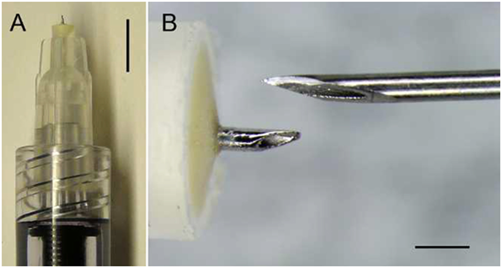
FIGURE 1. Microneedle for SCS injection. Low-magnification view of amicroneedle at the end of a syringe (A) and high-magnification comparison of a microneedle (left) to the tip of a 30-gauge hypodermic needle (right). Scale bars: 5 mm (A) and 500 lm (B) (1)
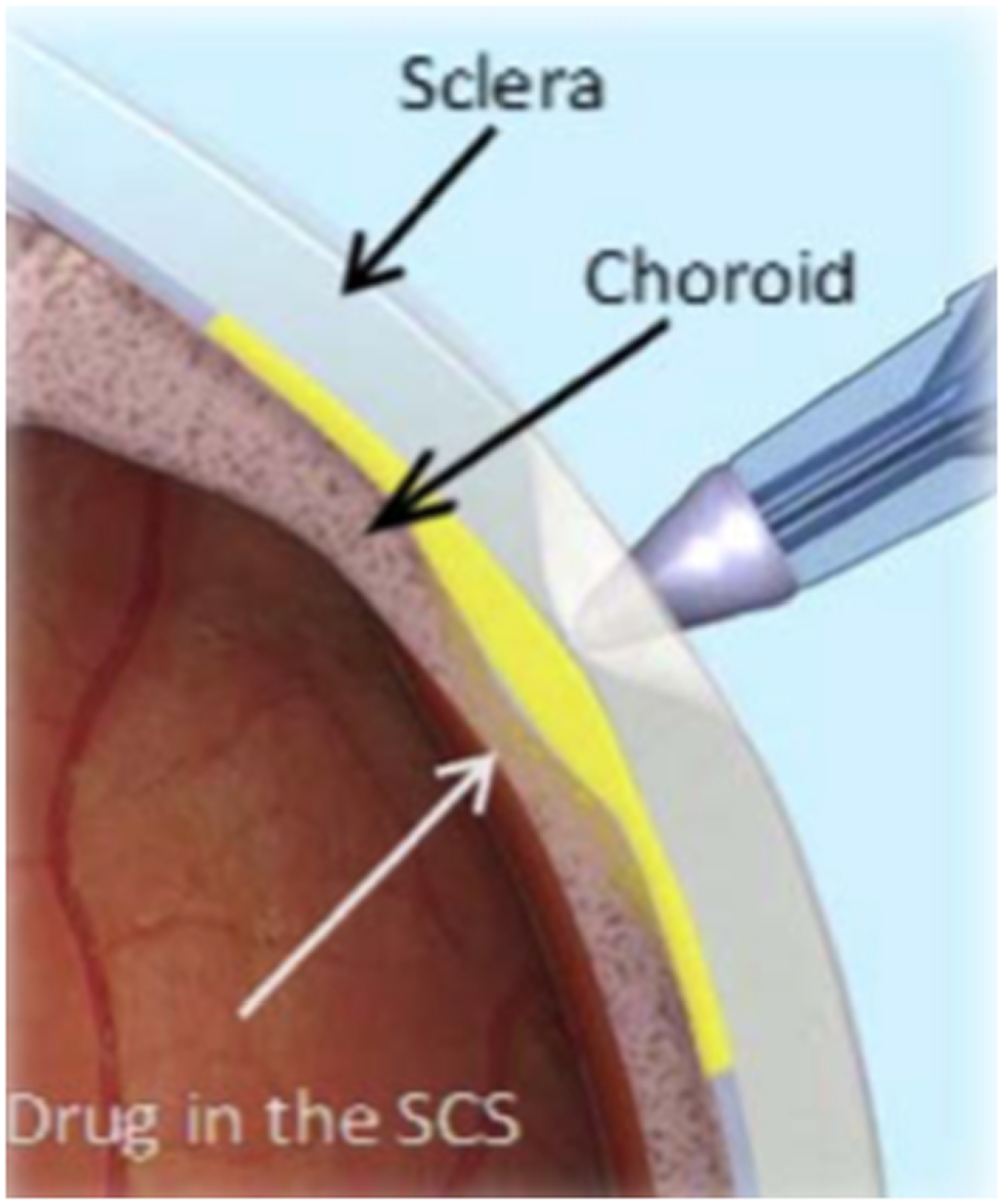
Fig2 Suprachoroidal Injection of Drug through Microneedle 3
The standard 30 Gauge needle with a bevel length of 1.7-1.8mm cannot be used for suprachoroidal injection for obvious reasons as the intended depth of suprachoroidal injections is only 1.0mm. CLS BIO device is able to achieve this because their total needle measures 900 or 1100 μm.With the Clearside device , the needle is directed perpendicular to the sclera to the full depth of the needle till the hub holding the needle touches the sclera, further pressure is exerted to create a dimple and the drug injected. The drug (Triamcinolone Acetonide, Zuprata, CLS-TA) spreads instantaneously through the potential SCS3.
Numerous authors (conference presentations, anecdotal reports and personal communication) have tried to circumvent the problem of long bevel length of 30 Gauge needle by creating a microneedle made by passing a 30 G Needle through a large bore needle (26 G Needle) long enough to have a short bevel exposed and joining the two needles together. This methodology being non-standardized has its obvious inconsistencies and sterility issues.Different workers have injecteddrugsand tamponade materialsin the SCS using a specially developed illuminated micro catheter that needs to be inserted through a partial thickness sclerotomy and is an invasive procedure with its learning curve.5,6
The aim of this paper is to test a novel technique of SCS injections developed by the author (AA) using the commercially available 30G needle obviating the need of a micro needle.
METHODS
Concept and Principle
- To reach the suprachoroidal space the intended average depth is 1.0mm
- Microneedles that are specially constructed with an extremely short bevel and overall short length can achieve this depth. However, microneedles are not commercially available.
- Author (AA) proposes to achieve this goal by entering the sclera obliquely with commercially available 30G needle, travelling intrasclerally assisted by a specially constructed device that allows with the bevel of the commercially available 30G needle. to eventually place the drug at the intended depth of 1.0mm. This concept is depicted below:
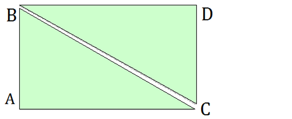
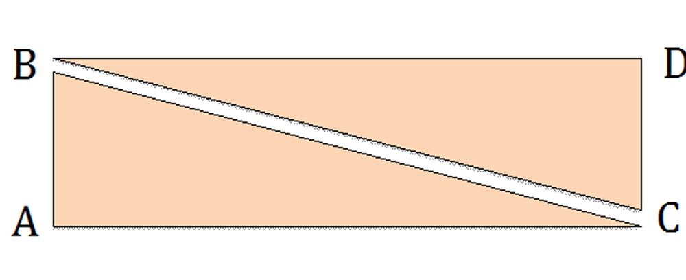
AB = 1.0mm
DC= 1.0mm
BD = Scleral Surface
AC= Suprachoroidal Plane
BA= Perpendicular direction of movement of Micro needle
BC= Proposed direction of movement of commercial 30G needle
Angle ACB = Angle DBC
IAs shown above, if the angle ACB is reduced the oblique length to which the 30 G needle has to travel along the path BC will become longer
If the angle of travel of the needle, angle ACB, can be fixed such that the distance BC is more than the bevel length, the needle tip that enters point C will be able to deliver the drug in the suprachoroidal space consistently and safely.
Novel Devices Used (Photographs not included as under Patent application process)
Two devices were tested on Goats eyes in a pilot study.
Device 1: The concept of device 1 was to create a dimple on the sclera using a forceps and then inject with the 30G needle, with the needle passing through a predetermined track whose angulation to the sclera was fixed
Device 2:The concept of device 2 was to run 30G needle through a fine track supported on an angulated base allowing defined entry of needle into the sclera.
Device 2 was selected for subsequent experimentation and clinical use.
Suprachoroidal Drug Injection Pump(Photographs not included as under Patent application process)
This is foot controlled, attaches to Vitrectomy machine and was developed with following aims:
- Injection of a viscoelastic material may induce movement of the hand and this can be dangerous to the underlying structures (choroid and retina).
- To test the ease and safety of injection of triamcinolone manuallyvs.Foot controlled pump.
Procedure
Device 2 was chosen for suprachoroidal Injections and manual injection technique appeared safe and effective for SCS injection of Triamcinolone.
The bevel of the 30 G Needle was inserted through the Device 2 such that the needle was intrascleral just past the bevel by a distance of 0.5mm. The correctness of the plane was identified by the ease of injection.If the bevel was still intrascleral, there was significant resistance noted with injection. The bevel was always kept pointing downwards.
After initial experimentation with Goats eyes, Triamcinolone was injected in the SCS after obtaining due consent from the patients.
Scanning Electron Microscopy of Available Triamcinolone
Tests were carried out on Zeiss EVO 50 Scanning Electron Microscope in the Textiles Department of the Indian Institute of Technology, New Delhi. The samples tested were:
- CLS-TA (Zuprata) Preservative free Triamcinolone Acetonide developed by Clearside Biomedical (alternative names CLS-1001; CLS-1003; CLS-1004; CLS-TA; suprachoroidal CLS-TA)
- Aurocort (Preservative free Triamcinolone Acetonide 40mg/ml (4% w/v) from Aurolab, Madurai, Tamilnadu)
- Corteye (Preservative free Triamcinolone Acetonide by Eyekare Kilitch Limited, Mumbai) from
- Tricort 40 (TriamcinoloneAcetonide 40mg/ml (4%w/V) + Benzyl Alcohol 0.9% w/v (as Preservative) and water for injection IP qs from Cadilla Phramaceuticals, Ahmedabad
Each sample was placed in an Ultrasonicator for 10 minutes to allow uniform admixture of the sample. 0.1cc of each was taken, diluted with distilled water in an Eppendorf tube. These were admixed again in an ultrasonicator. A drop of this was taken and placed on an aluminum tape, coated with a thin film of gold to obtain uniform conductivity. This was then placed in the SEM chamber and analysed. (Fig 2-5)
RESULTS
- Device 2 is superior to device 1 for SCS injections
- Triamcinolone can be safely and comfortably injected in the SCS manually.
- Injection of Healon can be safely done manually through the 30G needle into the SCS but is more controlled with the injection pump.
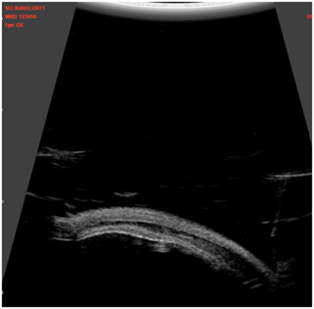
- We were able to demonstrate Triamcinolone by UBM in the Suprachoroidal space of Goats eyes. (Fig 6 ) It was found that Triamcinolone becomes undetectable on UBM after 3-5 minutes.
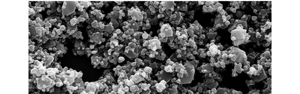
Fig 2: Zuprata (CLS-A) Scanning Electron Micrograph at 5,000 times magnification
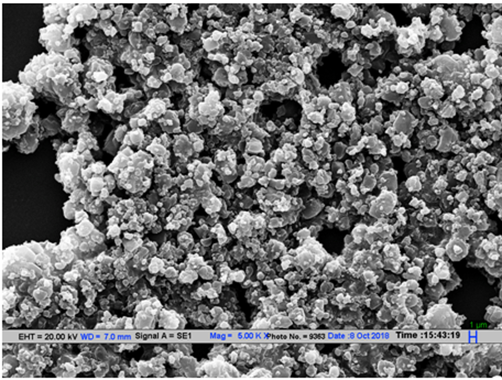
Fig 3: Aurocort SEM Scanning Electron Micrograph at 5,000 times magnification
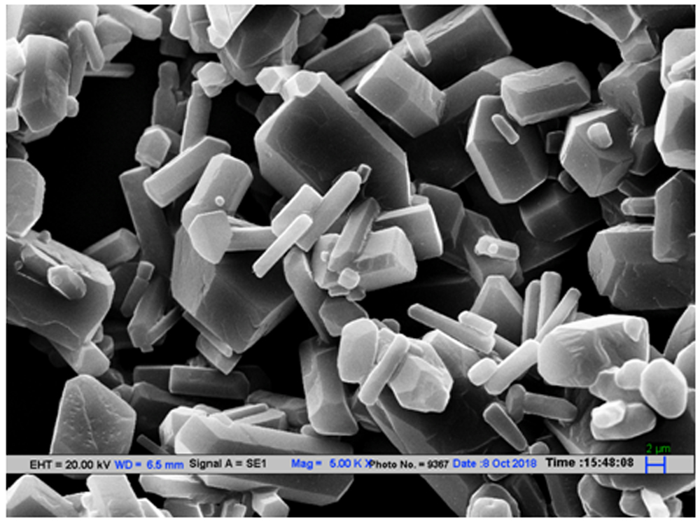
Fig 4: Corteye Scanning Electron Micrograph at 5,000 times magnification
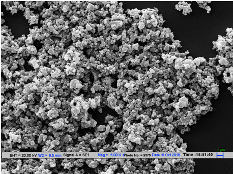
Fig 5 : Tricort:Scanning Electron Micrograph at 5,000 times magnification
Fig 6 Ultra Biomicroscopy Photograph showing Triamcinolone in the Suprachoroidal Space (Yellow arrow)
Suprachoroidal Injection Of Triamcinolone in Human eyes
Seven of the 10 eyes showed reduction of Central Foveal thicknessfollowing Suprachoroidal injection of Triamcinolone with either a qualitative symptomatic improvement or a demonstrable improvement of BCVA.
Suprachoroidal Injection of Ozurdex Implant
A pocket was created in the Suprachoroidal space of goat’s eye by injecting 0. 6 cc of Healon using the SCID.The linear separation created was measured using the UBM with an aim to have a separation of 8mm. The dummy Ozurdex implant that measures 6mm X 0.46mm, and simulates the real Ozurdex implant used for Intravitreal injections in humans, was injected into this pocket. There was egress of Healon from the injection wound. It was possible to demonstrate the Ozurdex implant in the Suprachoroidal space with the UBM. The sclera of the goat’s eye was then carefully dissected till the Suprachoroidal space and the injected Ozurdex implant retrieved.
DISCUSSION
With the anecdotal data and the limited Clearside data available for Suprachoroidal injection of Triamcinolone, it is clear that triamcinolone can be safely injected in the Suprachoroidal space. It is effective in this location for the retinochoroidal disorders (Macular edema due to Diabetic retinopathy, Central Retinal Venous Occlusion and non infectious Uveitis). It also effective for a longertime when compared with IVTA. Though the exact dose related effect data is not very clear but as per the data reported by CLS BIO it will be effective for 12 weeks4.This would enable the use of Triamcinolone in the suprachoroidal space without having to inject a steroid implant like Ozurdex in the Vitreous cavity.
Suprachoroidal Injection Device (SCID) (Device 2 was used in this study for all injections)
- Used here in goat’s eye has clearly shown that Triamcinolone and Healon can be safely injected in the Suprachoroidal space as confirmed by Ultrasound Biomicroscopy (UBM).
- The injected triamcinolone rapidly spreads both anteriorly and posteriorly from the point of injection and is detectable with difficulty with UBM after 5 minutes.
- Other than the Innovator (AA) , SCID was tested by Co author (SS, fellowship trained Retina consultant for its ease of using inthe Goat’s eyes. There was a minimal learning curve and the problems experienced have helped modify the device.
The SCID is Autoclavable and hence can be used repeatedly. It obviates the need of a special injector as the routinely available 30-gauge needle and syringe can be used. Potentially it will allow the injection of any drug in the SCS. The ease of injecting Healon through the SCID manually or machine aided can potentially be used to provide tamponade for a retinal detachment. Healon-5 has been recently shown to be effective in treating such cases by Mikhail M et al 6. They however, injected Healon -5 using a specially illuminated catheter, which has its own learning curve and complications.
Triamcinolone Acetonide and Scanning Electron Microscopy
- Zuprata (CLS-TA) Scanning Electron Microscopy of the sample showed particles varying in size from 1-7μm
- Aurocort (Preservative free Triamcinolone acetonide 40mg/ml (4% w/v)from Aurolab, Madurai, Tamilnadu) : Scanning Electron Microscopy of the sample showed particles varying in size from1-6 μm
- Corteye (Preservative free Triamcinolone Acetonide by Eyekare Kilitch Limited, Mumbai) Scanning Electron Microscopy of the sample showed particles varying in size from 1-12 μm
- Tricort 40 Scanning Electron Microscopy of the sample showed particles varying in size from 1-4 μm
While Zuprata (CLS-TA), Aurocort and Corteye can pass through a 30 G needle, Tricort needs to be injected through a 26-gauge needle. This is probably related to the particle size, number of particles of different sizes and the excipients used. Data is not available for the excipientsused by each company.
Suprachoroidal Injection of Healon
Healon acts as a spacer in the Suprachoroidal space. We were able to safely and easily inject Healon in the Suprachoroidal space using SCID both manually and with a specially developed injection pump using a 2cc syringe and a 30G Needle. Injection of methylcellulose was also attempted but was found to be difficult manually. We did not have access to Healon 5 as used by Mikhail M et al6. Injection of Healon through SCID will obviate the need for catheter based injection of Healon and may allow injection of Healon at the site necessary in cases of Retinal detachment. It will not only be less expensive but also involve minimal learning curve.
Pilot Study of Suprachoroidal Space Injection of Triamcinolone Acetonide (Aurocort)
Suprachoroidal Injection of 0.1ml (4mg) of Triamcinolone was done in a small sample study of 10 patients, with macular edema of diverse pathologies includingDiabetic Retinopathy, Central Retinal Venous Occlusion and a case of post Vitreoretinal Surgery with silicon oil injection for traumatic Vitreous Hemorrhage, Retinal detachment and retained Intraocular foreign body. Of the 10 eyes seven had reduction of Central foveal thickness of > 10%, three remained unchanged. There was no difficulty in injecting Triamcinolone in the SCS using the SCID and 30G needle.
Pilot Study of Suprachoroidal Injection of Ozurdex Implant in the Suprachoroidal Space in Goats Eyes
This is our preliminary data of Ozurdex injection under microscope in Goats eyes in the suprachoroidal space:A pocket was created in the SCS of goat’s eye by injecting 0. 6 cc of Healon using the SCID with 30G needle.The linear separation created was measured using the UBM with an aim to have a separation of 8mm. The dummy Ozurdex implant that measures 6 mm X 0.46mm, and simulates the real Ozurdex implant used for Intravitreal injections in humans, was injected into this pocket. Though, there was egress of Healon from the injection wound as the Ozurdex needle is larger bore, adequate Healon could be retained in the SCS for safe injection of the Ozurdex implant. The implant could be localized by the UBM and subsequently retrieved from the SCS after careful dissection of the Sclera under microscope.
CONCLUSIONS
This study shows the safety of using the novel Suprachoroidal Injection Device for Suprachoroidal Space Injection of triamcinolone in human eyes without the use of microneedle. However, larger human trials are needed to assess the safety and efficacy of Triamcinolone injection into the SCS using the SCID with the commercially available needles. The advantages are easy availability of the needle, ability to autoclave and reuse the device and obviating the need of a microcatheter. Use of SCID may allow us to inject Healon 5 in selected cases of Retinal detachment that will respond to local tamponade. SCID may also pave way for injecting drug implants into the Suprachoroidal space.
REFERENCES
- Chen M, Li X, Liu J, Han Y, Cheng L. Safety and pharmacodynamics of suprachoroidal injection of triamcinolone Acetonide as a controlled ocular drug release model. J Control Release. 2015; 203:109–117.
- Samirkumar R Patel, Damian E. Berezovsky,2 Bernard E. McCarey et al:Targeted Administration into the Suprachoroidal Space Using a Microneedle for Drug Delivery to the Posterior Segment of the Eye. Invest Ophthalmol Vis Sci. 2012;53: 4433–4441.
- Goldstein DA,Do D,Noronha G,Kissner JM et al.Suprachoroidal Corticosteroid Administration: A Novel Route for Local Treatment of Noninfectious Uveitis. Transl Vis Sci Technol.2016 Dec 14:5(6): eCollection 2016 Dec
- Zuprata: Report Store: Plasma Intelligence :Informa: Report code: Accessed September 2017
- Olsen TW, Feng X, Warner K, et al. Cannulation of the suprachoroidal space: a novel drug delivery methodology to the posterior segment. Am J Ophthalmol. 2006; 142:777–787.
- Mikel Mikhail, Ehab N. El-Rayes, Kentaro Kojima et al. Catheter-guided suprachoroidal buckling of rhegmatogenous retinal detachments secondary to peripheral retinal breaks. Graefes Arch Clin Exp Ophthalmol (2017) 255:17–23
Leave a Comment