

Dr.Raju S, R09402, Dr.Rajesh Ramanjulu
Surgery for the management of large macular holes is an established procedure.
Internal limiting membrane peeling is considered an essential step in macular hole surgery and is associated with better closure rates of patients with macular hole. However, Type 1 closure was difficult to achieve in patients with large macular holes. Michalewska et al1,2 in 2009 described the inverted internal limiting membrane (ILM) flap technique as an effective surgical technique for treating large idiopathic full thickness macular holes (MHs) and myopic MHs.They reported that the innovative technique has better type 1 closure rate than conventional ILM peeling for large idiopathic MHs. The better anatomic result was also associated with significant functional improvement in visual acuity.
In the original technique as described by them in the article 1 “The ILM was grasped with vitreous forceps and peeled off in a circular fashion on an area of about two-disc diameters around the macular hole. After circumferential peeling, the peeled-off ILM was not removed completely from the retina, but left still attached to the edges of the macular hole. In this way, a rolled fragment of the peeled-off ILM was “hanging” into the vitreous cavity. A piece of the ILM slightly bigger than the size of the macular hole was cut at the edge of the macular hole with a vitreous cutter and left on the surface of the macular hole. Then the ILM was gently massaged over the macular hole from all sides. The ILM became inverted, so that the surface normally directed toward the vitreous body was turned toward the RPE.
With time various modifications were used to the above technique and various adjuvants were used to keep the inserted ILM with the macular holes. Perflurocarbons liquids and Viscoat are being used as stabilising agents to increase the success of the stuffing of ILM into the hole.
When we had started doing the technique as described by Michalewska in 2011, we noted that there was RPE discoloration in the area of the macular hole in the post-operative period in few patients. This RPE damage we thought happened due to mechanical injury during the ILM stuffing step during surgery. Hence, we thought of eliminating this step during surgery. We share the anatomical and visual outcomes of minimally invasive inverted flap technique of peeling of internal limiting membrane (ILM) for macular holes with minimum diameter more than 600 μm.
METHODS:
Patients with very large idiopathic macular holes with minimum diameter of more than 600 μm(Range 600 to 1470 μm) were included in this retrospective study. Patients included in the study were operated between January 2012 to November 2017. They were operated either by Dr Raju S in a private set up or by Dr Rajesh R in a tertiary referral setup. This study includes the pooled data of the patients of both the authors. All patients underwent comprehensive ophthalmologic examinations, including measurement of best correctedvisual acuity (BCVA) intraocular pressure, slit-lamp bio-microscopy of the anterior and posterior segments. Diagnosis of Full thickness macular hole (FTMH) was confirmed on SD-OCT (OTI, OPKO health technologies, USA) or Topcon Maestro 3D OCT; (Topcon Corporation,Tokyo, Japan). The duration of the symptoms of MH was noted. The minimum and maximum hole diameters were measured. The patients with proliferative diabetic retinopathy, retinal detachment,age-related maculopathy, macular edema, or traumatic were excluded. All of these patients had undergone a minimally invasive inverted flap ILM peeling technique. The primary endpoint was the rate of hole Type 1 closure at 6 months after surgery. The secondary outcome measure was the change in best-corrected visual acuity at 6 months after surgery.
The surgical technique involved peeling of the ILM after staining under fluid with 0.05% of Brilliant Blue G (Ocublue plus, Aurolab, INDIA) The peeling was done in a radial fashion for about two-disc diameters around the hole and a rim of the ILM was left attached to the edge of the hole all around.(Figure 1, 2)No extra manipulations were done to mechanically stuff the macular hole with the ILM remnants. The peeled ILM with attachment at the edges of the hole were maintained carefully during the Fluid air exchange, care was taken not to aspirate the ILM flap. The ILM funnel was allowed to invert back on the retina during fluid air exchange such that a flap of the ILM covered the macular hole which could be in any direction. Air was exchanged with non-explansile gas either 20% SF6or 10 % C3F8. Patients were instructed to maintain prone positioning in the post-operative period at least for 4 days.
RESULTS:
Total of 66 patients were included in the study. There were 40 male patient and 26 female patients. Mean age was 57 ± 13 yrs. (range 48 to 72) Mean duration of symptoms were 5 ± 3 months (range 2 to 8 months). Mean macular hole diameter was 856 ± 77μm(608 to 1450) the primary end point of Type 1 macular hole closure was achieve in all the patients.Remodelling of the closure seemed to continue as long as 12 months after surgery. All patients had improvement in visual acuity of at-least 2 lines on Snellen’s Chart compared with baseline.Of the 66 patients 42 patients (64%) had a final vision between 6/24 to 6/18, 19 patients had a final BCVA between6/12 to 6/9 p (29%), 5 patients had a final BCVA of 6/36. (7%) Patients who had good visual improvement were found to have a restored External limiting membrane on OCT(Figure 5, 6) This restoration was sometimes seen in even patients with very large macular hole > 1000 microns(Figure 3. 4)
Discussion:
Inverted ILM peeling technique is a promising innovation in the management of large macular hole which hither to had Type 2 closure, i.e reduction in the hole size with out complete approximation of the hole edges. There are several mechanisms that have been proposed to explain the hole closure that occurs following use of the inverted ILM flap technique. The Müller cell fragments in the flap of the peeled ILM are thought to induce glial cell proliferation which fills the gap of MHcreating a scaffold for tissue proliferation and which in turn supports MH closure. It may also create a cellular environment that encourages photoreceptor proliferation which finally may help in improving postoperative anatomic and functional outcome. This hypothesis has support from the histopathologic findings that suggest a basement membrane is required for cell proliferation. ILM being a basement membrane, allows muller cell proliferation, and large MHs fill with tissue over time. In our case series too the cellular modulation seemed to continue till about 1 year following surgery. While the technique originally described by Michalewska1 required that the ILM flap be placed into the macular hole by mechanical manipulation. However, since this step can potentially damage the RPE at the base of the hole due to mechanical trauma it would be prudent to exclude this step if possible. Hence, an initial pilot study was done to see if the minimally invasive technique would work. Based on the promising results seen in the patients with even a large macular hole of 1100 microns we decided to try this technique in other patients with large macular hole. (Figure 3, 4a, 4b)The fact that all the patient who had large macular holes had type 1 closure proved that this technique is good enough for treating large macular holes of > 600 microns.
CONCLUSION:
Based on our results we conclude that the need to stuff the ILM mechanically into the macular holeand also the use of adjuvants like viscoelastic, per-flurocarbon liquids to aid this manoeuvermay not be mandatory. Minimally invasive inverted ILM flap technique can be considered even for large macular holes of more than 600 μm in diameter to achieve good anatomical and functional results.
References:
- Michalewska Z, Michalewski J, Nawrocki J. Macular hole closure after vitrectomy: the inverted flap technique. Retina Today 2009;3:73–74.
- Michalewska Z, Michalewski J, Adelman RA, Nawrocki J. Inverted internal limiting membrane flap technique for large macular holes. Ophthalmology 2010;117:2018–2025.
FIGURES
FIGURE 1
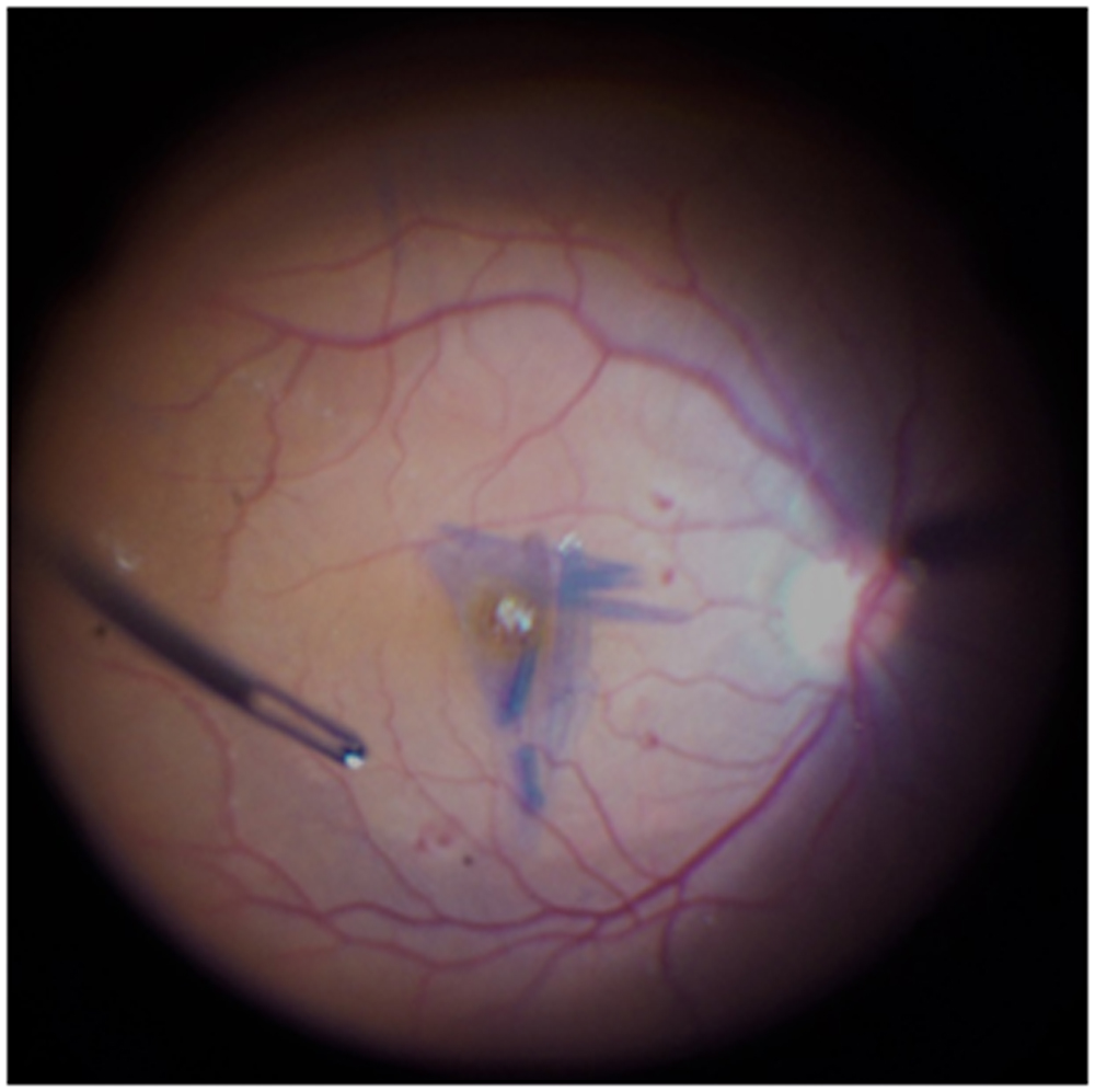
Figure 1: Intra operative photograph showing the ILM peeled in a radial fashion around the macular hole with attachment left at the edge of the macular hole.
FIGURE 2
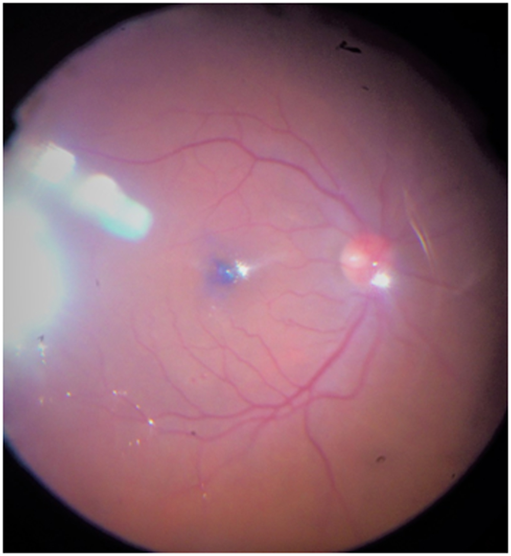
Figure 2 : Intra operative photograph showing ILM flap covering the hole after fluid air exchange
FIGURE 3
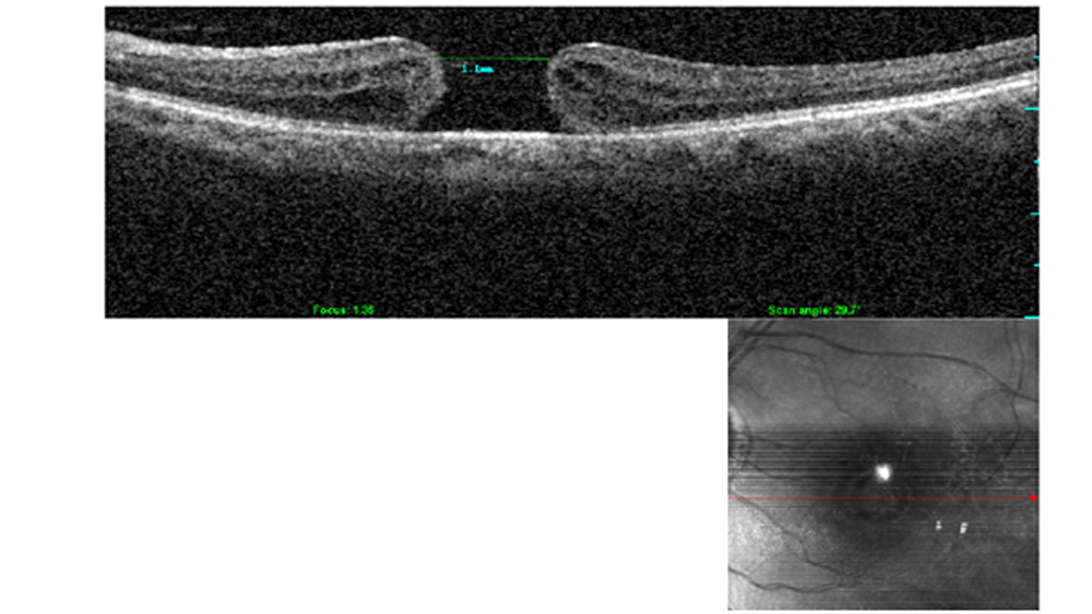
Figure 3 : OCT of Patient No.1 with a Macular hole with minimum linear diameter of 1100 µ and with a presenting best corrected vision of 4/60
FIGURE 4a
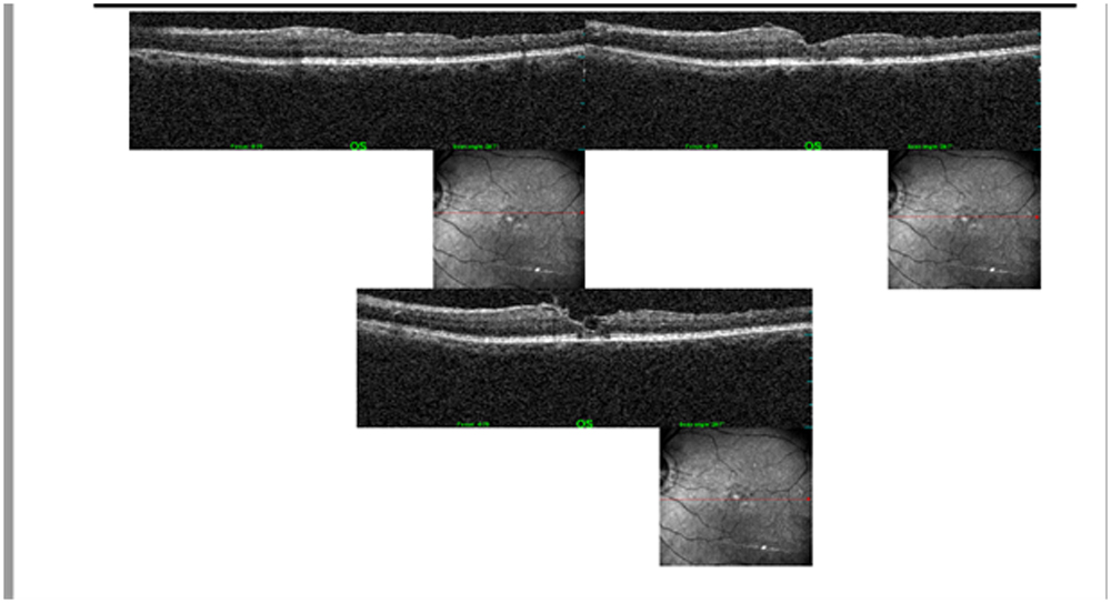
FIGURE 4b
FIGURE 4b
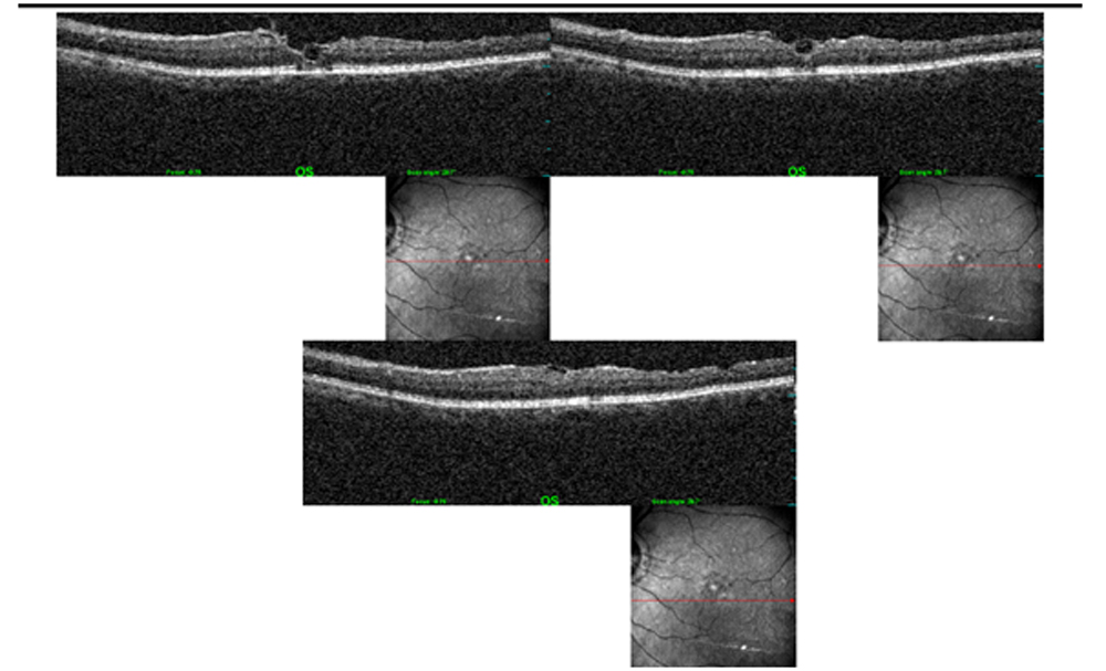
Figure 4a, b: Post-operative OCT images at 5 yr Follow up shows type 1 closure and the patient had a BCVA of 6/12p
FIGURE 5
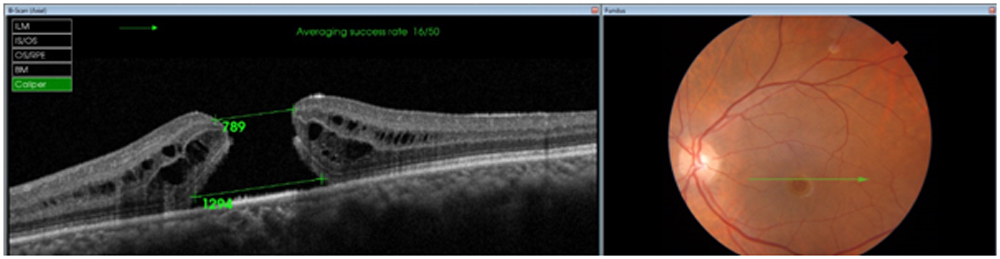
Figure 5: Preoperative OCT images of patient No 2 with a minimum hole diameter of 789µ and a basal diameter of 1294µ
FIGURE 6
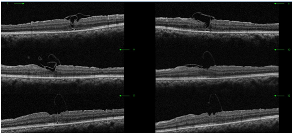
Figure 6: Post-operative OCT images of the patient No 2 showing multiple radial scan through the macular hole which shows type 1 Closure along with the ILM flap bridging the area of the macular hole. This patient had a BVCA of 6/9p at the end of 6 months follow up.
Leave a Comment