

Dr.Priyanka AgarwaL, P19286, Dr.Shroff Cyrus Minoo, Dr.Daraius Shroff, Dr.Charu Gupta
Abstract
Purpose: To describe features of peripapillary pachychoroid syndrome using multimodal imaging including OCT angiography (OCTA).
Methods: Prospective observational case series of 6 patients. Multimodal imaging was performed with detailed OCTA analysis.
Results: Imaging patterns of 12 eyes of 6 patients were analyzed. Middle age & male predominance with hyperopia was seen. B scan & EDI OCT showed thicker nasal macular choroid. The fluid was noted overlying the dilated pachyvessels. OCT A & enface showed dilated choroidal vessels, dark spots & dark areas in choriocapillaris segmentations in the nasal quadrant. Filamentous network was seen in 1 eye, indicating pachychoroid neovasculopathy. FFA showed hyper-reflective dots & ICG showed hypercyanescence with choroidal hyper permeability.
Conclusion: B scan OCT and OCT A is imperative in diagnosing the peripapillary pachychoroid syndrome. Dilated choroidal vessels, dark spots & dark areas in the peripapillary area are important findings
Introduction
Pachychoroid diseases are a spectrum of 4 different disease groups. These groups are essentially the stages of the disease itself, as the increased severity in the previous group leads the patient to the next group of disease. The spectrum comprises the following 4 groups: Pachychoroid pigment epitheliopathy (PPE). Central serous chorioretinopathy (CSC), Pachychoroid neovasculopathy (PNV) and Polypoidal choroidal vasculopathy (PCV). These share the distinctive choroidal features including focal or diffuse choroidal thickening associated with dilated Haller layer vessels (pachyvessels) with thinning of the overlying inner choroid and choroidal hyperpermeability.Initially, it was discussed in 2013 when Freund and colleagues described pachychoroid pigment epitheliopathy1 which was considered as forme fruste of CSC.
Phasukkijawatana et al described a new entity – peripapillary pachychoroid syndrome in which pachychoroid features surround the optic nerve and are associated with intraretinal and/or subretinal fluid and optic nerve head edema in some eyes2. Associated findings include serous pigment epithelial detachment, choroidal hyperpermeability, and pachyvessels.
Imaging of the choroid by traditional photographic and angiographic modalities is impeded by the relative opacity of the RPE to visible light. The absorption spectrum of melanin overlaps with both the excitation and emission spectra of fluorescein. Although this enables pre-RPE and sub-RPE components of vascular lesions to be distinguished, it prevents visualization of the choroid by fluorescein angiography (FA). The greater transparency of the RPE to longer wavelengths enables choroidal vessels to be delineated better by ICGA, but, because of vertical summation, angiographic features cannot be localized to their respective tissue layers. OCTA gives accurate depth resolved images of the different vascular layers noninvasively. Combining this with long-wavelength swept-source OCT (SS-OCT) enables the choroid and the choroid–sclera interface to be imaged at greater depth using shorter acquisition times providing new insights and a precise structural and functional analysis of the choriocapillaries and choroid.
The purpose of this study was to analyze this entity using multimodal imaging including swept source OCTA. We examined the various changes occurring at different levels and 3 dimensionally especially at choriocapillaris and choroid level. Our aim was to see whether this novel non-invasive imaging OCTA will help us to know this entity differently and will support in treating patients.
METHODS:
It is a prospective observational case series. The study adhered to the tenets of the Declaration of Helsinki. A total of 12 eyes 6 patients from the outpatient department of Shroff Eye Centre, were studied in detailed.
Patients with choroidal thickening, both diffuse and localized, associated with intraretinal/subretinal fluid in the nasal macula extending from the temporal margin of the disc were reviewed. Treatment naïve eyes which fulfilled above mentioned criterion were enrolled prospectively. A written consent was taken and the images were analyzed by our team of retina specialists.
Patients were excluded from this group if they demonstrated other causes of macular edema like diabetic macular edema or retinal vascular occlusion. Any clinical suspicion or imaging evidence of choroidal neovascularization due to other etiologies was excluded from the study. Conditions like optic pit with subretinal fluid were also excluded. Individual eyes were excluded if they had undergone intravitreal antiangiogenic treatment, photodynamic therapy, any of which might alter choriocapillaris and choroidal changes, or if media opacity or poor fixation prevented acquisition of scans of sufficient quality.
Demographic details were recorded. Snellen visual acuity, axial length, and refraction were noted. A comprehensive ocular examination was done using indirect ophthalmoscopy and slit lamp bio microscopy using 90D.
All patients underwent noninvasive imaging using swept source OCT angiography (Zeiss Plex Elite 9000 (California, USA). The software was updated with automated segmentation and removal of motion and projection artifacts. New vessels were looked into detail with segmentation analysis. Enface OCTA analysis was done at deep plexus, choriocapillaries and choroid slab. Structural Enface OCT was also analyzed. EDI OCT was evaluated on swept source OCT (Zeiss Plex Elite 9000 (California, USA) and choroidal thickness was compared in all patients. Bscan OCT was done and evaluated in all cases. Conventional multimodal imaging included color fundus picture, fluorescein angiography, fundus autofluorescence and ICGA (Heidelberg Engineering, Heidelberg, Germany) were also done. A comparative descriptive analysis was done and details were noted.
- Choroidal thickness measurement:
Choroidal thickness measurements were performed using EDI-OCT, and the caliper tool provided with the review software (Zeiss Plex Elite 9000 (California, USA). Choroidal thicknesswas defined as the perpendicular distance between Bruch membrane and the choroidal scleral junction.
Choroid thickness is different in different ages, refraction value, axial length.
In our case series, we have measured and defined thick choroid with a thickness of 300 µm and higher. Also, we followed the measurements taken on the following positions: subfoveal,1500 µm nasal and temporal to foveal centre, 3000 µm and 250 µm temporal to Bruch membrane originating at the temporal margin. (Fig 1). The brightness and contrast of the image were adjusted using the in-built adjustment tool, to visualise the choroidal scleral junction tool.
- Analysis of Juxtapapillary structures:
The temporal margin of the disc was evaluated in the Macula OCT ((Zeiss Plex elite 9000 (California, USA). Presence of intraretinal fluid, subretinal fluid, and its extent was taken into account. Associated RPE atrophy, any changes in ellipsoid zone (EZ) and external limiting membrane (ELM) were seen. Presence and extent of pachyvessels and also their correlation with fluid was noted. Also,the extent of pachyvessels whether they are diffused or localised were evaluated.
- OCT angiography analysis:
All study eyes underwent OCT A (Zeiss Plex elite 9000 (California, USA). The scan speed is 100,000 A-scans per second with an axial resolution of 6.3 µ m. It is based on the principal of OMAG.
Superficial and deep plexus were evaluated for the presence of hyper reflective dots (HRD) on Bscan OCT. Grey scale image was evaluated to quantify the HRD. Zones of reduced angiographic signal were interpreted as hypo flow areas in both the plexus. Choriocapillaris layer was studied for presence of areas of choriocapillaris attenuation (dark spots) and dark areas were noted whether they are corresponding to fluid.
Enface OCTA was evaluated for the presence of dilated choroidal vessels or pachyvessels. Pachyvessels were identified as groups of diagonally orientated hypo reflective Haller layer features with club-shaped origins at the posterior pole and large and constant calibre for the entire length of the vessel.
Also automated and manual segmentation was done to see the presence of a new vessel in peripapillary area and elsewhere. The pattern of new vessel was also studied.
STATISTICAL ANALYSIS:
Descriptive statistics was used to obtain mean and standard deviations. The tests used were unpaired t-test for comparing the mean in two groups. A p value of 0.05 was set as a threshold of significance.
RESULTS:
Total of 12 eyes of 6 patients, with peripapillary choroidal thickening with intraretinal /subretinal fluid extending from the temporal disc margin to the macula, multimodal imaging patterns were analyzed
Mean age of patients was61.7+_7.47 (range 40-70 years). In our study, a male preponderance was observed.Manifest refraction was available in all patients and nearly all were hyperopic with a mean refractive error of 1.03+_0.78 dioptres. Visual acuity in our patients and in both the eyes was good. Most patients had a Snellen visual acuity of 6/12 or better.
The mean subfoveal choroidal thickness was 362.67+_49.44. As the choroidal thickness was evidently thicker in the peripapillary region we performed the additional analysis of the ratio of nasal to temporal choroidal thickness.
The mean choroidal thickness measured at 1500 µm nasal to fovea was 407.25+_32.66. The mean choroidal thickness 1500 µm temporal to fovea was 276+_24.07.
The ratio of nasal macula choroidal thickness to temporal choroidal thickness at 1500 µm and was found to be extremely significant. (p<0.0001). Mean of the ratio of nasal to macular choroidal thickness at 3000 µm from fovea was also highly significant (p<0.0001)
Mean choroidal thickness at 250 µm temporal to Bruch membrane origin at the temporal disc margin was 196.33+_44.26
At juxta papillary area the retinal structures were studied on the OCT Bscan. In all eyes subretinal fluid / intraretinalfluid was present extending to the temporal disc margin (fig2). In this area, there was associated RPE atrophy with disruption of ellipsoid zone and ELM in 10 of the 12 eyes. But, in subfoveal area associated RPE atrophy was seen in 6 eyes only. The extent of pachyvessels were usually seen from peripapillary area to sub foveal area. The thickened nasal macular choroid was seen to be associated with pachyvessels and thinning of the overlying inner choroid. (fig2) The pachyvessels were more prominent in the nasal macula overlying the fluid than temporal sides of the macula in all the eyes. However, 2 patients had an additional temporal pachyvessels.
Hyper-reflective dots (HRD) were seen in all patients. Inone patient we tried to count them on reverse grey scale and were 4 in number. These were seen overlying the fluid. (fig 3)
On analysis of the different vascular layers on OCTA the superficial plexus was normal in all eyes. The deep plexus showed hypoflow areas in the peripapillary area corresponding to the intraretinal fluid in 10 of the 12 eyes. Hypoflow area seen on OCTA correlated well to the honeycomb appearance seen in the same area on the structural enface OCT in the same eyes (Fig 4)
Analysis of the choriocapillaris layer showed dark areas corresponding to the neurosensory detachment in all eyes. Hypoflow areas (dark spots) were seen corresponding to areas of choriocapillaris attenuation overlying dilated pachyvessels in 10 of the 12 eyes. Dark spots were seen in two patterns, fine diffuse areas and larger areas. The larger dark spots correlated well to hypocyanesence seen on ICGA. Some hyperflow spots were seen surrounding the larger dark areas and interspersed between fine diffuse areas. New vessel was seen in one patient. It was a filamentous pattern of vascular network with immature vessels which was suggestive of pachychoroid neovasculopathy. It was seen at choriocapillaries layer. There was no leak seen in FFA and even ICGA showed a plaque in the same patient.
The choroidal slab showed dilated choroidal vessels (pachyvessels) in all patients. Pachyvessels in the peripapillary area were seen in all cases. Pachyvessels were better defined on the structural enface OCT compared to OCTA. Enface OCTA showed dilated choroidal vessels or pachyvessels. They were seen as diagonally oriented, large calibre vessel markedly collected towards the Peripapillary area. (fig 5)
Of the 12 eyes, FA, FAF was available in all. Peripapillary RPE alterations were identified in all eyes. There was corresponding mottled, mixed autofluoroscence seen in all eyes with pigmentary gravitational tract in two eyes only.
On FA, all eyes demonstrated late hyperfluorescent with focal leakage in four eyes in Peripapillary area and diffuse leakage in two eyes.In six eyes questionable leak or merely late staining was seen. ICGA was available in 10 eyes and illustrated peripapillary dilated choroidal vessels with hyperpermeability seen. (fig 6)
Representative one cases is shown in fig7 which shows 50 years old male presented with intraretinal/ subretinal extending till temporal of optic nerve. There was associated PED seen. FA did not show any leakage, merely staining was seen. OCTA shown at choriocapillaries shows dark areas with choriocapillaries attenuation. Enface OCTA shows pachyvessels, with dilated Haller layer.
Discussion
We studied patients presenting with features of peripapillary choroidal thickening associated with intraretinal and /or subretinal fluid extending from the temporal disc margin into the macula.
Previously or in literature have shown that visualization of choroid both anatomically and functionally was difficult as it was partly obscured by the RPE. Sequential developments in OCT, such as enhanced depth imaging, swept-source, and dye-less angiography have allowed the choroid to be imaged functionally with depth resolution not achievable with dye angiography. Using these techniques, it has been clear that in pachychoroid disease the spatial distribution of RPE changes, neurosensory detachment, and neovascularization seems to correlate with localized choroidal thickening attributable to dilatation of Haller layer vessels and thinning of the choriocapillaris and Sattler layers4,5,6.
In this study, we expand our vision and looked into detail a new entity Peripapillary pachychoroid and assessed the choroidal thickness at various positions and its relation to other structural aspects in Oct angiography and structural OCT.
Our analysis demonstrated that the nasal macular choroid was significantly (p<0.0001) thicker than the temporal macular choroid in these eyes. Various studies have shown that macular choroidal thickness is normally greatest in sub foveal position followed by the temporal and nasal positions7,8. Rather in our study we took measurements at various positions (fig 1) and found to be maximum thickness at nasal macula. This was appreciably different from the patient we identified, in which nasal macular choroid was disproportionately thicker and was associated with pachyvessels as compared to other normal or typical pachychoroid spectrum eyes. Phasukkijwatana et al2 also studied 31 eyes and found that nasal macular choroid was more thickened than temporal and subfoveal choroidal thickness, however in their study they did not comment on any changes in OCTA and enface imaging in these eyes.
Our study has shown remarkably low flow areas at deep capillary plexus correlating well in enface OCTA also. This suggests that loss of homogeneity in flow signals, which were localized to foci of intra retinal fluid We found these areas more evident in deep capillary plexus and called them as “low flow areas”. However, Teussink et al9 in his study suggested that these low flow areas are localized to neurosensory detachment at choriocapillaries. We also looked at these areas at choriocapillaries and found them and called as “dark area”. These dark areas we have noticed to be seen commonly in patients with chronic CSC. Nicolo et al10 have studied OCT angiography segments of the choriocapillaries in patients with CSC using binarization and found areas of apparent flow reduction.
In our study, we observed dilated choroidal vessels were well visualised in enface OCTA. Their configuration can be well studied in this. They were seen as diagonally oriented, large calibre vessel markedly collected towards the fovea. In our patients we found these pachy vessels can be seen to be more collected at Peripapillary area.
In one eye we found the new vessel which was not seen in FA and ICGA.
We also looked for HRDs and quantify them in one patient and we found that they are more collected overlying the fluid. Studies have shown them to be more seen in inflammatory conditions, vascular disorders.
We did not found definite leakage on FA in these eyes. Balaratnasingam C et al11 has suggested that intraretinal cystic fluid may originate from the congested choroid through regions of peripapillary atrophy and associated atrophy of the RPE and ELM that normally serve as barriers for fluid to enter the retina. In our study we found associated, atrophy of the RPE and ELM and may be thesejuxtapapillary atrophic areas may facilitate fluid entry into the retina. A recent study by Lee et al12 described the presence of lamina cribrosa defects or disinsertions as a potential source of intraretinal fluid extending from the disk without leakage on FA in patients with pachychoroid disorders and patients with glaucomatous optic neuropathy.
Our findings suggest that wherever choroidal hyperpermeability is observed, excessive leekage probably originates from unhealthy choriocapillaries rather than from sattler’s or Haller’s layers,this hyperpermeability is then lost in a zone where unhealthy choriocapillaries yields to atrophy.
Limitation of this analysis includes the small sample size, quantitative observation of choripcapillaries attenuated areas. Nevertheless, we are unable to draw much of statically analysis, we present a number of cases that were imaged comprehensively and exhibit findings which are corroborating with the pathology. Further studies needed to see the changes following treatment in such scenario at choriocapillaries and choroid level.
We conclude that although the clinical features of pachychoroid spectrum shows considerable variability,this is a new pathology which can be better visualised on both OCTA and enface OCTA.With these recent imaging modalities one can distinguish it with similar presenting etiologies and can help in future treatment and follow up.
REFERENCES:
- Warrow DJ, Hoang QV, Freund KB (2013) Pachychoroid pigment epitheliopathy. Retina 33(8):1659–1672
- Phasukkijwatana et al. Peripapillary pachychoroid syndrome. Retina 0:1-16,2017
- Lehmann M, Bousquet E, Beydoun T, Behar-Cohen F (2015) Pachychoroid: an inherited condition? Retina 35(1):10–16.
- Dansingani KK, Balaratnasingam C, Naysan J, Freund KB. En face imaging of pachychoroid spectrum disorders with sweptsource optical coherence tomography. Retina 2016; 36:499–516.
- Dansingani KK, Balaratnasingam C, Klufas MA, et al. Optical coherence tomography angiography of shallow irregular pigment epithelial detachments in pachychoroid spectrum disease.
- Choi W, Mohler KJ, Potsaid B, et al. Choriocapillaris and choroidal microvasculature imaging with ultrahigh speed OCT angiography. Am J Ophthalmol 2015; 160:1243–1254.
- Shin JW, Shin YU, Lee BR. Choroidal thickness and volume mapping by a six radial scan protocol on spectral-domain optical coherence tomography. Ophthalmology 2012; 119:1017–1023.
- Margolis R, Spaide RF. A pilot study of enhanced depth imaging optical coherence tomography of the choroid in normal eyes. Am J Ophthalmol 2009; 147:811–815.
- Teussink MM, Breukink MB, van Grinsven MJJP, et al. OCT angiography compared to fluorescein and indocyanine green angiography in chronic central serous chorioretinopathy. Invest Ophthalmol Vis Sci 2015; 56:5229–523.
- Nicolo M, Rosa R, Musetti D, et al. Choroidal vascular flow area in central serous chorioretinopathy using swept-source optical coherence tomography angiography. Invest Ophthalmol Vis Sci 2017; 58:2002–2010.
- Balaratnasingam C, Lee WK, Koizumi H, et al. Polypoidal choroidal vasculopathy: a distinct disease or manifestation of many? Retina 2016; 36:1–8.
- Lee JH, Park HY, Baek J, Lee WK. Alterations of the lamina cribrosa are associated with peripapillary retinoschisis in glaucoma and pachychoroid spectrum disease. Ophthalmology 2016;123
Figures
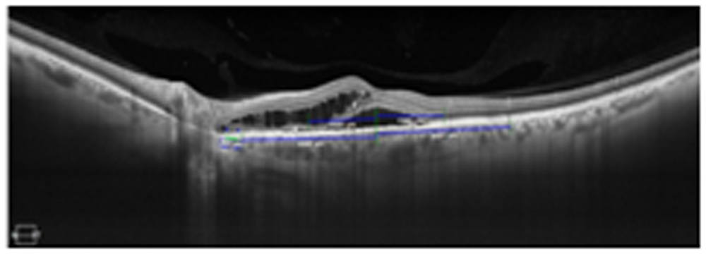
Fig 1: OCT shows various positions taken to calculate the choroidal thickness used in this study
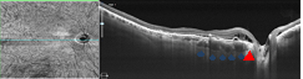
Fig 2: OCT image of one patient showing intraretinal/subretinal fluid. There is associated RPE atrophy (red arrow) seen at juxta papillary area. Also, localised thickening of choroid is seen. Pachyvessels can be seen overlying the fluid (blue arrowhead).
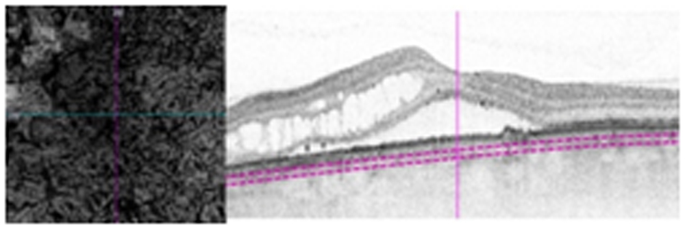
Fig 3: Panel A showing enface OCTA illustrating the pachyvessels with corresponding Bscan OCT showing HRD
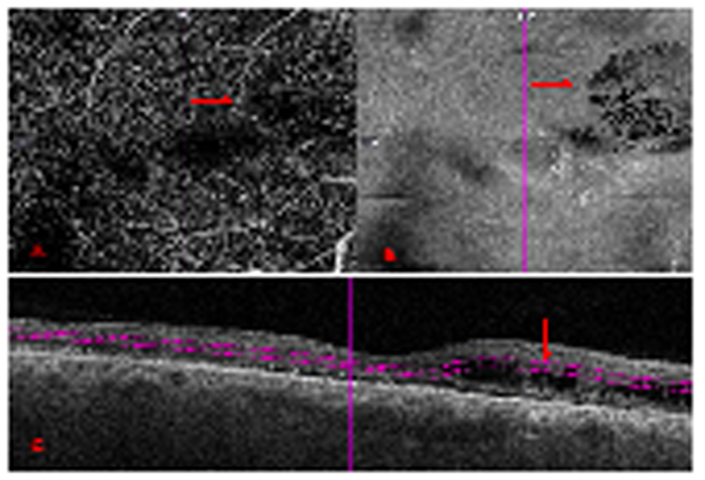
Fig 4: Panel A shows OCTA showing hypoflow areas with corresponding enface OCTA (panel B) depicting honeycomb appearance. Corresponding Bscan OCT showing intraretinal fluid.
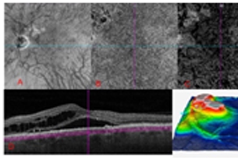
Fig 5: PanelA shows structural OCT at choroid segment showing dilated vessels at peripapillary area. Panel B showing OCTA depicting attenuated areas. Panel C enface OCTA showing dilated Hallers layer. Corresponding OCT and 3D image depicting involved area mainly at Peripapillary region.
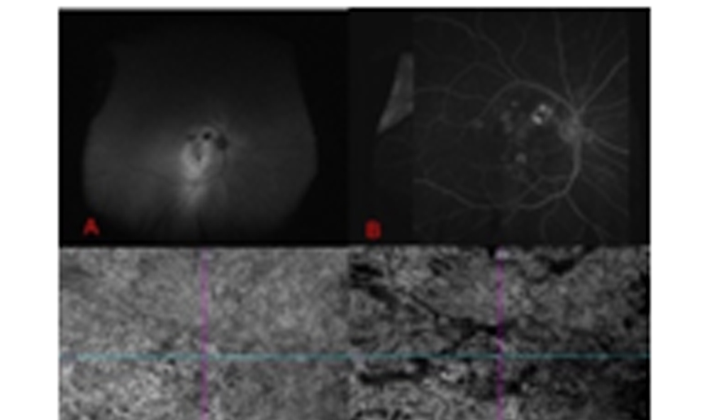
Fig 6: Panel A showing AF depicting hyperAF gravitational tract. Panel B showing localized leak at Peripapillary area. Panel C &D showing OCTA and enface OCTA depicting dilated Hallers layer.
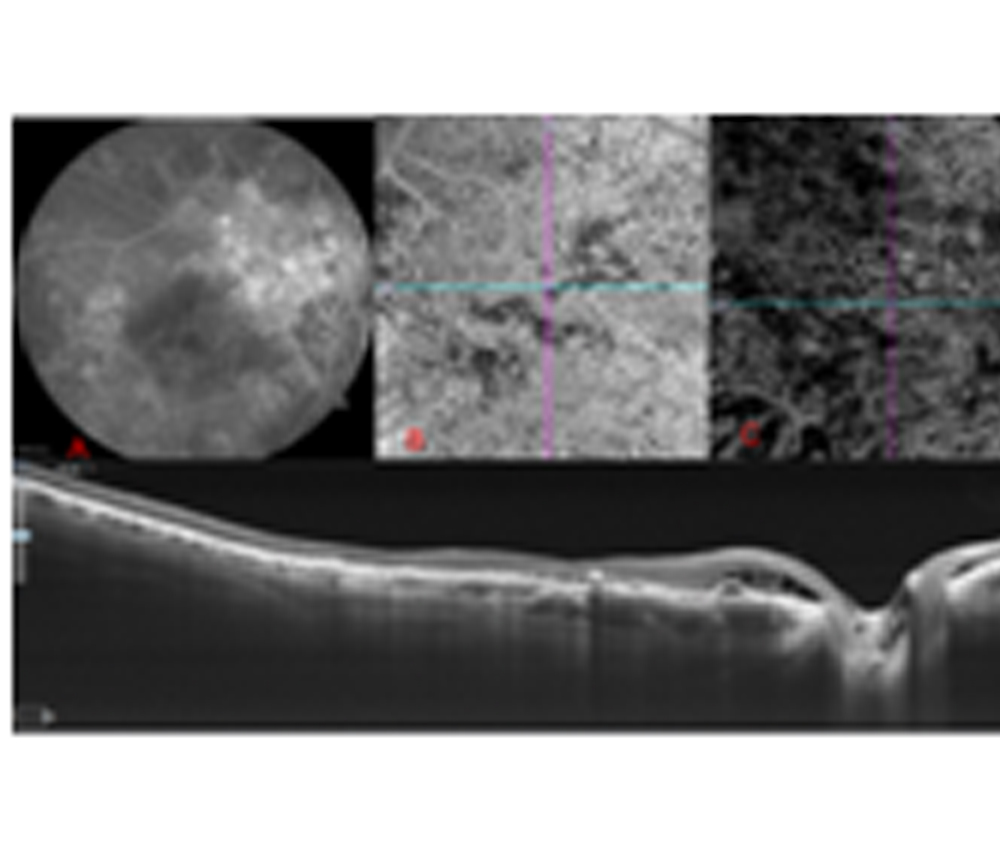
Fig 7: Panel A shows FA showing diffuse staining at Peripapillary area. Panel B shows OCTA at choriocapillaries showing dark area and Panel C shows dilated Haller layer. Corresponding OCT (Bottom) shows intraretinal/subretinal fluid with small serous PED.
Leave a Comment