

Dr.ABHISHEK ANAND,Dr.Ashraya Nayaka
Abstract
Suprachoroidal Buckling is a novel technique popularized by Dr.Ehab N El Rayes for peripheral buckling, macular Buckling and Delivery of drug and drug devices in suprachoroidal space. Assessing and exploring the suprachoroidal space is a potential game changer, however, it requires a learning curve to do the same. Initial experiences are multivaried with various limitations for beginners. The author wants to share his experience of few cases of mainly peripheral suprachoroidal buckling, delivery of suprachoroidal drugs at Pars plana, near the equator and around vascular arcade and few case of Suprachoroidal Macular Buckling. The author wants to share his case experiences, initial hiccups like in establishing the right suprachoroidal space, visualization of the cannula tip, simultaneous use of chandelier light and alternative long term fillers suitable for Price sensitive Indian Subcontinent.
Introduction
Scleral Buckling is an established procedure both for simple and complicated rhegamatogenous retinal detatchment. However, with the advent of three port Pars PlananVitrectomy introduced by Maachemer in 1985 and with gradual evolution of smaller gauge instrumentation, sutureless surgery, advanced fluidics, high cut rates and true visualization Buckling is gradually getting out of favour with
Its just not the gradual evolution of the Pars Plana viterectomy but the perils associated with exoplants also acts as barrier with every passing day as worldwide surgeons get more and more accustomed to PPV. Extensive dissection with postoperative ocular surface disorder and discomfort is one of the most common complaint. Buckle extrusion, scleromalacia and superadded infection are the most common long term complications. it may result in some vision-threatening complications such as mechanical ocular motility disturbance secondary to an extraocular muscle imbalance, corneal contour changes, anterior segment ischemia, and chorioretinal circulatory disturbances due to the direct depression of the sclera with the buckle and also depending on the size and location of buckle placement1,2,3
Suprachoroidal buckling (SCB) technique is a new approach developed by Ehab N El Rayes and Oshima for treating rhegmatogenous retinal detachment (RRD). SCB uses a specially designed catheter to inject and place a suprachoroidal filler (long-lasting hyaluronic acid solution) in the suprachoroidal space. This filler indents the choroid alone and creates a suprachoroidal buckling effect thereby closing retinal tears and supporting the overlying retina. The sclera itself is not involved in the indentation4. It avoids all perils of buckling as it is minimally invasive, requires a small opening in conjunctiva for the introduction of the suprachoroidal catheter. All the disadvantages of placing a long term silicone exoplant is also avoided..
Macular Buckling for Myopic Traction Maculopathies is an age old procedure. However, after seeing its golden days in 80s and 90s it lost in favor to PPV and with the advent of Internal Limiting Membrane Peeling. Macular Buckling has again saw its renaissance with various case series from around the world showing Macular Buckling with or without ILM peeling being superior to PPPV alone. Macular Buckling with exolant either Morin –Devin T implant or Ando Plombe Implant or Sponge implant requires a long learning curve and all the complications of a large exoplant. The indentation effect achieved with SCB can also be used as an alternative to episcleral macular buckling for managing myopic traction maculopathy (MTM)5.SCB can be used alone or in conjunction with vitrectomy.
Surgical Procedure
SCB technique uses two devices to create a suprachoroidal buckle.
- EL-Rayes Flex-tip Catheter (MedOne Surgical,
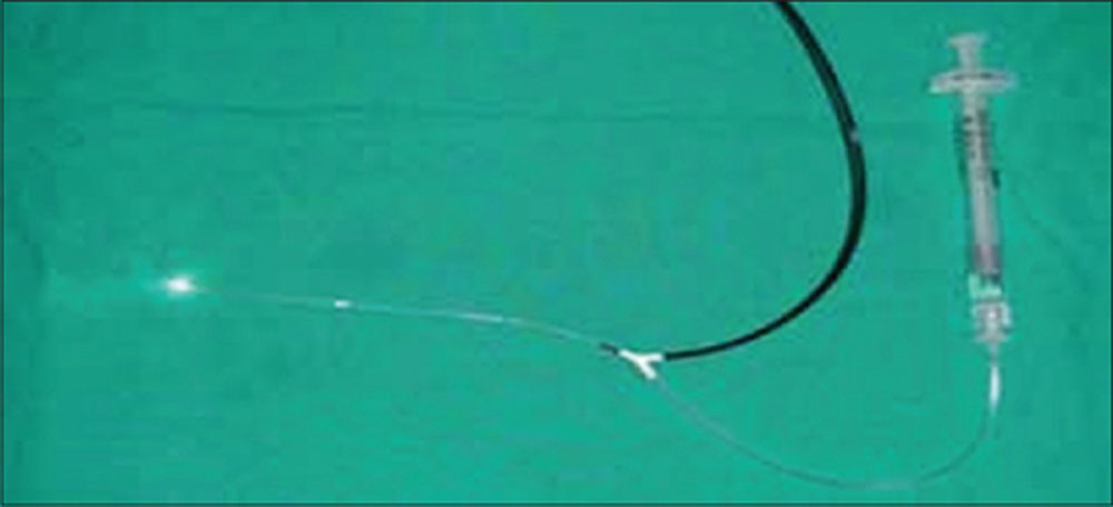
Suprachoroidal catheter for suprachoroidal filler injection. One arm is connected to a light source and the other is connected to the filler material
This 450-μm catheter has a dual injection/illumination capability. It can be connected to a regular bright illumination light source that illuminates a 31-gauge light fiber when placed in the suprachoroidal space for identifying the tip of the inserting cannula. The other end of the Y connector on the catheter is connected to high-pressure tubing to deliver stabilized (natural entangled) long-lasting HA solution (Healon GV or Healon V, Abbott Medical Optics Inc., or RestylanePerlane, Q-med) to the injection port of the catheter when placed in the suprachoroidal space. This works for both posterior pole retinal pathologies and peripheral breaks.
El-Rayes curved suprachoroidal cannula
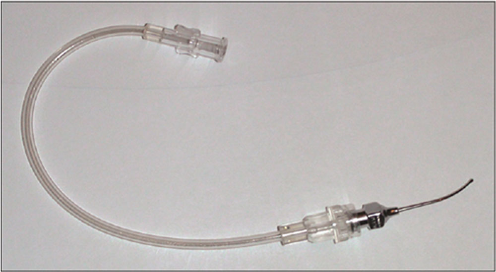
The suprachoroidal cannula notices the olive tip acting as a gentle choroidal depressor
Recently, as an alternative to the illuminated tip catheter, a nonilluminated 20-gauge curved solid cannula with an olive tip for injecting the filler can be used, thus creating a SCB effect. The advantage of the olive tip is that it acts as a choroidal depressor. However, this cannula can be used for peripheral pathology only; it cannot be guided into the postequatorial region.
Filler substance
Different materials are used to create the suprachoroidal buckle.
- RestylanePerlane (20 mg/ml) which is a high purity gel containing nonanimal HA stabilized with minimal modification resulting in a long-lasting three-dimensional gel network with excellent safety profile can be used. This filler is known to the past 8-12 months in dermal tissues and is used for restoring space and contour by its gel strength. The strength of the gel depends on the total number of cross-linkings between HA disaccharides (both chemical and natural entanglement)
- Shorter acting substances such as Healon5, 23 mg/ml, (Abbott Medical Optics, Santa Ana, CA, USA), which has less bonding, may also be used for treating peripheral breaks since it remains for 2-3 weeks in the suprachoroidal space which is enough to create chorioretinal adhesion after treating the break.
- Dr. Filler(20mg/ml) – A cheap substitute to Perlane and can last for upto 6 months in suprachoroidal space. This filler has been used by us for the first time from all over the world and seems to have excellent safety profile.
- The issue of “how long” (duration) is no longer a problem because the stabilization, bonding technology, and higher cross-linking solve the duration issues and create a gel of superior strength. Therefore, depending on the nature of the pathology, we can choose the type that we will inject.
Surgical Technique for Rhegmatogenous Retinal Detachment
Suprachoroidal Buckling can be used as Primary treatment for Rhegmatogenous Retinal Detachment or in conjunction with Pars Plana Vitrectomy for supporting inferior retina.
Primary RRD repair with SCB- 25 Gauge chandelier (Alcon) was put aat 12’O clock position. Approximate position of the breaks were established under visualization with Non contact wide angle visualization system . 1 clock hour peritomy was done in the concerned quadrant. Under direct visualization with 25 Gauge illumination retinal cryopexy of the break was done and position of the break was marked with surgical Pen marker. In the meridian of break 4 mm from the limbussuprachoroidal space was accessed through gentle dissection under high magnification. After assessing the suprachoroidalspace ,viscodissection of the suprachoroidal space was done. External Subretinal fluid drainage was done to lower the intraocular space for making potential space of 0.5-1 ml of suprachoroidal space. Medone olive tipped suprachoroidalcanula was introduced in the suprachoroidal space under visualization with wide angle viewing system. The tip of the canula is used to produce slight indentation on the choroid to confirm the approximate position of the tip. After reaching the approximate position for injection, the assistant is asked to gently push the filler to prodiuce an oval indentation beneath the break keeping a careful watch on optic nerve head pulsations.
Multiple breaks can be assessed at the same time and even posterior breaks and radial tears can be assessed likewise.
SCB in conjunction with PPV- cases needing support of inferior retina especially breaks around 6’o clock can be supported with SCB. Inferior peripheral retina can be supported in the same way. As described above the process is same. For supporting 180degree of inferior retina Olive tipped canula is passed medially and laterally from the 6’0 clock access point while injecting slowly to produce a moderate indentation.
Surgical Technique for Myopic Traction Maculopathy
MTM is schisis-like thickening in the outer layers of highly myopic eyes with posterior staphyloma, also known as myopic foveoschisis. Other findings may include inner retinal fluid, foveal detachment, and lamellar or even full-thickness macular holes with progressive retinal detachments6,7.Management of MTM is based on eliminating epiretinal traction forces by means of vitrectomy with or without internal limiting membrane peeling8,9. Episcleral macular buckling is a well-established technique that supports the posterior scleral wall, releasing the traction forces on the vitreoretinal interface (VMI) in the area of the staphyloma. There is a long learning curve, and it is very challenging to master the technique of locating and fixing the macular buckle in extremely thin sclera and deformed staphyloma.
The SCB technique can bypass the difficulties of conventional scleral buckling in the macular area by direct controlled delivery of a buckle-like effect (indentation) from the suprachoroidal space in the area of staphyloma. The choroidal indenting effect at the macula relieves all forms of traction at the VMI.
In macular buckling, the surgical procedure for all patients is a standard three-port, 25-gauge pars planavitrectomy and dissection of the posterior hyaloid. Then, the same technique of SCB as for RRD is adopted. Care is taken while attempting to cross the edges of the posterior staphyloma as the choroid is extremely thin, especially at the superior edge of the staphyloma. Before crossing this site, a small amount of filler is injected to dissect and lift the choroid at the edge, before pushing the catheter into the area of the posterior staphyloma. Once the catheter is placed in the suprachoroidal space underneath the target tissue (foveoschisis or macular hole), the filler is injected to dissect the thin choroid toward the retina, creating an indentation effect bypassing the staphyloma. This effect restores the normal contour of the choroid or even indents it to a convex configuration supporting the retina. The illuminated tip of the catheter is seen with ease in patients with foveoschisis and in patients with macular holes associated with shallow posterior pole detachment . Fluid/air exchange is used before filler injection if the subretinal fluid is extensive enough to obscure the catheter tip light.
DISCUSSION
The advantages of this technique over established scleral buckling procedures include faster surgery, less invasive, temporal indentation, no risk of strabismus/diplopia, no postoperative pain, and a quieter eye at immediate postoperative follow-up. No ischemic choroidal changes occurred, may be due to cushion-like effect created by the indentation causing less mechanical pressure or deformation to the choroidal circulation as compared to silicone implants.
The complications observed were minimal which included penetration of the catheter directly through the pars plana into the vitreous cavity, penetration of the choroid, and the retina in the meridian of the primary tear. These did not result in hemorrhage or redetachment due to thin structure of the catheter, and the penetration site can be easily treated with laser retinopexy. Hyperpigmentation at the edge of indentation due to RPE heaping at the edge of the dome can occur, which usually decreases with time. Another potential complication is choroidal hemorrhage, which is also a risk of the standard buckling procedure.
In conclusion, based on favorable results obtained from animal experiments and preliminary clinical trials, SCB procedure with the suprachoroidal catheter is technically safe and feasible. Since the physiologic adhesion between the choroid and sclera is loosened, no serious complications related to the insertion of the catheter and injection of HA solution into the suprachoroidal space was encountered. This technique represents a great addition to surgical inbox for repair of selected cases of retinal detachment. Further study is recommended to evaluate the choice of fillers for SCB according to the pathology and desired length of indentation.
REFRENCES
1.Sewell JJ, Knobloch WH, Eifrig DE. Extraocular muscle imbalance after surgical treatment for retinal detachment. Am J Ophthalmol 1974;78:321-3
2.Smiddy WE, Loupe DN, Michels RG, Enger C, Glaser BM, deBustros S, et al. Refractive changes after scleral buckling surgery. Arch Ophthalmol 1989;107:1469-71.
3.Yoshida A, Feke GT, Green GJ, Goger DG, Matsuhashi M, Jalkh AE, et al. Retinal circulatory changes after scleral buckling procedures. Am J Ophthalmol 1983;95:182-8.
4.El Rayes EN, Oshima Y. Suprachoroidal buckling for retinal detachment. Retina 2013;33:1073-5.
5.El Rayes EN. Supra choroidal buckling in managing myopic vitreoretinal interface disorders: 1-year data. Retina 2014;34:129-35.
6.Gaucher D, Haouchine B, Tadayoni R, Massin P, Erginay A, Benhamou N, et al. Long-term follow-up of high myopic foveoschisis: Natural course and surgical outcome. Am J Ophthalmol 2007;143:455-62.
7.Shimada N, Ohno-Matsui K, Yoshida T, Sugamoto Y, Tokoro T, Mochizuki M, et al. Progression from macular retinoschisis to retinal detachment in highly myopic eyes is associated with outer lamellar hole formation. Br J Ophthalmol 2008;92:762-4.
8.Kwok AK, Lai TY, Yip WW. Vitrectomy and gas tamponade without internal limiting membrane peeling for myopic foveoschisis. Br J Ophthalmol 2005;89:1180-3.
9.Spaide RF, Fisher Y. Removal of adherent cortical vitreous plaques without removing the internal limiting membrane in the repair of macular detachments in highly myopic eyes. Retina 2005;25:290-5.
Leave a Comment