

Dr.Amit Porwal,Dr.Jitendra Nenumal Jethani,Dr.Parthvi Shah,Dr. Meghna Shrishrimal
Aim:
Introduction:
Cataract surgery is one of the most efficient and safest surgical interventions but it is dependent on the surgeon’s skill and experience especially in difficult cases like intumescent cataracts. Surgery in intumuscent cataracts poses a challenge in many ways as the anterior chamber is shallow. Due to high intralenticular pressure extension of the capsulorhexis to the periphery and occurrence of Argentinian Flag sign is high.[1]
Gimbel and Neuhann first described capsulorhexis in 1987 as a central, circular and curvilinear opening in the anterior capsule.[2] An intact capsulorhexis is very crucial in achieving an uneventful cataract surgery as it offers good stability and centration of IOL and prevents extension of radial tears.
Several approaches are described in literature to achieve a central circular capsulorhexis in intumescent cataracts and to avoid the extension of capsulorhexis towards the periphery like use of trypan blue dye, 2 stage capsulorhexis , aspiration of fluid cortex by 30 gauge needle , capsulorhexis using microcapsulorhexis forceps and femto assisted capsulorhexis.[3]
Here we describe a simple preoperative procedure to decrease the intralenticular pressure and to deepen the anterior chamber by doing Nd Yag laser capsulotomy. The use of a single central disruptive pulse of energy can maintain an equal distribution of vector forces, preventing uncontrolled splitting of the anterior capsule to the periphery. When Nd YAG laser is done in the presence of an intact anterior chamber, the intraocular pressure guarantees a safer pressure balance compared to other techniques.
Materials and Methods:
eyes were selected in this prospective study done from May 2018 to September 2018. Mean age of patients was 57.6 ± 5.2 years. Patients with any other ocular pathology like lens induced glaucoma, uveitis or any history of ocular trauma and any posterior segment pathology on B-scan ultrasonography were excluded. Preoperative evaluation comprised of demographic details, visual acuity, slit lamp examination, fundus evaluation of the fellow eye, B scan of the eye to be operated, IOL power calculation, physician fitness to rule out any systemic ailment and an informed consent. After Dilatation of pupils with tropicamide plus eye drops, Intra ocular pressure (IOP) was recorded by Icare TA01i Tiolat OY, Helsinki, Finland and Anterior Chamber depth using Anterior Segment Optical Coherence Tomography (OCT) (Carl Zeiss Visante OCT 1000) was measured pre and post Nd YAG laser anterior capsulotomy. Preoperative anterior YAG capsulotomy was performed in all eyes with 1 shot of 1.2 mJ & a gush of fluid was noted. Patient was taken up for surgery within 10 minutes of laser after giving a peribulbar block.
A manual small incision cataract surgery was performed in all patients by a single surgeon. Intraoperatively post staining of the capsule with trypan blue, a cruciate opening was noted on the capsule with everted edges (FIG 1). The capsulorhexis was done with Utratas capsulorhexis forceps taking one of the flaps of the opening created by laser. The ease of performing a complete central circular capsulorhexis was noted. The surgical comfort was assessed.
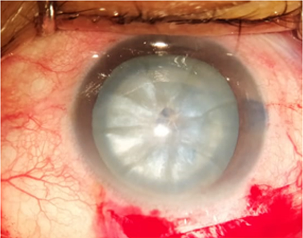
FIG.1: Cruciate opening in Anterior Capsule stained with Trypan Blue.
Results:
Pre YAG mean IOP was 13.5 ± 2.3 mmHg (Range: 9-18), mean AC Depth was 1.66 ± 0.28 mm (Range: 1.04-1.93). Post YAG mean IOP was 11.9 ± 1.9 mmHg (Range: 8-15), mean AC Depth was 1.93 ± 0.3 mm (Range: 1.35-2.28). There was a mean increase in AC Depth by 0.24 mm (FIG.2) and a mean decrease in IOP by 1.61 mm post laser (FIG.3). On two tail t test, the change in AC depth and IOP change was significant (p<0.05). (FIG. 4,5) No intraoperative complications were noted except for rhexis extension in 1 eye. (FIG.6). High level of surgeon’s operative comfort was there
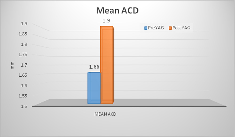
FIG.2: THE MEAN ANTERIOR CHAMBER DEPTH INCREASES POST YAG
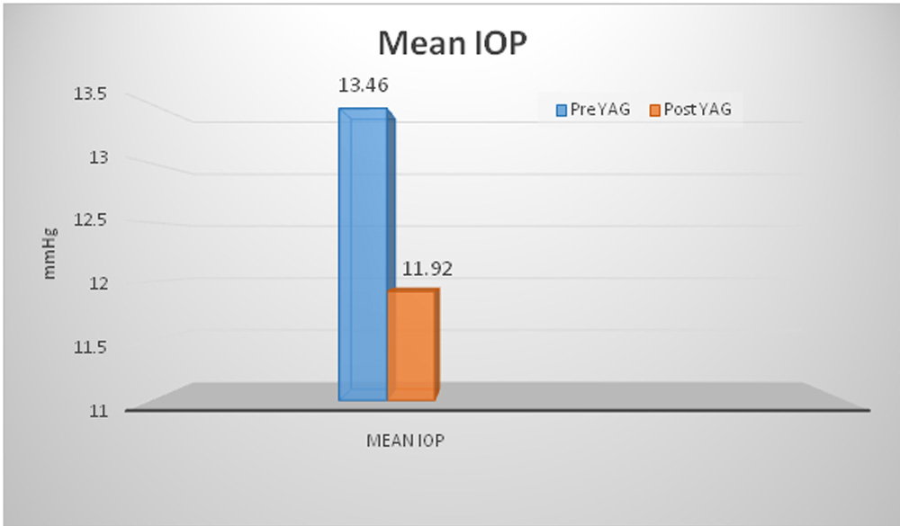
FIG.3: THE MEAN IOP DECREASES POST YAG
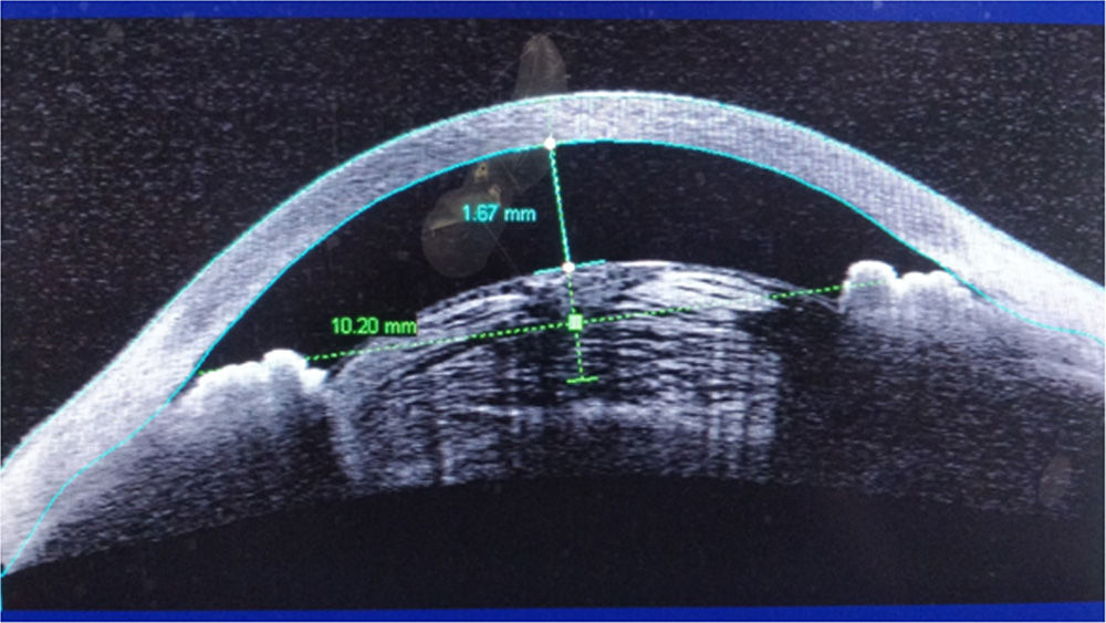
FIG.4: PRE YAG AS OCT
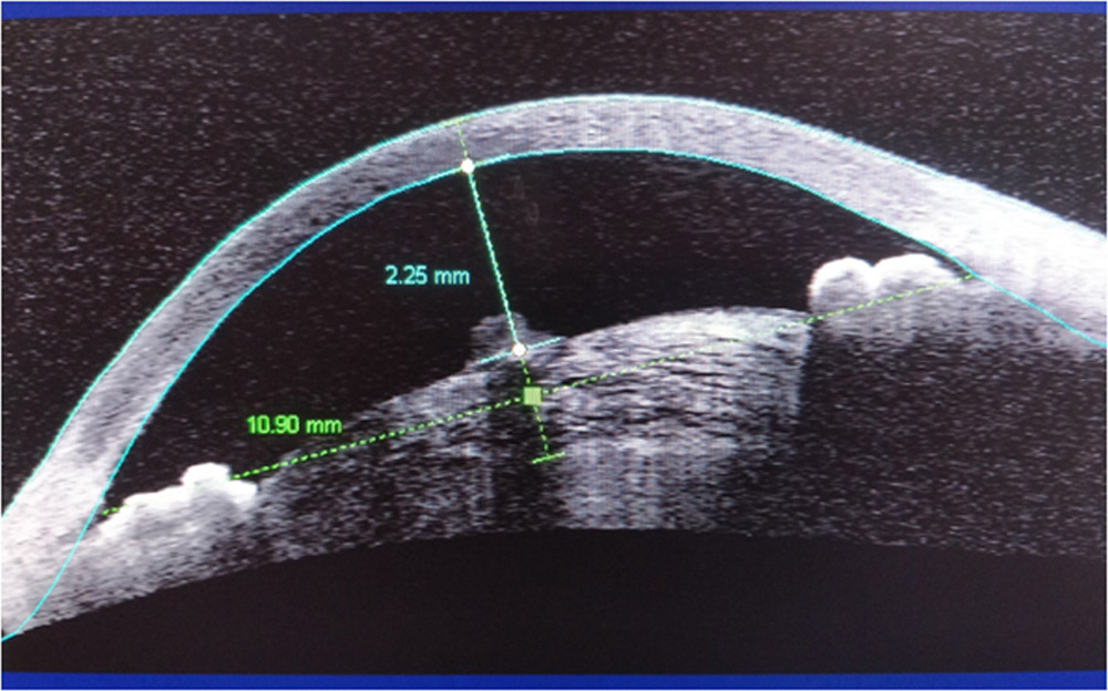
FIG.5: POST YAG AS OCT
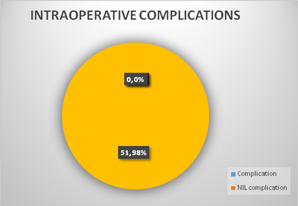
FIG.6 RATE OF INTRAOPERATIVE COMPLICATIONS
Discussion:
In the third world countries the incidence of white cataracts is fairly high and accessibility to expensive infrastructure like femto laser is difficult. In these situations, creation of a stable manual capsulorhexis is of utmost importance to deliver results. Despite all studies and efforts to minimize intraoperative complications in intumescent cataract, the complication that might take place within seconds of puncturing of the anterior capsule is that the high intralenticular pressure expands the initial capsule incision in an uncontrolled way with the development of the Argentinean flag sign.
In a study by
Gavris et al in 2003, on 70 eyes with intumescent cataract, capsulorhexis skidding frequency was 11.43%, and in one case, 1.43% respectively, the posterior capsule rupture occurred.[4]
For this purpose, Gimbel and Willerscheidt originally suggested the two-stage CCC approach with a small capsulorhexis or a multipuncture of the anterior capsule before capsulotomy
[2]After emulsification of the nucleus, the small CCC was enlarged to the desired diameter. With this technique, CCC was achieved in 57 eyes (95%)
[5]but other authors reported a 28.3% incidence of incomplete capsulorhexis, [6] within a very low success rate in white cataracts with a liquefied cortex (68%) compared to those with a solid cortex[4].
Newton Kara-Junior et al. compared the results of the 1-stage versus the 2-stage capsulorhexis in intumescent white cataracts and found anterior capsule tears in 23.07% of the cases in which the 1-stage capsulorhexis was performed (11 cases) and no ruptures of the anterior capsule were evidenced in which capsulorhexis was done in 2 stages (13 cases) [7]
Coelho et al said that preoperative Nd YAG laser anterior capsulotomy represents a safe and easy option for white intumescent cataracts after conducting a study in 11 eyes.[8]
Chan et al described the successful preoperative use of Nd YAG laser capsulotomy in the presence of an intact anterior chamber.[5].
In our experience our approach has certain advantages as compared to other approaches especially in a developing country where high end machineries are not readily available. In a high work load centre capsulorhexis followed by anterior Nd YAG capsulotomy saves a lot of time.
The author’s technique reduces the need for repeated entry of instruments in the Anterior chamber. A perfect central opening is achieved in a single attempt. Not only the complication rate is least, but there is a significant increase in the anterior chamber depth and which in turn increases the ease and comfort of surgery and reduces the surgical time.
Conclusion:
he technique for managing intumescent cataracts as described by us has the advantage of familiarity of the steps of standard Small incision cataract surgery while ensuring enhanced safety and consequently better surgical outcomes.
References:
- Dhingra D, Balyan M, Malhotra C, Rohilla V, Jakhar V, Jain AK.Indian J Ophthalmol. 2018 Sep;66(9):1304-1306.
- Gimbel HV, Willerscheidt AB. What to do with limited view: the intumescent cataract. J Cataract Refract Surg. 1993;19(5):657-61. Comment in: J Cataract Refract Surg. 1994;20(3):365-6.
- Gavriș M, Mateescu R, Belicioiu R, Olteanu I. The manual capsulorhexis with the Utrata forceps. Rom J Ophthalmol 2017 Jan-Mar;61(1):18-22.
- Gavris M, Popa D, Caraus C, Gusho E, Kantor E. Facoemulsificarea in cataracta alba, intumescenta. Oftalmologia. 2004;48(2):81–87.
- Chan DD, Ng AC, Leung CK, Tse RK. Continuous curvilinear capsulorhexis in intumescent hypermature cataract with liquefied cortex. J Cataract Refract Surg. 2003;29(3):431-4.
- Hausmann N, Richard G. Investigations on diathermy for anterior capsulotomy. Invest Ophthalmol Vis Sci. 1991;32(7):2155-9.
- Kara-Junior N, de Santhiago MR, Kawakami A, Carricondo P, Hida WT. Mini-rhexis for white intumescent cataracts. Clinics. 2009;64(4):309–312.
- Coelho RP, Paula JS, Silva RN, Garcia TV, Martin LFPreoperative Nd:YAG laser anterior capsulotomy in white intumescent cataracts: report of 11 casesArq Bras Oftalmol 2009 Jan-Feb;72(1):113-5.
Leave a Comment