

Dr.Sonal Vyas,Dr.Ashish Mahobia,Dr.Sangeeta Bawankure,Dr.Shamali Kohade
INTRODUCTION
Descemet membrane detachment (DMD) is a well known cause of surgery related corneal oedema after intraocular surgery. Though rare cases of spontaneous reattachment have been reported1,but they areprolonged, unpredictable, and can lead to fibrosis, shrinkage, and wrinkling of the detached Descemet’s membrane, which might prevent reattachment. Surgical intervention to promote attachment remains the preferred approach for most patients. There is no gold standard of treatment for DMD, various modes of intervention like reposition with air, gas or viscoelastic, suturing the DM to the cornea have been described, the superiority of one particular modality over the other and the appropriate timing of intervention are unclear.2,3,4,
In this study, we report our descemetopexy procedure results by using isoexpansile perfluoropropane (14% C3F8) gas in post cataract surgery Descemet’s membrane detachment.
MATERIAL AND METHOD
This retrospective non‑comparative interventional study was conducted at SBH Eye Hospital Raipur between January 2016 to April 2018. Institutional review board approval was obtained and retrospective review was performed of cases that had undergone descemetopexy with isoexpansile perfluoropropane (14% C3F8) for the treatment of DMD after cataract surgery.
The data retrieved from the medical records included age, sex, the eye operated, type of initial surgery, interval between descemetopexy and initial cataract surgery,pre operative and post operative visual acuity and corneal oedema. Best corrected visual acuity (BCVA) was assessed using snellen’s visual acuity chart and converted tologMARnotations for analysis. DMD was identified on slit lamp examination as separation of the DM from the posterior stroma with an area of corneal oedema overlying it. Anterior segment OCT was performed to delineate the exact location and extent of DMD.
Descemetopexy was performed under topical or local anaesthesia in the operating room under all aseptic precaution. A small peripheral corneal paracentesis was made in an area of attached DM.Isoexpansile C3F8 was injected intracamerally using 30 gauge needle mounted on a 2 ml disposable syringe.All patients were explained the importance of posture and supine posture was advised to facilitate continuous DM tamponade with gas. Topical antibiotic and tapering dosed of topical steroid were given to the patients in post operative period along with homatropine eye drop to keep pupil dilated.
The patients were examined at 1 day, 1 week, and after 4 weeks. Visual acuity, slit lamp examination, IOP measurements and AS OCT was done at each visit.
RESULTS
Statistical analyses were performed with Statistical Package for Social Sciences software (SPSS version 16). Paired t test was applied to compare the vision logMAR and corneal oedema pre and post . For all statistical analyses, P < 0.05was considered as significant and p<0.001 as highly significant.
16 eyes of 16 patients were included in the study. The mean age of the 8 men and 8 women was 68.62+6.58years. Right eye was involved in 6 (37.5%)and left in 10 (62.5%) patients. 3 (18.75%) patients had SICS and rest 13(81.25%) patients had phacoemulsification. The majority 12(75%) of DM detachment was noted at the site of the main incision, while 3 (18.75%) occurred at the side port. Visual axis was involved in all cases. The mean interval between cataract surgery and descemetopexy (Interim period) was 54.90 +121.63 days and is detailed in table 1.
Table 1. Duration between cataract surgery and descemetopexy
| Interim Period | No. Of eyes |
| Less than or equal to 1 week | 3 |
| >1 week to 1 month | 11 |
| >1 months | 2 |
At post operative 1 month, complete DM reattachment was observed in 15 of 16 (93.75%) patients while One patient showed persistent detached DM so advice for repeat descemetopexy or endothelial transplant. Mean BCVA was 1.32 ± 0.66 logMAR prior to descemetopexy which was improved significantly to 0.28 ± 0.32 logMAR after descemetopexy (P < 0.001, Table 2, figure 1). There was significant improvement in corneal oedema postoperative as shown in table 3, figure 2. Intra ocular pressure was raised in 1 patient due to pupillary block which was managed medically.
Table 2. Comparison of vision logMAR pre and post (n = 16)
| Mean | S.D | P value | |
| Vision logMAR PRE | 1.32 | 0.66 |
<0.001** |
| Vision logMAR POST | 0.28 | 0.32 |
Paired t test
**p<0.001 highly significant
Table 3. Comparison of corneal oedema pre and post (um)
| Mean | S.D | P value | |
| Corneal oedema PRE | 935.00 | 73.72 |
<0.001** |
| Corneal oedema POST | 573.68 | 94.42 |
Paired t test
**p<0.001 highly significant
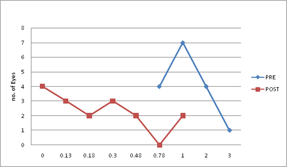
Fig 1. Vision (logMAR) improvement pre and post\
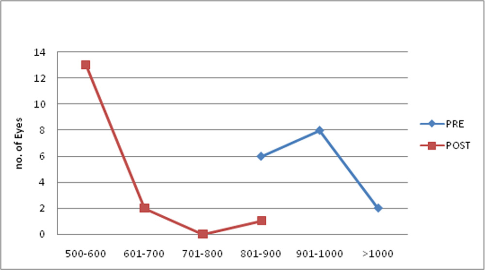
Table 3. Comparison of corneal oedema pre and post (um)
|
Mean |
S.D |
P value |
|
|
Corneal oedema PRE |
935.00 |
73.72 |
<0.001** |
|
Corneal oedema POST |
573.68 |
94.42 |
Paired t test
**p<0.001 highly significant
Fig 2. Corneal oedema (um) pre and post
DISCUSSION
Descemet membrane detachment is a known post operative complication of anterior segment surgery. Improper techniques of instrument entry into the eye, inadvertent injection of saline or viscoelastic and use of blunt instruments are the most likely causes of DM detachment.
There are various studies, reports and case reports available in the literature for management of DMD. Anterior chamber injection of gas or air as the primary management strategy has been already described 2,3,4,5 We have used isoexpansile 14% C3F8 gas as it stays longer within the anterior chamber, keeping Descemet’s membrane longer adhere to cornea and our results are comparable to previous studies.
Garget al 4 showed that anatomical reattachment of DM was seen in 71.64% and functional improvement in visual acuity in 74.63% of treated eyes; however Lucena et al 6
and Shah et al have reported 100% success rate. In our study we found 93% anatomical success rate and significant improvement in post operative BCVA (<0.001). Only one patient was having persistent detached Descemet and was planned for repeat injection or DSEK. One patient developed pupillary block glaucoma which was managed medically.
This study has some limitations. It s a retrospective study that has its inherent limitations. Sample size is less. Our study did not compare the results of C3F8 with air or any other intervention or observation. Another limitation is that we did not analyse the endothelial toxicity of C3F8.
CONCLUSION
The results in this series suggest that descemetopexy with isoexpansile 14% C3F8 for DMD after cataract surgery gives reasonably successful anatomical and functional outcome. It should always be tried before planning for a major surgery like keratoplasty.
REFERENCES
1. Minkovitz JB, Schrenk LC, Pepose JS. Spontaneous resolution of an extensive detachment of Descemet’s membrane following phacoemulsification. Arch Ophthalmol 1994;112:551–2
2. Donzis PB, Karcioglu ZA, Insler MS. Sodium hyaluronate (Healon) in the surgical repair of Descemet’s membrane detachment. Ophthalmic Surg 1986;17:735–7.
3.Chaurasia S, Ramappa M, Garg P. Outcomes of air descemetopexy for Descemet membrane detachment after cataract surgery. J Cataract Refract Surg 2012;38:1134–9.
4. Garg J, Mathur U, Acharya MC, et al. Outcomes of descemetopexy with Isoexpansile Perfluoropropane after Cataract Surgery. J Ophthalmic Vis Res 2016;11:168–73..
5. Jain R, Murthy SI, Basu S, Ali MH, Sangwan VS. Anatomic and visual outcomes of descemetopexy in postcataract surgery descemet’s membrane detachment. Ophthalmology 2013;120:13661372.
6. Lucena Ada R, LucenaDda R, Macedo EL, Ferreira Jde L, de Lucena AR. C3F8 use in Descemet detachment after cataract surgery. Arq Bras Oftalmol 2006;69:339‑343.
7. Shah M, Bathia J, Kothari K. Repair of late Descemet’s membrane detachment with perfluoropropane gas. J Cataract Refract Surg 2003;29:1242‑1244.
Leave a Comment