

ABSTRACT
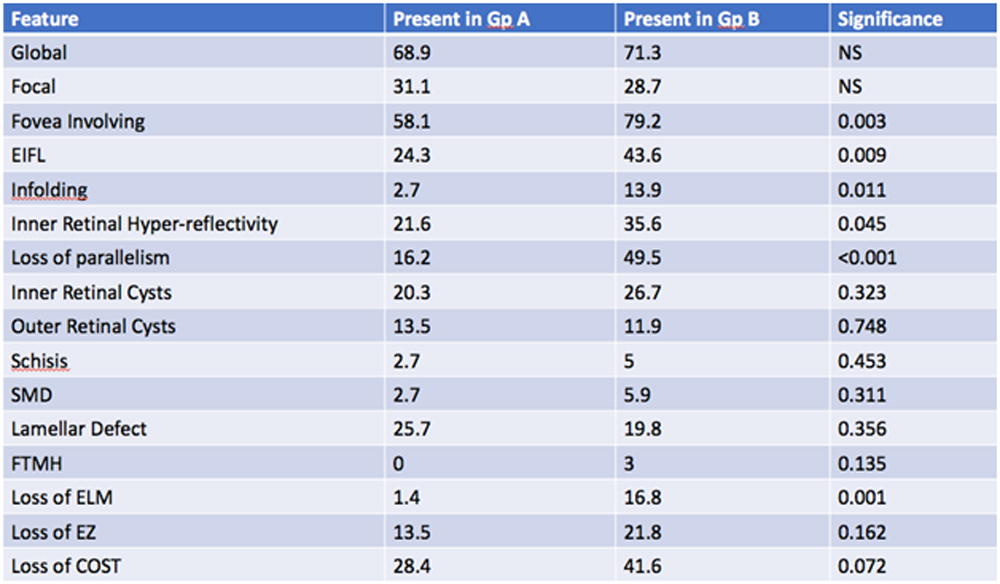
This study included 175 eyes with ERMs to assess the SDOCT findings associated with poor visual acuity. The tomographic features evaluated included the extent of ERM, foveal involvement, presence of Ectopic Inner Foveal Layers(EIFL), Retinal In folding, Inner Retinal Hyper-reflectivity, loss of parallelism, lamellar defects and the outer retinal layers. 74 eyes were in group A (vision better than 20/40) and 101 eyes in group B (equal to or worse than 20/40). Foveal Involvement, Presence of EIFL, Infolding of the retina, Inner Retinal Hyper-reflectivity, loss of parallelism was associated with poorer vision. The presence of cysts or lamellar defects was not associated with poor vision. 16.8 % eyes with poorer vision was associated with ELM loss and only 1.4 % were in group A. A qualitative evaluation of ERM morphology reveals certain characteristics which are associated with poor vision and these may have prognostic value in deciding the timing and outcomes of surgery for ERM.
INTRODUCTION
Since its introduction, Spectral Domain OCT(SD-OCT) has revolutionized retinal diagnosis, management and prognostication. It has been invaluable in defining pathogenetic mechanisms, understanding structural anomalies and classification and quantification of various disease entities. But the most significant group of disease which have been brought to light and characterized would be the vitreo-macular interface anomalies, of which idiopathic epiretinal membranes would be the most directly observable entity clinically. SD OCT has howeveridentified the presence of different types of epiretinal proliferation.LikeLamellar Hole associated epiretinal proliferation(LHEP) and tractional epi-retinal membranes. It has thrownup various newer tomographic internal ultrastructural findings like EIFL which have bearing on visual acuity and prognosis after surgery. Moreover, newer classification systems have emerged incorporating the newer features discovered. The use of these biomarkers should definitely assist in in making the correct surgical decision as to when to intervene and how the retina would recover after a surgical intervention. This work would look atsome of the new tomographic appearances and their possible association with poor visual acuity and possibly derive some information regarding ideal timing of surgery and outcomes.
METHODOLOGY
175 consecutive patients who presented with tractional epi-retinal membranes between January 2016 and January 2018 were included in the study.They underwent SD OCT evaluation using the dense macular scan protocol in the Heidelberg Spectralis OCT Heidelberg Germany. Patients with lamellar hole associate epiretinal proliferation were excluded from the study.Any eye where clear SD OCT were not obtainable were excluded. All ocular co-morbidities like macular edema due to diabetic retinopathy or other vascular disease were excluded. The following topographic features were evaluated.
- Extent of ERM and foveal involvement
- Presence of Ectopic Inner Foveal Layers(EIFL)
- Inner Retinal Hyper-reflectivity beneath the ERM
- Tissue between ERM and the retinal surface
- Retinal In-folding
- Loss of differentiation and parallelism
- Lamellar defects and cyst
- Outer retinal health.
The eyes were categorized into two groups. Group A had eyes with visual acuity better than 20/40 and group B has Visual acuity equal to or worse than 20/40. The presence of various tomographic features in the two groups were evaluated.
60 eyes underwent standard ERM surgery with ILM peeling and were followed up for a minimum period of 6 months. Sub group analysis included assessingthe features that were associated withpoor visual recovery after surgery and those associated with poor structural recovery.
Statistical analysis was done by SPSS v20. Pearson/Spearman’s chi-square test and Fisher exact test were used for comparison of discrete variables as appropriate. The t-test was used for comparison of continuous variables. All p-values were two – tailed and a value of less than 0.05 was considered statistically significant.
RESULTS
There were 175 eyes in the study of which 74 eyes were in group A (VA better than 20/40) while 101 in group B (VA 20/40 or worse)
Table 1 :
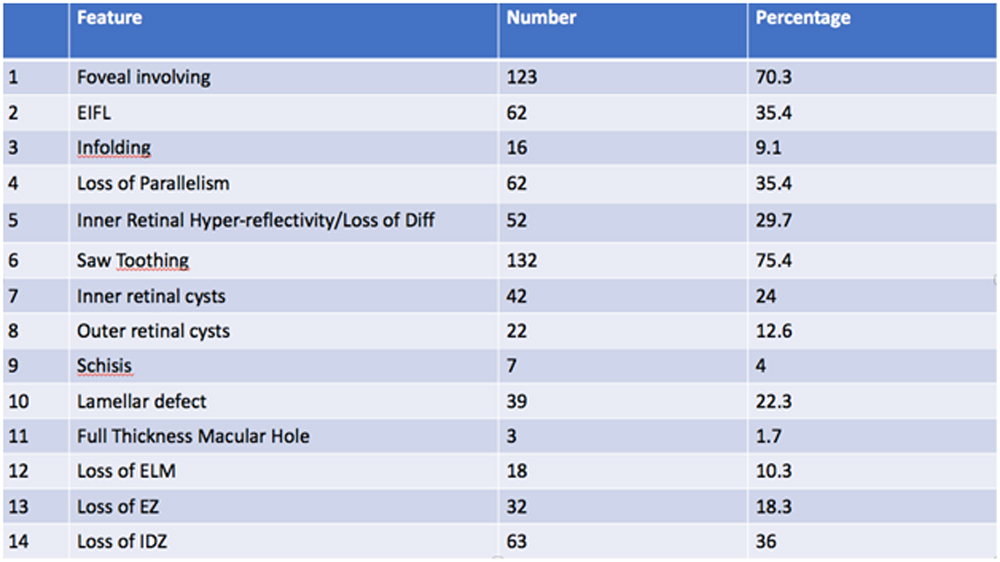
Table 2:
The mean follow-up was 6.2 months. The average best corrected distance visual acuity (BCDVA) of the study cohort was 0.31±0.29 log MAR, 20/41 Snellen equivalent. The whole group was divided into good visual acuity group (BCDVA better than 20/40) and bad visual acuity group (BCDVA 20/40 or bad). Out of 175 patients 101 (57.7%) was in bad visual acuity group.
60 patients underwent PPV + ERM peel + ILM Peeling + tamponade. 52 eyes were phakic and 8 were pseudophakic at the time of surgery. 38 of the 52 phakic eyes received combined surgery. The other 14 phakic eyes received simple ERM peeling surgery. Mean preoperative IOP was 13.1 ± 4.2 mmHg. 8 patients presented an elevated IOP (>21 mmHg) 7 days postoperatively and needed topical antihypertensive treatment. But in the subsequent follow-up visits mean IOP was reduced to 12.6 ± 4.3 mmHg that did not differ significantly from the IOP preoperatively.
Sub group Analysis done based on visual recovery or structural recovery after 6 months. 36 of the 60 eyes had a good visual acuity recovery with an improvement of at least two lines or a visual acuity of 20/30 or better
Table 3:
| ELM | Post-operative VA | Total | X2 | df | p | ||||
| Good | Bad | ||||||||
| N | % | N | % | N | % | 5.625 | 1 | 0.018 | |
| Present | 36 | 100 | 12 | 50 | 48 | 80 | |||
| Absent | 0 | 0 | 12 | 50 | 12 | 20 | |||
| total | 36 | 100 | 24 | 100 | 60 | 100 | |||
37 eyes recovered near normal contour after surgery.10 eyes had significant improvement of the contour, but not normalwhile 13 eyes had retained their abnormal contour after 6 months and had less than 100 microns decrease in central foveal thickness.
DISCUSSION
SD OCT hasallowed for the identification of many features including recently described one likely EIFL and central bouquet anomalies. These features may often be associated with extent of anatomical distortion by the tractional forces. These may also correspond to the visual and structural recovery after removal of the obvious tractional element.Descriptively we have divided the changes into those based on location and extent of the ERM. Global ERMs were more common than Focal ERMs. Foveal involvement of the ERM was found to be significantly associated with poorer visual acuity as expected.
The next group of changes studied were those affecting the inner and mid retinal elements. EIFL has been described as a persistence of the inner retinal elements in the foveolar area. This probably represent a centripetal pull of retinal layer at the center. This was described by Govetto(1) . The presence of continuous ectopic inner foveal layers in ERMs is a newly described OCT finding associated with significant vision loss and is an essential element of a novel OCT-based grading scheme of ERMs that may influence visual prognosis(1).
The intraretinal changes are different in iERMs and dERMs, as increased expression of CEIFLs in iERMs vs ICs in dERMs. The CEIFLs are associated with worse anatomical and functional outcomes in iERMs, whereas GFAP expression in peeled ILMs is higher in dERMs(2).
The presence of EIFL should be considered a negative prognostic factor for postoperative anatomical and functional recovery(3).
ERM stages were defined based on the SD-OCT staging system as stage 1: negligible morphological or anatomical disruption, retinal layers, and foveal pit are identified; stage 2: characteristic stretching of the outer nuclear layer, absence of foveal depression, retinal layers are identified; stage 3: continuous EIFL crossing the central foveal area, absence of foveal depression, retinal layers are identified; and stage 4: anatomical disruption of the fovea, continuous EIFL crossing the entire foveal area, retinal layers are distorted(4).The SD-OCT staging system according to the presence of EIFL is effective for grading retinal damage and visual loss in eyes with ERM(4)
We would like to describe the presence of increases inner retinal hyper-reflectivity underneath the ERM associated with a loss of differentiation of retinal layers. This feature was described in 52 eyes(29.7%) and was a key feature that persisted after ERM removal and was associated with poor structural recovery after surgery. We hypothesize that the increases reflectivity may be attributed to structural changes due to increased crowding on tissue or even due to gliotic changes extending into the inner layers from the surface anomaly.
Another feature we would like to describe is retinal Infolding. This is seen as a folding on itself of the mid retinal layers to such an extent that later in the disease process there is an obliteration and disappearance of the ONL and a villi like appearance. This feature was seen in 16 eyes (9.1%). Again, the presence of infolding was significantly associated with poor structural recovery and persistence of abnormal shale after ERM removal.
Other features like presence of inner and outer retinal cysts, schisis, sub macular detachment and macular defect were not significantly associated with poorer visual acuity.
Subgroup analysis was done in the operated 60 patients. The average preoperative BCDVA was mean preoperative BCVA was logMAR 0.50 ± 0.26, Snellen equivalent 20/80 which improved significantly to a mean of logMAR 0.18 ±0.35, Snellen equivalent 20/30 at the 6-month follow-up. Mean CFT measured with OCT was 532±105.5 mm at baseline, which was significantly reduced at the final follow-up to 352±42.9 mm. we compared the structural recovery of the ERM peeled cases. In that 13 patients showed poor anatomical recovery that is change in the CFT less than 100 microns or poor foveal contour. In this group of patients, the incidence of ectopic inner foveal layer, infolding, inner retinal hyper-reflectivity and loss of differentiation was more compared to the rest of the patients where the structural recovery was relatively good.
The pre-operative features in these eyes were EIFL, infolding, loss of parallelism & Inner Retinal Hyper reflectivity & loss of differentiation.
Features like surface wrinkling, saw-toothing, sub ERM tissue, cystic and lamellar changes & sub macular detachment should disappearance in all eyes after surgery and probably represent a milder less significant sequela of traction than the previous mentioned symptoms.
Although CFT may be useful for evaluating the impact of ERM on baseline VA, it is probably not useful for predicting postoperative VA(5). The preoperative OCT characteristics are more important. Intact preoperative inner segment ellipsoid (ISe) band is associated with a better postoperative VA than a disrupted preoperative ISe band in both idiopathic and secondary ERMs(6). The longer photoreceptor outer segment (PROS) length is also reported to be a good prognostic factor for the postoperative VA(6,7). It has also been shown that the integrity of outer photoreceptor cell layer as well as of ELM is related with better postoperative VA.Patients with preoperative inner retinal deformation were found to have significantly improved long-term visual outcomes after epiretinal membrane removal in a study of 114 patients(6). The same study showed that inner retinal irregularity index, a marker of the inner retinal deformation, was associated with visual improvement after adjusting for age, gender, and other optical coherence tomography findings.
In a study of 263 eyes with ERM, Govetto reported that the presence of ectopic inner foveal layers negatively correlated with the presence of central bouquet tractional abnormalities(8). Visual acuity was highest in association with the cotton ball sign and lowest in the acquired vitelliform lesion group. The group concluded that the cotton ball sign, foveolar detachment, and acquired vitelliform lesion may comprise a continuum in the same clinical spectrum and may represent subsequent stages of CB abnormalities.
Mathews evaluated the change in foveal contour in eyes with idiopathic epiretinal membrane (ERM) before and four months following pars plana vitrectomy with internal limiting and epiretinal membrane peeling(9). They reported that in eyes with ERM and no foveal depression, the majority did not regain foveal depression following surgery even though retinal thickness improved.
Massin studied 62 consecutive patients operated for idiopathic ERM(10). Macular thickness reduced from 419 µm to 300 µm, but returned to normal only in three eyes. However this did not preclude improvement in visual acuity, which improved from 20/60 to 20/40.
Inner retinal hyper reflectivity is a change that could possible reflect crowding of tissue, reactive gliotic changes that alter reflectivity while Infolding occurs due to mid retinal centripetal changes. Both resulting in a loss of parallelism and progressively in the loss of ability to differentiate tissue layers. EIFL occurs when tissue bunching happens at the foveolar center. These changes do not merely represent a mechanical distortion as evidenced by the lack of recovery after release of surface tractional changes.
In this study, the main determinant for good visual recovery after ERM surgery was found to be the intactness of the external limiting membrane.
The limitation of the study would be a lack of longer follow up and the surgical decision making being based on not on the tomographic features but on other considerations like visual acuity and visual symptoms.
These would indicate that idiopathicepiretinal membranes is not just a surface pathology. Though by definition a surface disease, it has structural repercussions in the mid retinal and outer retinal layers. Changes in the outer retinal layers have effect on visual acuity and recovery after surgery. Changes in the mid retinal layers seem to have impact on the structural recovery after surgery.
REFERENCES
- Govetto A, Lalane RA, Sarraf D, Figueroa MS, Hubschman JP. Insights Into Epiretinal Membranes: Presence of Ectopic Inner Foveal Layers and a New Optical Coherence Tomography Staging Scheme. Am J Ophthalmol. 2017 Mar;175:99–113.
- Romano MR, Ilardi G, Ferrara M, Cennamo G, Allegrini D, Pafundi P, et al. Intraretinal changes in idiopathic versus diabetic epiretinal membranes after macular peeling. PLoS ONE [Internet]. 2018 May 8;13(5). Available from: https://www.ncbi.nlm.nih.gov/pmc/articles/PMC5940191/
- Govetto A, Virgili G, Rodriguez FJ, Figueroa MS, Sarraf D, Hubschman JP. FUNCTIONAL AND ANATOMICAL SIGNIFICANCE OF THE ECTOPIC INNER FOVEAL LAYERS IN EYES WITH IDIOPATHIC EPIRETINAL MEMBRANES: Surgical Results at 12 Months. Retina Phila Pa. 2017 Nov 16;
- Doguizi S, Şekeroğlu MA, Ozkoyuncu D, Omay AE, Yilmazbaş P. Clinical significance of ectopic inner foveal layers in patients with idiopathic epiretinal membranes. Eye. 2018;1–9.
- Stevenson W, Prospero Ponce CM, Agarwal DR, Gelman R, Christoforidis JB. Epiretinal membrane: optical coherence tomography-based diagnosis and classification. Clin Ophthalmol Auckl NZ. 2016 Mar 29;10:527–34.
- Jeon S, Jung B, Lee WK. LONG-TERM PROGNOSTIC FACTORS FOR VISUAL IMPROVEMENT AFTER EPIRETINAL MEMBRANE REMOVAL. RETINA [Internet]. 2018 Sep 26 [cited 2018 Oct 8];Publish Ahead of Print. Available from: https://journals.lww.com/retinajournal/Abstract/publishahead/LONG_TERM_PROGNOSTIC_FACTORS_FOR_VISUAL.96466.aspx
- Kinoshita T, Imaizumi H, Miyamoto H, Katome T, Semba K, Mitamura Y. Two-year results of metamorphopsia, visual acuity, and optical coherence tomographic parameters after epiretinal membrane surgery. Graefes Arch Clin Exp Ophthalmol Albrecht Von Graefes Arch Klin Exp Ophthalmol. 2016 Jun;254(6):1041–9.
- Govetto A, Bhavsar KV, Virgili G, Gerber MJ, Freund KB, Curcio CA, et al. Tractional Abnormalities of the Central Foveal Bouquet in Epiretinal Membranes: Clinical Spectrum and Pathophysiological Perspectives. Am J Ophthalmol. 2017 Dec 1;184:167–80.
- Mathews NR, Tarima S, Kim D-G, Kim JE. Foveal contour changes following surgery for idiopathic epiretinal membrane. Invest Ophthalmol Vis Sci. 2014 Nov 13;55(12):7754–60.
- Massin P, Allouch C, Haouchine B, Metge F, Paques M, Tangui L, et al. Optical coherence tomography of idiopathic macular epiretinal membranes before and after surgery. Am J Ophthalmol. 2000 Dec 1;130(6):732–9.
Leave a Comment