

Dr.Pavan Shroff, P17064, Dr.Apoorva A G, Dr.Guruprasad Ayachit, Dr.Shrinivas M Joshi
Abstract:
Title: Unraveling the Macular mysteries using Intraoperative Optical Coherence Tomography.
Purpose: To study the influence of Intraoperative Optical Coherence Tomography (iOCT) on management of Dense Vitreous Hemorrhage (VH).
Methods:
Prospective, cross-sectional study between December 2017 & May 2018.
Inclusion criteria:
- Pars Plana Vitrectomy (PPV) in VH where pre-operative OCT was not possible
- VH due to any cause
- Any age
iOCT was done for all the cases during PPV. Any macular pathology detected was treated. Surgeon’s Feedback grading was acquired. Time taken for iOCT was noted.
Results:
Out of 26 eyes, 7 eyes had CME, 7 had ERM, 2 had Macular hole & 1 had ERM with foveal atrophy. iOCT influenced diagnosis & management of 15 eyes. Surgeon’s grading showed iOCT had an impact on diagnosis & management of macular pathologies in VH. The mean iOCT time was 3.46 minutes.
Conclusion:
iOCT has a role in intraoperative diagnosis & management of the macular pathologies in dense VH. Screening all PPV cases of VH with iOCT is recommendable. Increase in surgical time due to iOCT is not significant.
Keywords:
Intraoperative Optical Coherence Tomography, Intraoperative,iOCT, Vitreous Hemorrhage, Diabetic Retinopathy, Vitrectomy, Dense Vitreous Hemorrhage.
Introduction:
Vitreous Hemorrhage (VH) is a reversible cause of blindness with an incidence of 7 cases per 100,000 population.[1]Diabetic Retinopathy (DR), retinal tears and Retinal Vein Occlusion (RVO) are among the common causes of VH. Vitreous Hemorrhage clears at a rate of 1 percent per day and is managed conservatively unless indicated by its etiology like Retinal detachment.[1] However, non-clearing VH is a more frequent indication for surgery.[2] Dense VH, which is a more likely indication for surgery, poses difficulty in evaluation of the retina. Ultrasound helps in picking up Retinal Detachment (RD) but there is no way to know the status of macula pre-operatively.
Incidence of Macular Holes (MH) has been estimated to be around 33 in 10,000 population over the age of 55 years.[3]It is not uncommon to find a coexistent macular pathology while operating a patient for another condition.[4] Finding a macular pathology during a Pars Plana Vitrectomy (PPV) for VH sounds similar to finding a pale disc after operating for a Mature Cataract. It is likely that a macular pathology be missed or raise doubts about its diagnosis during a PPV.[5]Making a decision intraoperatively, which may involve a surgical maneuver, and not having a documentation on OCT is also something that a surgeon would less prefer. Use of gases as tamponading agents after a macular surgery causes hindrance in acquisition of immediate post-operative OCT.These points have led to realization about the need for being able to look into the macula intraoperatively and document it.
Optical Coherence Tomography (OCT) has effectively established its role in the diagnosis of various macular conditions and beyond.[6,7,8]This technology has now entered into the Operating Room and is likely to help surgeons make better, more confident decisions. Recent work in research has shown the effectiveness of intraoperative Optical Coherence Tomography (iOCT) in visualizing macular pathologies like MH, Epiretinal Membrane (ERM), Macular Edema (ME) and Vitreo-macular Traction (VMT) and performing efficient macular surgery.[9,10,11,12]Better visualization of the macula intraoperatively using iOCT leads to the idea of screening all cases of PPV for dense VH and its justification. In this study, we make an attempt to find the prevalence of macular pathologies discovered during PPV for dense VH and the role of iOCT in decision making intraoperatively.
Methods:
This is a prospective, cross-sectional, single center study on the role and feasibility of iOCT in PPV for dense VH. Patients presenting with dense VH due to any cause and in whom it was not possible to acquire OCT for macular status were included in the study. All the patients were explained about the study and written informed consent was obtained.
All the patients underwent standard small gauge (23g or 25g) vitrectomy. During PPV, vitrectomy was first performed followed by assessment of the macula by the surgeon. Then iOCT was acquired using aspheric macular lens. After iOCT guided diagnosis, macular surgery was performed as needed and iOCT was then repeated. The total time of surgery and the time taken for iOCT was noted. Immediately after surgery, the surgeon graded the role of iOCT as per the Surgeons’ feedback grading.
iOCT Scanning System:
In this study, iOCT was done using Zeiss RESCAN 700 OCT Scanning System mounted on a Zeiss OPMI LUMERA 700 microscope. iOCT images were obtained after vitrectomy and was repeated after the macular maneuver, if done. 10 mm * 10 mm cubic scans were obtained after placing the aspheric macular lens.
Surgeons’ Feedback Grading:
| Grade | Feedback |
| Grade 1 | Not useful |
| Grade 2 | Useful for Diagnosis |
| Grade 3 | Useful, Altered/Added a surgical intervention |
| Grade 4 | Enhanced performance of a surgical intervention. |
Results:
For the 26 eyes in the study, the mean age was 59.6 years, ranging from 30 to 80 years. Twenty of 26 eyes (76.92%) had Proliferative Diabetic Retinopathy (PDR) as the cause of VH and 3 eyes (11.54%) had Retinal Vein Occlusion (RVO) with neovascularization leading to VH. Presumed Peripheral Exudative Hemorrhagic Chorioretinopathy (PEHCR) (n=1, 3.85%), presumed Retinal Artery Macroaneurysm (RAM) (n=1, 3.85%) and Posterior Vitreous Detachment (PVD) (n=1, 3.85%) induced Retinal Tear were the causes for the rest 3 eyes.
Seven of 26 eyes (26.92%) were found to have Macular Edema (ME), 7 eyes (26.92%) were found to have ERM, and 2 eyes (7.69%) had MH while 1 eye (3.85%) had ERM with foveal atrophy. ERM peeling was done for 4 eyes (15.38%), Internal Limiting Membrane (ILM) peeling was done for 7 eyes (26.92%), ERM peeling with ILM peeling was done for 3 eyes (11.54%) and ILM peeling with Intravitreal Bevacizumab injection was done for 1 eye (3.85%).
Representative Case Discussion:
Case 1:
A 65 years old male presented with non-clearing vitreous hemorrhage in Right eye since 8 months. PPV was done for the Right eye and his macula appeared normal on assessment by surgeon. When subjected to iOCT, the images revealed a thin ERM on the macula. ERM peeling was then done and complete ERM peeling was confirmed on iOCT.
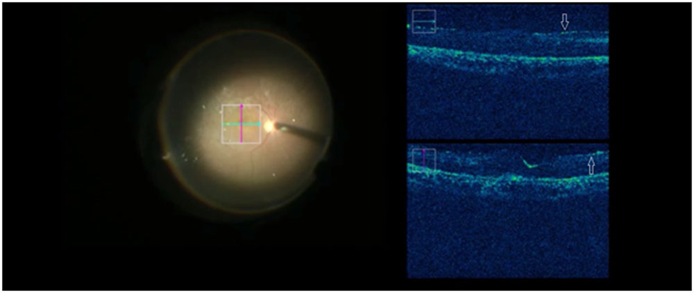
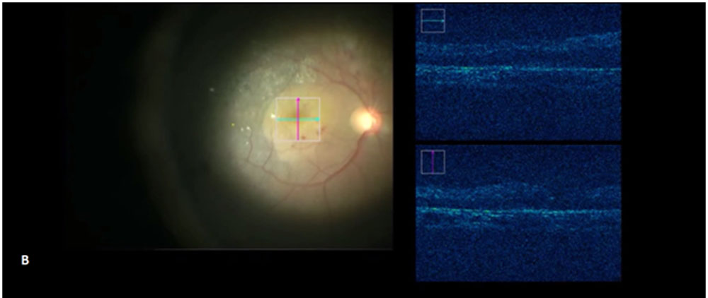
Figure 1 The intraoperative OCT reveals a thin ERM in a 65 years old male being operated for dense VH. The images are what was seen on the Zeiss Callisto Eye relaying images from the surgeons’ eye piece and RESCAN 700. (A) On the left side of the image, the live surgeons view is seen with a superimposed macular grid indicating the area of the scan. To the right, a thin ERM (arrows) can be seen which was picked up efficiently by the intraoperative OCT. (B) To the left, surgeons live view shows the macula after the ERM and ILM peeling while to the right side, intraoperative OCT shows complete removal of ERM.
Case 2:
A 62 year old male presented to us with dense vitreous hemorrhage due to PDR in his Right eye. He was operated in his Right for the same. After vitrectomy, macular edema was thought to be present but iOCT images revealed that the macula was dry and ILM peeling was avoided.
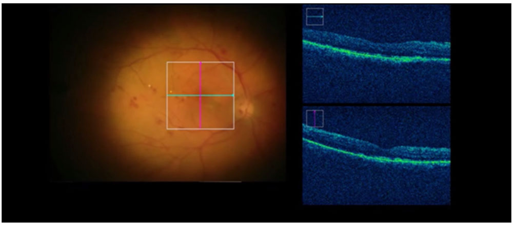
Figure 2 Intraoperative OCT shows a normal foveal contour which was first thought as having macular edema
iOCT guided diagnosis and Impact on Decision-making
Out of 26 eyes, a total of 17 eyes (65.38%) had macular pathology of which 4 eyes (15.38%) having ME and 4 eyes (15.38%) having thin ERM were identified through iOCT guidance. One eye was diagnosed to have foveal atrophy by iOCT imaging. In one eye, suspicion of ME was ruled out by iOCT.
As per the Surgeons’ feedback, in 16 eyes (61.54%) iOCT was found useful for diagnosis (helped confirm the diagnosis or altered it). In 11 eyes (42.30%) iOCT was useful in intra-surgical decision-making by altering/adding an intervention. The mean time taken for iOCT was 3.46 minutes.
Discussion:
Dense VHis one disease which needs surgical treatment and the surgeon is often unaware of the macular status prior to the surgery. Recent studies have shown the efficacy of iOCT in studying structural changes in the retina during macular surgeries for MH, ERM and vitreo-macular traction.[13,14,15,16] However, there is no study establishing the role of iOCT in screening cases of PPV for dense VH. The prevalence of macular pathology discovered during PPV for dense VH has also not been studied. The technology of iOCT has the potential to help surgeons with the intraoperative diagnosis and decision-making for immediate macular intervention.
This study shows that eyes undergoing PPV for dense VH had a significant prevalence of macular pathology. Around 65% of the eyes were found to have some macular pathology and 57.69% of the eyes needed some intervention for the macula. Higher prevalence of macular pathology and being able to intervene for the same immediately means iOCT can save thepatient from the morbidity of a possible secondary procedure for the otherwise undiagnosed/missed macular pathology.
A thin ERM is likely to be missed intraoperatively. Identification of the ERM and its characteristics is an advantage of iOCT and provides opportunity of ERM peeling. In this study, eight eyes (30.77%) had ERM of which 4 eyes (15.38%) were identified and peeled through iOCT guidance. The long term visual outcomes of ERM peeling done for iOCT guided ERMs diagnosed needs to be compared with a comparison group where no iOCT was done.
Structure and shape of a normal fovea varies from a shallow pit with continuity of the inner nuclear layer to a complete separation of inner layers.[17] Clinical appearance of a normal fovea can at times be deceiving. Diagnosing a ME intraoperatively can be a mere guess work. Intravitreal anti-VEGF is widely used for ME. ILM peeling has also been described for ME due to Diabetic Retinopathy.[18] Confirmation of ME and providing with a treatment is a benefit from iOCT. In this study, seven eyes (26.92%) had ME of which 4 eyes (15.38%) were diagnosed through iOCT guidance. One eye (3.85%) was ruled out of having ME by iOCT.
A confident diagnosis and treatment has an impact on the patient counselling and education about the condition they have. The confirmation of diagnosis provided by the OCT in the outdoor patient department would be welcome in the OR. There is no data comparing the surgeons’ diagnosis with the iOCT guided diagnosis. This study showed that the surgeons found iOCT helpful in making or ruling out a diagnosis in a startling 61.54% of cases and led to surgical decision alteration in 42.30% of cases. Acquiring iOCT images during a surgery is a quick process and does not significantly add to the surgical time.
The limitations of this study is long term follow-up of macular status and the impact of iOCT on the long term management of the macular pathology. Long term visual outcomes and a lack of a comparison group are additional limitations.
This study justifies the role of iOCT for screening all cases of PPV for VH and establishes importance of iOCT in intraoperative diagnosis and decision-making. The feasibility of iOCT in terms of ease and time taken is also demonstrated. However more studies are needed to establish the long term benefit of iOCT for macular pathologies.
References:
- Spraul CW, Grossniklaus HE. Vitreous Hemorrhage. Survey of ophthalmology. 1997;42(1):3.
- Flynn Jr HW, Chew EY, Simons BD, Barton FB, Remaley NA, Ferris III FL, Early Treatment Diabetic Retinopathy Study Research Group. Pars plana vitrectomy in the Early Treatment Diabetic Retinopathy Study: ETDRS report number 17. Ophthalmology. 1992 Sep 1;99(9):1351-7.
- Mester V, Kuhn F. Internal limiting membrane removal in the management of full-thickness macular holes. American journal of ophthalmology. 2000 Jun 1;129(6):769-77.
- Demetriades AM, Gottsch JD, Thomsen R, Azab A, Stark WJ, Campochiaro PA, De Juan Jr E, Haller JA. Combined phacoemulsification, intraocular lens implantation, and vitrectomy for eyes with coexisting cataract and vitreoretinal pathology. American journal of ophthalmology. 2003 Mar 1;135(3):291-6.
- Ehlers JP, Griffith JF, Srivastava SK. Intraoperative OCT during Vitreoretinal Surgery for Dense Vitreous Hemorrhage in the PIONEER Study. Retina (Philadelphia, Pa.). 2015 Dec;35(12):2537.
- Panozzo G, Gusson E, Parolini B, Mercanti A. Role of OCT in the diagnosis and follow up of diabetic macular edema. InSeminars in ophthalmology 2003 Jan 1 (Vol. 18, No. 2, pp. 74-81). Taylor & Francis.
- Regatieri CV, Branchini L, Duker JS. The role of spectral-domain OCT in the diagnosis and management of neovascular age-related macular degeneration. Ophthalmic Surgery, Lasers and Imaging Retina. 2011 Jul 1;42(4):S56-66.
- Medeiros FA, Zangwill LM, Bowd C, Vessani RM, Susanna Jr R, Weinreb RN. Evaluation of retinal nerve fiber layer, optic nerve head, and macular thickness measurements for glaucoma detection using optical coherence tomography. American journal of ophthalmology. 2005 Jan 1;139(1):44-55.
- Dayani PN, Maldonado R, Farsiu S, Toth CA. Intraoperative use of handheld spectral domain optical coherence tomography imaging in macular surgery. Retina (Philadelphia, Pa.). 2009 Nov;29(10):1457.
- Asahina Y, Tachi N, Asahina Y, Yoshimura K, Ueta Y, Hashimoto Y. Six-month postoperative outcomes of intraoperative OCT-guided surgical cystotomy for refractory cystoid macular edema in diabetic eyes. Clinical Ophthalmology (Auckland, NZ). 2017;11:2099.
- Ehlers JP, Itoh Y, Xu LT, Kaiser PK, Singh RP, Srivastava SK. Factors associated with persistent subfoveal fluid and complete macular hole closure in the PIONEER study. Investigative ophthalmology & visual science. 2015 Feb 1;56(2):1141-6.
- Leisser C, Hirnschall N, Hackl C, Döller B, Varsits R, Findl O. Diagnostic precision of a microscope-integrated intraoperative Oct device in patients with epiretinal membranes. European journal of ophthalmology. 2018 May;28(3):329-32.
- Ehlers JP, Xu D, Kaiser PK, Singh RP, Srivastava SK. Intrasurgical dynamics of macular hole surgery: an assessment of surgery-induced ultrastructural alterations with intraoperative optical coherence tomography. Retina. 2014 Feb 1;34(2):213-21.
- Ray R, Barañano DE, Fortun JA, Schwent BJ, Cribbs BE, Bergstrom CS, Hubbard III GB, Srivastava SK. Intraoperative microscope-mounted spectral domain optical coherence tomography for evaluation of retinal anatomy during macular surgery. Ophthalmology. 2011 Nov 1;118(11):2212-7.
- Wykoff CC, Berrocal AM, Schefler AC, Uhlhorn SR, Ruggeri M, Hess D. Intraoperative OCT of a full-thickness macular hole before and after internal limiting membrane peeling. Ophthalmic Surgery, Lasers and Imaging Retina. 2010 Jan 1;41(1):7-11.
- Dayani PN, Maldonado R, Farsiu S, Toth CA. Intraoperative use of handheld spectral domain optical coherence tomography imaging in macular surgery. Retina (Philadelphia, Pa.). 2009 Nov;29(10):1457.
- Tick S, Rossant F, Ghorbel I, Gaudric A, Sahel JA, Chaumet-Riffaud P, Paques M. Foveal shape and structure in a normal population. Investigative ophthalmology & visual science. 2011 Jul 1;52(8):5105-10.
- Gandorfer A, Messmer EM, Ulbig MW, Kampik A. Resolution of diabetic macular edema after surgical removal of the posterior hyaloid and the inner limiting membrane. Retina (Philadelphia, Pa.). 2000;20(2):126-33.
Leave a Comment