

Dr.Puneeth Isloor,Dr.Shrinivas M Joshi,Dr.Guruprasad Ayachit,Dr.Apoorva A G
Abstract
PURPOSE:To study anatomical and visual outcomes ofMultilayered Inverse ILM peeling in large Macular holes (MH)and Macular hole associated Retinal detachments(MH-RD)
METHOD:Prospective study of 45 eyes of 44 patients.Pre-op Minimum Linear Diameter(MLD) and Basal Diameter(BD) noted. Twenty five gauge pars plana vitrectomy and posterior vitreous induction done.ILM flaps were peeled and placed in a multilayered manner in the MH and under PFCL in MH-RDs. post-op.BCVA at week, 2 months and 3 months & type of macular hole closure noted.
RESULTS:7/45 eyes had MH-RD.Mean age 55.6 ± 16.7 years. M:F -14 :31 (put as 1: 2. Decimal).Mean MLD 761.2 ± 400.8 µm and BD 1392 ± 427.2 µm.Mean pre-op Logmar BCVA improved from 1.4 ± 0.5 to 1.1 ± 0.5 (p value?) at 3 months post-op. Forty one (91.1%) had type 1 hole closure while 3(6.6%) had type 2 closure.2/3 eyes with type 2 closure had good vision. Only Eyes withtype 1 closure had significant improvement in vision at 3 months(p<0.001).
CONCLUSION:Inverse ILM peeling improves outcomes in MH and MH-RD and provides . good anatomical and functional outcomes.Always attempt surgery in all cases of large macular holes irrespective of the size
Key words :Multilayered Inverse ILM Peeling , Large macular holes , Macular hole associated retinal detachments , Type 1 closure
Introduction
Macular hole formation begins with the degeneration of the inner retina at the Muller cell cone-vitreous cortex interface. The macular defect enlarges due to the contraction of the internal limiting membrane (ILM) because of Muller (Glial) cell migration on to the ILM.[[i]]The earliest reports of macular holes being closed by vitrectomy was that of Kelly and Wendel.[[ii],[iii]]
The closure rate of macular holes has been found to be increased with the use of ILM peeling.[[iv]] Large macular holes(>400um) proved to be a challenge and the success rates were suboptimal and hence the inverted ILM flap technique was developed.[[v],[vi]] It is believed to prevent the post operative flat open type of closure in large macular holes(>400 microns) with improved outcomes .[[vii]]The visual recovery that occurs after the surgery in large macular holes is somewhat limited despite having multiple surgical techniques to improve the anatomical closure . However there is definitely some improvement of vision if hole closure is attempted.[[viii], [ix],[x]]
The original technique has been subjected to a lot of modifications as flap retroversion was seen in 14-20% of cases during fluid air-exchange and this led to failure of hole closure.[[xi],[xii]]
A limitation is thatthe patient has to undergo repeated surgeries to reposition the flap to cover the Macular hole and this can be cumbersome. [ix]
This study aims to analyze the visual and anatomical outcomes of Multilayered Inverse ILM peeling in MH and MH-RD
Materials and methods
It was a prospective interventional study conducted at a tertiary eye hospital in South India spanning a period of one year from May 2017- June 2018.Study was conducted according to the tenets of declaration of Helsinki and was approved by the institutional review board (IRB). Written informed consent was obtained from all patients. 45 eyes of 44 Patients having a large MH(>400 µ)and (MH-RD)(acronyms already used in introduction. ) were included. Both Idiopathic and traumatic macular holes were included in the study. Eyes with severe media opacity which precluded a high quality OCT image were excluded from the study. Best Corrected Visual Acuity(BCVA) was recorded pre-operatively Slit lamp biomicroscopywas done using 78 D . OCT was done using 30 degree objective; high resolution protocol and 6mm scans across the macula were obtained .On the OCT , Minimum Linear Diameter and Basal Diameter were noted pre-operatively at the line scan passing through the fovea in all cases on the Spectralis Heidelberg platform (Heidelberg Engineering, Heidelberg) using the built- in calipers. The measurement was agreed upon by two observers. In case of inter- observer variability, average of two values was noted.
All eyes underwent 25 gauge pars plana vitrectomy with induction of posterior vitreous detachment. Brilliant Blue Green (BBG)was used to stain the ILM.ILM flaps were peeled using the pinch and peel technique using the ILM forceps and placed one over the other in a multilayered fashion in the macular hole (Intra-operative OCT guided) under PFCL.The flaps were made in a petalloid fashion around the macular hole and the petals were stuffed into the macular hole in an atraumatic manner.The edges of the macular hole was gently massaged with the extrusion soft tip to aid the closure.The same technique was carried out in MH-RD after flattening the posterior pole with PFCL. A draining retinotomy was done in the peripheral retina to drain the subretinal fluid.Fluid Air – exchange was done.Non-expansile C3F8 gas(15%) was injected.. Prone position was advised for 1 week (number of hours) ,patients were followed up at 1 week and 1 month post operatively .BCVA,IOP, type of closure on OCT were noted on all post-operative visits
Data was entered into Microsoft excel data sheet and was analyzed using SPSS version 22 (IBM SPSS Statistics, Somers NY, USA) software. Categorical data was represented in the form of frequencies and proportions. Chi-square test or Fischer’s exact test (for 2×2 tables only) was used as test of significance for qualitative data. Continuous data was represented as mean and standard deviation. Independent t test or Mann Whitney U testwas used as test of significance to identify the mean difference between two quantitative variables and qualitative variables respectively.
Results-
GENERAL PROFILE- 45 eyes of 44 patients were included. M:F-0.45:1 . Mean age of the patients was 55.6 ± 16.7 years .40/45 eyes were idiopathic macular hole ,4/45 eyes were traumatic macular holes and 1/45 eyes was a post surgical macular hole.7/45eyes had MH-RD. The rest were purely large macular holes .
Table 1 shows the profile of the patients included in the study including the lens status of these patients.
VISUAL ACUITY AND HOLE DIMENSIONS
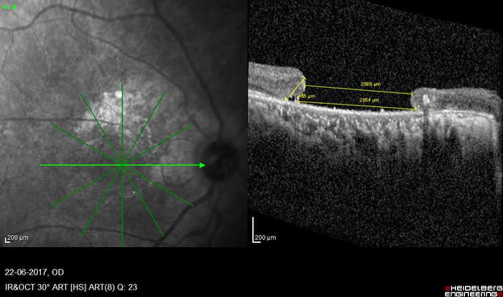
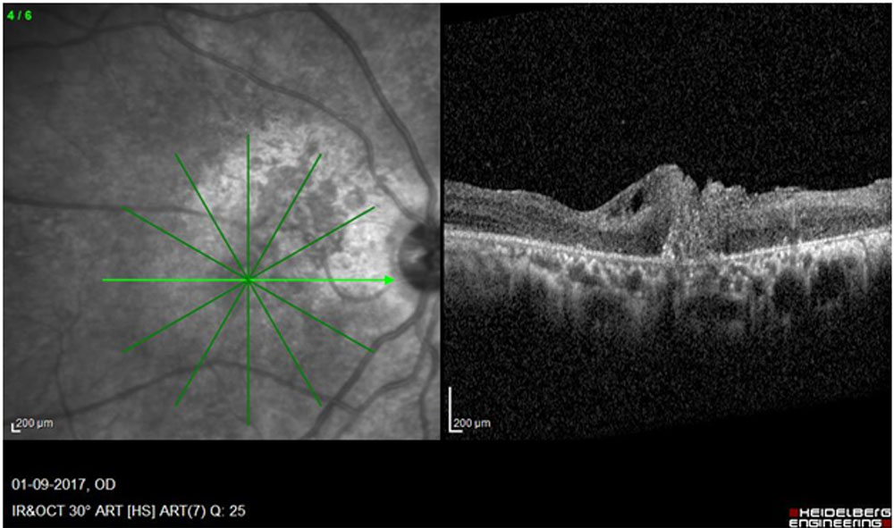
Mean pre- operative BCVA was 1.4 ± 0.5 Mean Minimum Linear Diameter was 761.2 ± 400.8 µm and the Mean Basal Diameter was 1392 ± 427.2 µm.However the largest MLD in our study was that of a 40 year old female patient with MLD of 2586 um and BD of 2854 um.(Figure 1 ).Pre and Post operative vision were however the same i.e 1/60 or logmar 1.77 despite achieving type 1 closure
HOLE CLOSURE-
41/45 eyes ( 91.1% ) had type 1 hole closure while 3/45(6.6%) had type 2 closure and 1/45(2.2% )did not show closure. 2/3 eyes with type 2 closure had improvement of vision, though not statistically significant. One patient with type 2 closure improved from CF to 1/60 i.e from logmar 2 to logmar 1.77,while another patient with type 2 closure improved 3/60 to 6/60 i.e from logmar 1.07 to logmar 1.0.Eyes with type 1 closure showed a significant improvement in vision at 1 month(p<0.001) )post operatively ,while those with type 2 closure did not show a statistically significant change in vision.(Table 2)
Discussion
The ILM has always been believed to be the causative factor in the pathology of the macular hole .The tangential traction of the ILM has been implicated in the pathology of macular holes and conventional ILM peeling relieves this traction.However, conventional ILM peeling is insufficient to close large macular holes and mostly results in flat open type of closure. The closure rate is 86–100% with the use of ILM staining agents,[[xiii]]and has improved compared to 68% when pars plana vitrectomy was first introduced to close macular holes.Newer techniques are constantly being developed to improve closure rates .
Various mechanisms in the technique of closure of macular holes have already been described.The Peeled-off multiple flaps of ILM which contain numerous Müller cell fragments may induce gliosis.[[xiv],[xv]]While conventional ILM peeling itself induces gliosis , these flaps of multilayering technique can trigger more gliosis to create a stronger centripetal pull and result in hole closure.Proliferation of glial cells produces an environment conducive to the repositioning of photoreceptors in direct proximity to the fovea.[5]
Surface tension effect of the gas bubble seals the large macular hole more effectively due to the multiple flaps over it. We hypothesise that the ILM flaps probably prevent further seepage of fluid into the hole with the momentary changes of position of the patients keeping the edges of hole dry and closed ,thus providing a higher chance of hole closure.
In our study ,we have used the multi-layered ILM flaps in a petalloid fashion to close the large macular holes which showed improved outcomes both functionallyand anatomically. While majority of the eyes showed type 1 closure and also a statistically significant improvement in visual acuity,those with type 2 closure also showed improvement in vision , despite it not being statistically significant . It is noteworthy that this technique can be easily perfomed under PFCL in a patient with MH-RD to achieve good results. The patient who did not show closure failed to comply with post operative positioning in the first week post operatively re-iterating the importance of immediate post operative positioning.
To the best of our knowledge and after a thorough review of literature , no other study published till date has data on this sample size of Large macular holes and macular hole associated retinal detachments with regard to outcomes using this petalloid multi-layered inverted ILM flap technique and with the aid of intra-operative OCT.
We also report the first use of intra-operative OCT for this technique and ensure stuffing of the hole with the flaps in real- time i- OCT assistance. Studies have previously been conducted which assess surgery induced ultra-structural alterations [[xvi]].In this study Macular hole base area and volume increased after surgery ,but apex area decreased. In our study we have not measured the Macular hole dimensions intraoperatively , but just used it to confirm the position of the flaps before performing fluid air exchange .We have used pre operative and post operative OCT for measuring the dimensions . In a study conducted by Borrelli et al [[xvii]] , Intra-operative OCT was used to confirm the position of the ILM flaps .However this study consisted of only 3 samples.There are some limitations such as the fact that multifocal ERG could not be used to analyze the functional outcomes of all these patients as most of them refused electrophysiologic studies due to financial constraints .
CONCLUSION
Multilayered ILM peeling is an excellent technique and results in type 1 closure in majority eyes. Intraoperative OCT is a valuable tool in this technique and aids in visualization and aids in stuffing the hole. Multilayering provides optimized results in terms of visual acuity in very large holes as evidenced by our study .
REFERENCES
[i]A Kumar, G Prakash, Rajender P Singh.Indocyanine green enhanced maculorhexis in macular hole surgery.Indian Journal of Ophthalmology.2002.50(2):123-126
[ii] . Kelly NE, Wendel RT. Vitreous surgery for idiopathic macular holes. Results of a pilot study. Arch Ophthalmol. 1991;109: 654e659.
[iii] Wendel RT, Patel AC, Kelly NE, et al. Vitreous surgery for macular holes. Ophthalmology. 1993;100:1671e1676
[iv]Eckardt C, Eckardt U, Groos S, et al. Removal of the internal limiting membrane in macular holes. Clinical and morphological findings [in German]. Der Ophthalmologe. 1997;94: 545e551
[v]Michalewska Z, Michalewski J, Nawrocki J. Macular hole closure after vitrectomy: the inverted flap technique. Retina Today. 2009;3:73e74.
[vi] . Michalewska Z, Michalewski J, Adelman RA, Nawrocki J. Inverted internal limiting membrane flap technique for large macular holes. Ophthalmology. 2010;117:2018e2025.
[vii]Chen Z, Zhao C, Ye JJ, Wang XQ, Sui RF. Inverted Internal Limiting Membrane Flap Technique for Repair of Large Macular Holes: A Short-term Follow-up of Anatomical and Functional Outcomes. Chin Med J 2016;129:511-7.
[viii] Imai M, Iijima H, Gotoh T, Tsukahara S. Optical coherence tomography of successfully repaired idiopathic macular holes. Am J Ophthalmol 1999;128:621–627
[ix] Michalewska Z, Michalewski J, Nawrocki J. Diagnosis and evaluation of macular hole with the HRT 2 retina module [in German]. Ophthalmologe 2007;104:881–888.
[x] . Michalewska Z, Michalewski J, Cisiecki S, et al. Correlation between foveal structure and visual outcome following macular hole surgery: a spectral optical coherence tomography study. Graefes Arch Clin Exp Ophthalmol 2008;246:823–830.
[xi]Z. Michalewska, J. Michalewski, K. Dulczewska-Cichecka, and Nawrocki, “Inverted internal limiting membrane flap technique for surgical repair of myopic macular holes,” Retina, vol. 34, no. 4, pp. 664–669, 2014.
[xii] S. Kuriyama, H. Hayashi, Y. Jingami, N. Kuramoto, J. Akita, and M. Matsumoto, “Efficacy of inverted internal limiting membrane flap technique for the treatment of macular hole in high myopia,” American Journal of Ophthalmology, vol. 156, no. 1, pp. 125–131.e1, 2013.
[xiii] Shukla D, Kalliath J, Neelakantan N, Naresh KB, Ramasamy K. A comparison of brilliant blue G, trypan blue, and indocyanine green dyes to assist internal limiting membrane peeling during macular hole surgery. Retina 2011;31:2021‑5. doi: 10.1097/ IAE.0b013e318213618c.
[xiv]Winkler J, Hagelstein S, Rohde M, Laqua H. Cellular and cytoskeletal dynamics within organ cultures of porcine neuroretina. Exp Eye Res 2002;74:777– 88.
[xv]. Fernandez-Bueno I, Pastor JC, Gayoso MJ, et al. Müller and macrophage-like cell interactions in an organotypic culture of porcine neuroretina. Mol Vis [serial online] 2008;14:2148– Available at: https://www.molvis.org/molvis/v14/a252/. Accessed January 4, 2010.
[xvi]Ehlers JP, Xu D, Kaiser PK, et al. Intrasurgical dynamics of macular hole surgery: An Assessment of Surgery-Induced Ultrastructural Alterations with Intraoperative Optical Coherence Tomography. Retina. 2013.
[xvii]Borrelli E Palmieri M Aharrh-Gnama et al . Intraoperative optical coherence tomography in the full-thickness macular hole surgery with internal limiting membrane inverted flap placement .Int Ophthalmol. 2018.
Table 1 –Profile of subjects included in the study
| Count | % | ||||
| Age | <40 Years | 8 | 17.8% | ||
| 41 to 60 Years | 13 | 28.9% | |||
| >60 Years | 24 | 53.3% | |||
| Sex | Male | 14 | 31.1% | ||
| Female | 31 | 68.9% | |||
| Lens status | Phakic | 16 | 35.6% | ||
| Pseudophakic | 16 | 35.6% | |||
| Cataractous | 12 | 26.7% | |||
| Aphakic | 1 | 2.2% | |||
| Eye | Right | 24 | 53.3% | ||
| Left | 21 | 46.7% | |||
| Type of Macular hole | Idiopathic | 40 | 88.9% | ||
| Traumatic | 4 | 8.9% | |||
| Post Surgery | 1 | 2.2% | |||
Table 2-Statistics of Hole closure
| OCT | P value | ||||||
| Type 1 | Type 2 | ||||||
| Mean | SD | P value | Mean | SD | P value | ||
| Pre Op Logmar | 1.4 | 0.5 | 1.4 | 0.6 | 0.798 | ||
| Day 7 Logmar | 2.1 | 0.6 | <0.001* | 1.4 | 0.5 | 0.569 | 0.088 |
| Day 28 Logmar | 1.0 | 0.5 | <0.001* | 1.2 | 0.5 | 0.08 | 0.677 |
Figure 1
The figure shows the Pre-operative OCT of the patient with the largest MH in our study. And the post operative OCT showing type 1 closure.
Leave a Comment