

Dr.Brijesh Takkar, T14584, Dr.Atul Kumar, Dr.Pradeep Venkatesh, Dr.Anubha Rathi
Abstract
Purpose: To evaluate the effect of extra-retinal proliferative vitreoretinopathy (PVR) on retinal shortening in eyes with rhegmatogenous retinal detachment (RD) using ultrasound (USG) and objectively prove presence of intra-retinal PVR (iPVR).
Methods: This is a double masked pilot prospective controlled case series.Patients with total RD planned for vitreoretinal surgery were included in the study. USG was used to determine retinal length to choroidal length ratios (RCR) in all the quadrants. Group 1 included 10 patients with pre operativePVR more than Grade B while group 2 had 14 with PVR of grades A or B. Severe retinal shortening was defined as RCR less than 0.8. Primary outcome measures were severe retinal shortening and an early unexplained recurrence of RD within 15 days of surgery.
Results: Mean RCRs were significantly low in all the 4 quadrants of group 1 upon comparison with group 2. The mean RCR had a good negative correlation with number of quadrants of PVR (R=-0.66, p= <0.001). Overall, severe quadrantic retinal shortening was detected in 9 patients. In these 9 patients, 11 of the 36 retinal quadrants had severe retinal shortening in absence of extra-retinal PVR (ePVR). Six patients developed early unexplained RD, and all of these belonged to Group 1. Severe quadrantic retinal shortening had the highest odd ratio of developing early unexplained RD (OR=58, p=0.01).
Conclusions: Retinal shortening occurs both due to ePVR and iPVR, and iPVR occurs independently at least in some cases. Severe quadrantic retinal shortening indicates poor primary anatomical prognoses.
Keywords
Retinal detachment, proliferative vitreoretinopathy, retinal shortening, ocular ultrasound
Introduction
Despite all preventive efforts and retinal screening for lesions, rhegmatogenous retinal detachment (RRD) remains a very frequent, if not the commonest, indication for vitreoretinal surgery (VRS). Although VRS has had its generous share of conceptual and technical advancement, proliferative vitreoretinopathy (PVR) remains the surgeons nemesis and its prevention is a challenge. Although techniques have been discussed for tackling preoperative PVR, the long term results are not good.[1-3]
Recently, classification systems have been proposed to incorporate intra-retinal PVR (iPVR) as an independent entity.[3] The need is evident if one considers the role of iPVRin causing recurrent retinal detachment following VRS/scleral buckling procedures.[4]iPVR has been a subjective diagnosis and may be oblivious to the naked eye examination.[4,5]In this regard, a new technique was introduced recently to quantify retinal shortening or iPVR with the help of ultrasound (USG) imaging.[4] The technique involves measuring retinal to choroidal length ratios (RCR). In that study, only patients with advanced ePVR were imaged. During the study we noted,as also pondered by others, that the impact of epi-retinal PVR (ePVR) and sub-retinal PVR( sPVR) on retinal shortening or iPVR can’t be negated entirely, and they may occur simultaneously.[4,6]
In this study we compare RCR findings on USG between patients with and without ePVR. We aim to determine the extent of interaction between different forms of PVR in relation to retinal shortening. We also discuss the possibility of a critical RCR that may determine eyes prone to recurrent RRD.
Methods
Design: The study was conducted in accordance with the Declarations of Helsinki. Informed written consent had been obtained for surgery as well as the investigative procedures involved. This was a prospective investigative self controlledstudy conducted at a tertiary eye care center of northern India. Institute review board clearance had been obtained for the study. The methodology of this studyhas been briefly presented in figure 1 as a flowchart.
Patients: Consecutive patients undergoing primary VRS for total rhegmatogenous RD were included in the study. The exclusion criteria were similar to the parent study.5 These included history of other ocular disease (apart from cataract), media haze > Grade 1,[7] giant retinal tears and presence of choroidal detachment. All the patients underwent meticulous ocular workup with emphasis on duration of RD, and grade and type of PVR. PVR, including grade, ePVR and sPVR, was defined using silicone oil study classification system.[8] High myopia was defined as an axial length more than 26 mm or presence of posterior staphyloma. Optic disc was used as the landmark to clinically identify quadrant of PVR. This was done to have a common landmark between the 2 masked investigators for examination and USG.
Ultrasound and measurement of retinal shortening: The detailedmethodology of performing the USG B-scan has been presented in the parent study.[4] In brief, pre-operative USG B-scan of the involved eye was performed in all the patients and longitudinally sectioned images were obtained. Optic nerve head was present in each image.
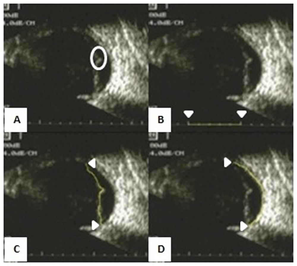
All the ultrasound based measurements were performed using “freehand line tool” of the ImageJ software (https://imagej.nih.gov/ij/). With the help of the software distance between 2 endpoints on a structure can be measured while moving the cursor’s locus along the contour of the structure (Fig 2).Thus the retina was measured starting from the optic nerve head till its point of fusion with the choroid. The choroid was measured similarly between these endpoints. If the oraserrata could not be localized in a USG image, the end point for choroid was determined by drawing a perpendicular from the end of the retina towards the choroid.[4]These measurements were done 5 timesfor each image and mean was taken after excluding measurements deviating more than 0.5 mm from the median for each measurement.Retinal length to choroidal length ratio (RCR) was calculatedfor each quadrant separately along with a mean ratio for each patient.
Surgery: Surgery was done on the day following ultrasound imaging in every case. Standard 3 port 25 gauge VRS was performed in all the patients.Encirclage was used as per surgeons choice. Fluid-air exchange with active fluid extrusion at 40 mm Hg air pressure, laser retinopexy, 360 degreeendolaser photocoagulation and C3F8 gas/silicone oil injection was done. Membrane peeling and sub retinal band removal were performed as needed, but no patient underwent relaxing retinectomy. The VRS was not combined with cataract surgery or lensectomy in any patient. Routine post operative care and head positioning for 7 days was advised to all patients.
Outcomes: All patients were followed till 3months. Patients who developed recurrent RD during this period for discernable reasons like missed retinal break, post operativeePVR, post operativesPVR and poor oil/gas fill were excluded from the study. Finally, 24 patients and 96 Images (~ 1000 measurements)were included for analysis. The study hence was designed to address if recurrent “unexplained” RD during first 15 days of surgery could be attributed to severe retinal shortening.
Analysis:This was a double masked study. Patient workup was performed by a single surgeon (ST) while all ultrasoundrelated measurements were done by another (BT). These authors were blinded to each other’s findings throughout the study period.
Data was entered intoMicrosoft excel sheets and statistical analysis was performed (SPSS software, version: 22). Retinal shortening was categorized as severe when RCR was measured to be less than 0.80. Fischer exact was used for parametric comparisons, and a two tailed p value <0.05 was defined as significant. Comparison was done between eyes with PVR > Grade B(Group 1) and <Grade B(Group 2).Therefore, patients with clinically visible ePVR and sPVR were categorized into Group 1 and Group 2 was used as control group.Mann –Whitney U test was done for comparison of means between these 2 groups. Pearson’s coefficient was used for assessing correlation amongst variables. RCRs were evaluated separately for eyes with unexplained recurrent RD. Odds ratio were calculated for variables associated with unexplained recurrent RD within first 15 days, and all the confidence intervals (CI) described here after are true for 95% of population.iPVR was later objectively identified in the eyes which hadsevere quadrantic retinal shortening in absence of pre operativeePVR in that quadrant.
Results
The mean age of the 24 patients was 49.58+16.76 years, 9 were female and the right and left eyes were equally affected, 12 cases each. Three cases were found to have high myopia. Mean duration of RD (defined from vision loss) was 2.76 months. 10 cases had PVR greater than grade B. Among these 10 patients, average number of quadrants with PVR was 2.2. All these cases had ePVR, while sPVR was present in one patient only. Encirclage was used in 13 cases. C3F8 gas was used as vitreous substitute in 8 cases while silicone oil was used in 16 cases.
The RCR’s of the 24 cases have been presented in Table 1. The mean RCR overall was 0.85 (Range: 0.69-1.00). Quadrant based RCRs ranged from 0.62 to 1.36. Overall 3 RCR values were found to be in excess of 1, all these 3 cases had bullous RD. The mean quadrant based RCRs in superior-temporal, inferior-temporal, superior-nasal and inferior-nasal quadrants were 0.86, 0.84, 0.88 and 0.84 respectively, thus slightly lower in the inferior quadrants. The mean RCR was 0.82 in the 3 patients with high myopia. The mean RCR had a good negative relation with number of quadrants with PVR (combined ePVR and sPVR), correlation coefficient being -0.66 (p= <0.001). However there was no statistically significant relation between mean RCR and age of the patient (R=-0.16, p=0.45) and duration of RD (R=-.06, p=0.78).
Group 1 (patients with pre operativePVR >Grade B) had 10 patients while group 2 had 14. The mean duration of RD was significantly more in group 1 than group 2 (4.1 months vs. 1.8 months, p=0.03). The mean RCR was significantly less in group 1 (0.8+/-0.07) as compared to group 2 (0.89+/-0.05) (p<0.001). All the quadrantic RCRs were also significantly lower in Group 1 than group 2 (p<0.05 for all quadrants). Overall 6 patients had unexplained RD within the first 15 days. All these patients belonged to Group 1.
The details of the patients with recurrent RD attributable to retinal shortening have been presented in Table 1 (cases 1,2,4,5,8,9). The mean RCR of these 6 patients was 0.78 whereas the mean number of quadrants with ePVR was 2.5. More interestingly, all these 6 cases had RCR less than 0.8 at least in 1 quadrant of retina, whereas 4 had in 2-4 quadrants (Table 1). Apart from these 6 patients, there were 3 more patients who had RCR less than 0.8 in at least in a single quadrant but didn’t develop unexplained RD in first 15 days (cases 3,6 and 7 in Table 1). One of these patients had thick-taut adherent posterior hyaloids, as noted during surgery and which may have lead to under estimation of RCR, one had a healthier mean RCR of 0.82 while the third developed RD in the second month of follow up.
Univariate analysis for factors associated with recurrent RD attributable to retinal shortening has been presented in Table 2. Presence of ePVR, mean RCR <0.8 or presence of RCR below 0.8 in 1 quadrant were significantly associated with occurrence of unexplained RD in first 15 days.Odds ratio were calculated to ascertain impact of these 3 factors on unexplained RD in first 15 days.Quadrantic RCR <0.8 had the highest odds ratio (57.57, CI=2.58-1279.96), followed by presence of ePVR (41.88, CI=1.95-897.66) and mean RCR <0.8 (34, CI= 2.43-474.57). Though the CI of all these variables were very large, the lower confidence bound was 2 or more in all cases.
Discussion
In the current study, we aimed to identify the impact of ePVR on retinal shortening using ultrasound based RCRs. We have found a strong statistically significant relationship between quadrantic RCR<0.8 and ePVR, and mean RCR correlated well with amount of ePVR.Retinal shortening has been quantified with ultrasound recently.[4] In that study it was speculated that as retina is an elastic structure, the RCR is likely to fall to some value below 1 after RD and that ePVR and sPVR are likely to coexist with iPVR, therefore the term mixed PVR.[3,4] Like the previous study, in the current study also we found unexplained early recurrent RD to have high odds of occurring in cases with quadrantic RCR below 0.8.
Though minute fall in RCR is likely to be related to elasticity of neuro-sensory retina (NSR) as discussed above, severe retinal shortening is more likely to be due to structural changes.This is reflected in significantly lower RCR in the group with advanced PVR and the negative correlation between number of quadrants with ePVR and mean RCR values. iPVR, with the help of immune markers like glial fibrillary acidic protein, has been proven to be due to glial proliferation within the detached NSR.[4,5] This is different from the traditional RPE cell theory advocated for ePVR.[9,10]However, one should remember that ePVR by virtue of fibrotic contraction would result in fall of RCR.[10] This reflects in strong association ePVR with severe retinal shortening below RCR of 0.8. During surgery, manual dissection or removal of this ePVR would result in favorable intra-operative RCR that should allow for flattening of retina along the choroidal contour and future surgical success. However, in cases where “enough” iPVR has set in, recurrent “unexplained” RD would occur defying the high fluid/air pressure used during surgery. This is more likely to occur as soon as patient positioning is stopped and before NSR-RPE micro-structure bonding has occurred.[4,11] That is why we chose 15 days as the cut off duration. After this time, post operative PVR can also come into play.
Further, to objectively identify iPVR and prove it as an independent cause of retinal shortening, we analyzed the location of ePVR vis. a vis. severely low RCRs. Data of all patients with at least 1 quadrantic RCR below 0.8 along with location of ePVR has been presented in Table 3. It can be seen that of the 36 quadrantic RCRs, in 11 cases (30%) there was severe retinal shortening in absence of ePVR. As this is a pilot technique, direct comparisons cannot be drawn with previous data.The lack of studies on iPVR is related to the subjective difficulty in its pre operative identification and need for immune-histochemistry.[4,5]However, we are not the first to speculate on importance of iPVR. A lot of work has been done by Pastor et al,3 who have emphasized the role of iPVR in staging PVR in general. The author group has evaluated previously published studies using PVR classification systems and has discussed the need for including iPVR in grading techniques.[3,4,12]The authors have also done a study on 60 patients of retinal shortening. In that study, iPVR was defined as inability to achieve intraoperative retinal flattening despite removing extra-retinal PVR after PFCL injection.[6]However only 9/60 patients had needed retinectomy.[6]
Our study has some limitations. We are taking a single dimension per quadrant into account for calculating RCR, and ideal would be retinal area to choroidal area ratio, but current technology eludes such an analysis. Our sample size was low (24 cases) and we didn’t have enough patients withsPVR (only 1).It would also be interesting to analyze the RCR values in eyes with early RD and no clinical PVR in a larger sized study as RCR is expected to fall below 1 in most cases (Table 1). Though a single surgical team operated, there was no fixed protocol on use of explants or vitreous substitutes. Patients with bullous RD do not seem to be good candidates for this imaging technique. In the older study, 7 of the 40 images analyzed needed perpendicular method (explained in methodology section to judge RCR.[4]In the current study, 11/96 images needed this method for calculating RCR. We believe if blinding is removed and the sonographer is aware of the clinical findings, this ratio will further lessen.We could not study the angle subtended by the detached retina on the choroid as a function of retinal shortening because the retina was curved in most cases and was not straight like a chord is to an arc.
Gold standard for presence of iPVR would be histo-pathological analysis, but as relaxing retinectomy was not done, such tissue was not available. In this regard, future study on pre operative planning of site of retinectomy based on lowest RCR can be undertaken whereretinectomy would increase intra-operative RCR andtackle retinal shortening, and also provide tissue for analysis. Thus deciding a critical threshold for planned retinectomy is an interesting surgical option and needs appropriate evidence before bringing to surgical practice.[4]
Conclusions
Ultrasound based calculation of retinal length to choroidal length ratio is a good method of documenting retinal shortening.While ePVR is an important cause of retinal shortening, in some cases iPVR may exist independent of it. Retinal shortening is an important indicator of poor anatomical prognoses in patients with RD. Severe retinal shortening in any single quadrant should prompt appropriate measures.
Declarations
Conflicts of interest: None
Funding sources: None
Acknowledgements: None
References
- Kolomeyer AM, Grigorian RA, MostafaviD,Bhagat N, Zarbin MA.. 360° retinectomyfor the treatment of complex retinal detachment. 2011;31:266-74.
- Shalaby KA. Relaxingretinotomies and retinectomies in the management of retinal detachment with severeproliferative vitreoretinopathy (PVR). Clin Ophthalmol. 2010;4:1107-14.
- Pastor JC, Rojas J, Pastor-Idoate S, Di Lauro S, Gonzalez-Buendia L, Delgado-Tirado S. Proliferative vitreoretinopathy: A new concept of disease pathogenesis and practical consequences. Prog Retin Eye Res.2016;51:125-55.
- Takkar B, Tripathy K, Azad SV, Venkatesh P, Chawla R. ObjectiveQuantification of Retinal Shortening: Sonographic Evidence of Intraretinal Proliferative Vitreoretinopathy. Ophthalmic Surg Lasers Imaging Retina. 2016;47(8):746-50.
- Pastor JC, Méndez MC, de la Fuente MA, Coco RM, García-Arumí J, Rodríguez de la Rúa E, Fernández N, Saornil MA, Gayoso MJ. Intraretinal immunohistochemistry findings in proliferative vitreoretinopathy with retinal shortening. Ophthalmic Res. 2006;38(4):193-200.
- Pastor JC, Rodríguez de la Rúa E, Martín F, Mayo-Iscar A, de la Fuente MA, Coco R, Bailez C, Mahave S. [Retinal shortening: the most severe form of proliferative vitreoretinopathy (PVR)]. Arch Soc EspOftalmol. 2003;78(12):653-657.
- No authors listed. A randomized trial of immediate vitrectomy and of intravenous antibiotics for the treatment of postoperative bacterial endophthalmitis. Endophthalmitis Vitrectomy Study Group. Arch Ophthalmol. 1995;113(12):1479-1496.
- Lean JS, Stern WH, Irvine AR, Azen SP. Classification of proliferative vitreoretinopathy used in the silicone study. The Silicone Study Group. Ophthalmology. 1989;96(6):765-771.
- Landiev I, Bringmann A, Wiedemann P. [Proliferative vitreoretinopathy–pathogenesis and therapy]. 2010;227(3):168-74.
- Pastor JC. Proliferative vitreoretinopathy: an overview. 1998;43(1):3-18.
- Kang HK, Luff AJ. Management of retinal detachment: a guide for non-ophthalmologists.BMJ : British Medical Journal. 2008;336(7655):1235-1240.
- Di Lauro S, Kadhim MR, Charteris DG, Pastor JC. Classifications for Proliferative Vitreoretinopathy (PVR): An Analysis of Their Use in Publications over the Last 15 Years. Journal of Ophthalmology. 2016;2016:7807596.
Figure Legends
Fig 1: Flowchart depicting the study methodology. * PVR was assessed as per Silicone oil study classification.8 VRS: Vitreoretinal surgery, RD: Retinal detachment, GRT: Giant retinal tear, CD: Choroidal detachment, PVR: Proliferative vitreoretinopathy, iPVR: Intraretinal PVR
Fig 2: Ultrasonic measurement of retinal shortening. (A) Ultrasound B-scan of an eye with retinal detachment and proliferative vitreoretinopathy. A retinal fold (encircled) with overlying adherent vitreous can be seen. (B) Figure depicts setting up a scale for further measurements. Note the yellow line, indicated by arrowheads, placed over the vector scan. This pixel distance was set as 10 mm, and future measurements were done accordingly. (C) Figure shows the process of retinal length measurement. The measuring yellow line has been drawn along the retinal contour, the end points being the optic disc and point of fusion of neuro-sensory retina with the RPE-choroid (arrowheads). In this case, the retinal length was measured as 12.44 mm after taking 5 readings. (D) Figure shows the process of choroidal length measurement. The endpoints used for retinal length (arrowheads) were used for measuring the choroidal length, drawn as a curved yellow line along its length. In this case, the choroidal length was measured as 15.95 mm after taking 5 readings. The retinal length to choroidal length ratio was hence measured as 0.78.
| Table 1: Summary of 24 cases analysed | |||||||||
| Age
(years) |
Duration
(months) |
Quadrants with PVR | Recurrent RD
within 15 days |
Superior –temporal RCR | Inferior-temporal RCR | Superior-nasal RCR | Inferior-nasal RCR | Mean RCR | |
| Case 1 | 48 | 3 | 3 | Yes | 0.79 | 0.62 | 0.73 | 0.63 | 0.69 |
| Case 2 | 52 | 2.5 | 4 | Yes | 0.80 | 0.75 | 0.71 | 0.71 | 0.74 |
| Case 3 | 55 | 2 | 1 | No | 0.81 | 0.78 | 0.82 | 0.84 | 0.81 |
| Case 4 | 72 | 4 | 1 | Yes | 0.81 | 0.75 | 1.00 | 0.77 | 0.83 |
| Case 5 | 56 | 1 | 3 | Yes | 0.80 | 0.83 | 0.81 | 0.77 | 0.80 |
| Case 6 | 66 | 5 | 1 | No | 0.68 | 0.76 | 0.91 | 0.74 | 0.77 |
| Case 7 | 60 | 0.75 | 0 | No | 0.87 | 0.63 | 0.97 | 0.80 | 0.82 |
| Case 8 | 64 | 1.5 | 3 | Yes | 0.71 | 0.79 | 0.84 | 0.78 | 0.78 |
| Case 9 | 10 | 12 | 1 | Yes | 0.85 | 0.79 | 0.86 | 0.88 | 0.84 |
| Case 10 | 60 | 0.75 | 0 | No | 0.93 | 0.91 | 0.98 | 0.88 | 0.93 |
| Case 11 | 60 | 1 | 0 | No | 0.84 | 0.81 | 0.81 | 0.83 | 0.82 |
| Case 12 | 23 | 9 | 4 | No | 0.86 | 0.81 | 0.88 | 0.80 | 0.84 |
| Case 13 | 30 | 1 | 0 | No | 0.91 | 0.89 | 0.97 | 1.10 | 0.97 |
| Case 14 | 27 | 6 | 0 | No | 0.83 | 0.86 | 0.91 | 0.85 | 0.86 |
| Case 15 | 45 | 1 | 1 | No | 0.81 | 0.90 | 0.89 | 0.88 | 0.87 |
| Case 16 | 47 | 2 | 0 | No | 1.15 | 0.93 | 0.89 | 0.85 | 0.96 |
| Case 17 | 65 | 1 | 0 | No | 0.85 | 0.81 | 0.91 | 0.82 | 0.85 |
| Case 18 | 52 | 1 | 0 | No | 0.84 | 0.81 | 0.83 | 0.89 | 0.84 |
| Case 19 | 22 | 0.33 | 0 | No | 0.91 | 0.89 | 0.88 | 0.90 | 0.89 |
| Case 20 | 45 | 0.25 | 0 | No | 0.91 | 1.36 | 0.95 | 0.80 | 1.00 |
| Case 21 | 59 | 2 | 0 | No | 0.88 | 0.91 | 0.85 | 0.87 | 0.88 |
| Case 22 | 76 | 7 | 0 | No | 0.92 | 0.82 | 0.98 | 0.99 | 0.93 |
| Case 23 | 42 | 0.25 | 0 | No | 0.86 | 0.85 | 0.93 | 0.83 | 0.87 |
| Case 24 | 54 | 2 | 0 | No | 0.97 | 0.93 | 0.88 | 0.88 | 0.91 |
| PVR: Proliferative vitreo-retinopathy (includes both ePVR and sPVR), RD: Retinal detachment, RCR: Retina length to choroidal length ratio | |||||||||
| Table 2: Factors associated with unexplained recurrent RD within first 15 days | ||
| Recurrent RD | P value | |
| Age> 50 years
Yes (14) No (10) |
4 (28%) 2 (20%) |
1.00 |
| Duration of RD > 3 months
Yes (7) No (17) |
3 (43%) 3 (18%) |
0.3 |
| Presence of ePVR
Yes (10) No (14) |
6 (60%) 0 (0%) |
0.001 |
| Mean RCR <0.8
Yes (5) No (19) |
4 (80%) 2 (11%) |
0.004 |
| RCR <0.8 in 1 quadrant
Yes (9) No (15) |
6 (67%) 0 (0%) |
<0.001 |
| RD: retinal detachment, ePVR: epi-retinal proliferative vitreo-retinopathy, RCR: retinal length to choroidal length ratio | ||
| Table 3: Relation between location of ePVR and severe quadrantic retinal shortening | ||||||||
| Superior-temporal | Inferior-temporal | Superior-nasal | Inferior-nasal | |||||
| RCR | ePVR | RCR | ePVR | RCR | ePVR | RCR | ePVR | |
| Case 1 | 0.79 | yes | 0.62 | yes | 0.73 | no | 0.63 | No |
| Case 2 | 0.80 | yes | 0.75 | yes | 0.71 | no | 0.71 | No |
| Case 3 | 0.81 | No | 0.78 | No | 0.82 | Yes | 0.84 | No |
| Case 4 | 0.81 | yes | 0.75 | no | 1.00 | no | 0.77 | No |
| Case 5 | 0.80 | yes | 0.83 | no | 0.81 | no | 0.77 | Yes |
| Case 6 | 0.68 | yes | 0.76 | no | 0.91 | no | 0.74 | No |
| Case 7 | 0.87 | no | 0.63 | no | 0.97 | no | 0.80 | No |
| Case 8 | 0.71 | no | 0.79 | yes | 0.84 | yes | 0.78 | Yes |
| Case 9 | 0.85 | no | 0.79 | yes | 0.86 | no | 0.88 | No |
| ePVR: epi-retinal proliferative vitreo-retinopathy, RCR: retinal length to choroidal length ratio | ||||||||
Figure 1
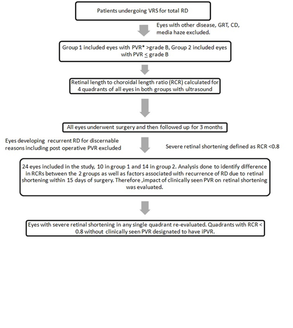
Figure 2
Leave a Comment