

Dr.Bhavagna Bandla,Dr.Thomas kuriakose
Introduction:
Ocular trauma causes lifetime disability in children, being an important cause of morbidity and acquired unilateral blindness (1). Pediatric ocular trauma account to 8%–14% of total injuries in children (2). Delayed presentation results in substantial damage to the ocular structures and poor visual outcome in these children.
Injury leading to loss of tissue and leaving corneal defects are rare. However, when injuries are not repaired immediately, can lead to tissue contracture and permanent defects which are surgically challenging to repair.
Penetrating injury to the eye can be by a sharp object or a projectile object and is further sub classified as penetrating (entry wound but no exit wound) or perforating (entry & exit wounds). (3)
Epidemiology:
- Annual incidence: 3.5 eye injuries/100,000 population
- Incidence: Males 6 times > females
- Age group: 10-30 years
Case Report:
We present a 6-year-old girl from Bangladesh, brought 1-month post trauma with edge of a tile to right eye which was playfully thrown at her. She initially complained of redness and pain and was treated with antibiotics, cycloplegics and antifungals at a local hospital. There was improvement in symptoms with topical treatment and she was referred to higher center for further management.
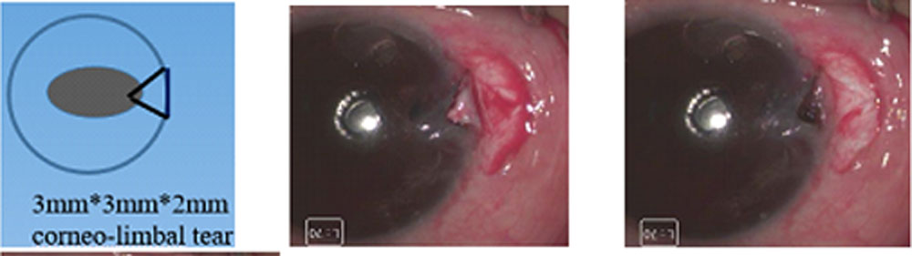
Examination of right eye revealed vision of 6/9, N6. Anterior segment examination showed circumcorneal congestion with 0.5+ cells and 1+ flare with no hypopyon. There was a 3mm vertical corneo-limbal tear plugged with iris tissue incarceration at 3 o clock position. Posterior segment showed few anterior vitreous cells with hyperemic disc with macular edema. There was no foreign body seen on computer tomography.
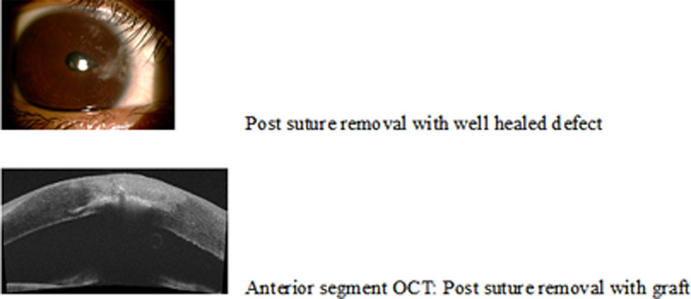
She underwent iris reposition with corneo-limbal tear suturing with 10-0 nylon black monofilament sutures under general anesthesia. Intraoperatively after the reposition of the iris, corneal tissue was found to be contracted with a triangular defect which prevented direct closure. (Fig 1,2). A corresponding limbus based partial thickness scleral flap was created and flipped over the defect & sutured. (Fig 3)
Post-Operative Follow Up:
| VISIT | 1 Week Post-Operative Period (OD) | 2 Month Post-operative Period (OD) |
| BCVA | 6/9 | 6/6 |
| Corneal Tear | Good anatomical closure Vascularization |
Good defect closure Vascularization |
| Posterior segment | Minimal Anterior Vitreous cells
Disc hyperemia |
Minimal Anterior Vitreous cells
Altered foveal reflex
|

First post-operative day 1-week post-operative visit 2 months’post-operative visit
Suture removal was done at 2 months’ post-operative period.
Discussion:
Type of injury, mode of injury, time lapse and tissues involved of the injured eye, all affect the outcome. A careful surgical procedure can retain a round pupil and complete iris diaphragm.
In children, vision should be monitored closely and if required amblyopia therapy should be initiated early. On delayed surgical intervention, iris tissue adheres with fibrin to wound edges within a few hours, fibrosis in few days causing wound contraction, preventing adequate wound closure.
The defects of 2mm and more need grafts to close.
Commonly used grafts are tenon’s graft with glue and contact lens, cornea, conjunctival flap, amniotic membrane, autologous sclera, homologous sclera. All grafts have risks of rejection, infection and foreign body reaction. Scleral collagen tissue having close resemblance with corneal tissue is a good substitute to cover such defects compared to other tissues. Autologous sclera when done with a “twist & turn” as described in our case can result in favorable outcomes.
Conclusion:
Injuries leading to loss of tissue, leaving corneal defects are rare. Injuries when not repaired immediately can lead to tissue contracture and can be surgically challenging to repair.
Approach to open globe injuries needs modifying techniques from the routine, with innovation & thinking on the feet.
Corneal tissue defects can be repaired not only with corneal grafts but also with autologous scleral hinge graft which can be a good alternative to close juxta limbal corneal tissue defects with good results, as in our case.
THERE IS ALWAYS TWIST AND TURN!
References:
- Epidemiology, clinical profile and factors, predicting final visual outcome of pediatric ocular trauma in a tertiary eyecare center of Central India. Singh S, Sharma B, Kumar K, Dubey A, Ahirwar K.
Indian J Ophthalmol. 2017 Nov;65(11):1192-1197. doi: 10.4103/ijo.IJO_375_17.
- Brophy M, Sinclair SA, Hostetler SG, Xiang H. Pediatric eye injury-related hospitalizations in the United
Pediatrics. 2006;117:e1263–71 - Kuhn F, Morris R, Witherspoon CD. Birmingham Eye Trauma Terminology (BETT): terminology and classification of mechanical eye injuries. Ophthalmol Clin North Am. 2002; 15:139–43.
- Korah, S. S. Selvin, Z. S. Pradhan et al., “Tenons patch graft in the management of large corneal perforations,” Cornea, vol. 35, no. 5, pp. 696–699, 2016.
- John B, Raghavan C. Open globe injuries-primary repair of corneoscleral injuries. Kerala J Ophthalmol. 2010; 22:225–34.
Leave a Comment