

Dr.Kshirsagar Sucheta, Dr. NILESH KAKADE,Dr. ROHIT MENDKE,Dr.(col) Madan Deshpande
INTRODUCTION
Development of retinal vessels starts early in fetal stage (12 weeks) and is completed by the end of full term pregnancy (40 weeks).(1)The process of vasculogenesis (formation of primordial or major vessels) starts around 14-15 weeks of gestation and angiogenesis (extension of vascular plexus with formation of capillaries and inner plexus) starts around 25-26 weeks of gestation.(2,3) The process of vascular remodeling and retraction continues throughout pregnancy and is still ongoing in full term neonates.(4)Previous studies from India (5) and America (6) reported the status of retinal vascularization in newborn (preterm as well as healthy term neonates) within one and four weeks of birth respectively. These studies suggest that there is considerable variability in the proportion of neonates of the same gestational age whose retinal vessels are mature. For example, some term neonates have immature retinal vasculature at birth whereas some preterm infants (28 weeks) have completely vascularized retinas at or shortly after birth. Factors which are associated with incomplete retinal vascularization are gestational age and longer exposure to supplemental oxygen (Jalali).
Physiologic hypoxia acts as a stimulus for the developing vasculature.(7)Pathological events during the antenatal or postnatal periods can affect the normal development of human organs, including retina.Inadequately administered supplemental oxygen with chronic hypoxemia can lead to poor growth and delayed development of a range of organs, and is well recognized as an important risk factor for retinopathy of prematurity (ROP).(8) ROP is emerging as an important cause of blindness in children in India and other middle income countries as neonatal care services expand.(9,10)Apart from prematurity and hyperoxia or fluctuating hypo / hyperoxia, other risk factors associated with ROP include sepsis, poor weight gain and transfusion with adult blood. (11–16)
Screening for ROP needs to be well organized and systematic, starting with the first examination a few weeks after birth. At each screening episode one of the following management decisions needs to be made: 1. further screening is required (and when), as the retinal vessels are not yet mature or ROP is present; 2. urgent treatment is required;3. no further screening is required as the retinal vessels are mature, or signs of ROP are definitely regressing. Most of the literature focusses on decisions 1 and 2, with less emphasis on management decision 3. The decision to discontinue screening is important, as discontinuing screening too soon means they may still develop ROP, and continuing screening when the risk is negligible puts additional stress on babies, their parents and the health system. This is particularly relevant in settings where neonatal care is suboptimal, where larger more mature infants also need to be screened than in settings providing excellent care (refs). The wider screening criteria required means that a high proportion of babies will be discharged from the neonatal unit before the date of the first screening. In these settings neonatal units are also often overcrowded which can also lead to early discharge. Under these circumstances the “safe to discontinue screening” decision becomes even more important.
It is assumed that the temporal retina matures at a post menstrual age of between 40 and 44 weeks (PMAi. e.gestational age plus chronological age).Several studies reporton development of retina in animals(17)and humans,(3,18,19)but these studies focus on the cellular processes occurring in retina rather than the retinal vasculature. In practice, detailed clinical examination of the retinal periphery is the only way to assess whether the retinal blood vessels are mature.. To the best of authors’ knowledge, there are no published studies on retinal vessel maturation in preterm infants with and without ROP.
METHODS
This retrospective cohort study was conducted to establish the PMA at which retinal vessels matured in preterm infants who did and who did not develop ROP, and whether the timing of maturation is associated withrisk factors for ROP. The study population was consecutive preterm infants enrolled in the ROP screening program of a tertiary eye care centre in MaharashtraState, western India.The criteria for screening are similar to those recommended by the government of India,(20) and include infants born before 34 week’s gestational age (GA) or a birthweight (BW) of <2000g. Larger, more mature infants are also screened at the discretion of the neonatologist.
Ethics
Written informed consent is routinely obtained from parents before ROP screening, as a part of hospital’s protocol. The institutional ethics committee granted a waiver for the study.
Sample size calculation:
Almost all full term infants born between 37 and 40 weeks of GA have mature retina within a week of birth.(5,6)Assuming a 7% difference between the proportion of preterm and full term infants attaining retinal maturity by 40 weeks PMA, 0.05 as error alpha, to achieve 80% power, the sample size was calculated to be 241. The sample size was increased by 25% to 300 to allow adjustment for confounders.
Electronic medical records of allpreterm low birthweight infants enrolled in the hospital’s ROP screening program between April 2014 and March 2018 were reviewed. All preterm infants who were screened and completed follow up when retinal vessel maturity had been confirmed were reviewed for eligibility.The following infants were excluded: unknown GA; missing data on risk factors; infants developing sight-threatening ROP or with asymmetrical disease e.g. mild ROP in one eye and no ROP in other eye; infants with mature retinal vessels at the first screening, as the PMA at maturation was unknown, and screening not undertaken using retinal imaging. Data on risk factors are routinely collected from neonatal records of all infants.
The first screening is often performed before day 30 of life of a preterm neonate or earlier for extremely premature infants as per Indian guidelines.(20)The hospital’s ROP program uses one of two digital wide field cameras (Retcam shuttle, Clarity MSA, USA and 3nethra Neo, Forus, Banagalore, India) for screening. One of the two trained and experiencedtechnicians performs imaging. The images are obtained under topical anesthesia and 5 standard images capturing the posterior pole, temporal, superior, nasal and inferior periphery are obtained. In order to the capture temporal periphery, the technician routinely turns the head and rotates the eye, using an infant scleral depressor,if required.Images are graded by one of the two trained and experienced ROP specialists. The hospital protocol defines ‘mature retinal vessels’ as vesselsseen at least one disc diameter (DD)anterior to the junction of Zone II and III temporally.
Infants were divided into subgroups of PMA at which they were discharged from further ROP screening as they had mature retinal vessels. The twelve subgroups of PMA at maturation started with 30-32 weeks and ended with 52-54 weeks. Delayed maturation was defined as maturation beyond 44 week’s PMA.
Data collected on risk factors for ROP i.e., GA, BW, oxygen supplementation, sepsis, anemia requiring blood transfusion, neonatal jaundice, bradycardia, respiratory distress syndrome, episodes of apneaweredefined according to guidelines from National Neonatology Forum of India.(21) Thesewere collected from discharge summaries or hospital records.In all neonatal units covered by hospital’s ROP screening program, GA is estimated using the date of the last menstrual period of the mother or by referring to pregnancy ultrasound and is confirmed using Ballard score.(22,23)If these were unavailable, GA was defined as indeterminate.
Data were entered into Microsoft Excel spreadsheet and transferred into STATA IC 14 (StataCorp, Tx) for analysis. Summary statistics were generated to establish mean GA, BW. Measures of association were studied using Chi 2 test and multiple logistic regression analysis.
RESULTS
388 infants were included in the study; mean GA, 32.2 weeks (SD ±2.3) and mean BW, 1576g (SD ± 425). 302 infants did not develop any ROP and 86 developed ROP not requiring treatment. Mean GA and BW in infants without ROP was 33.1 weeks (SD± 2.6) and 1708 grams (SD ± 460) respectively, and 30 weeks (SD± 2.1) and 1221g (SD± 309), respectively for infants with ROP.
Among the infants with ROP, PMA at diagnosis of ROP was <38 weeks in 66 (76.8%), 38.1-42 weeks in 16 (18.6%) and >42 weeks in 4 (4.6%). None developed ROP beyond 44 weeks of PMA.
Among the 302 infants without ROP, 77.2% had mature retinal vessels by 44 weeks PMA which was higher than those with ROP (47.7%)(p <0.001)(Table 1).
Table 1: Postmenstrual age at which retinal blood vessels matured in infants with and without retinopathy of prematurity
| PMA at retinal vessel maturation (weeks) | No ROP (n=302) | ROP (n=86) | Total N=388) | |||
| No ROP | % | N | % | N | % | |
| Maturation not delayed | ||||||
| 30-32 | 0 | 0 | 1 | 1.1 | 1 | 0.4 |
| 32.1-34 | 0 | 0 | 0 | 0 | 0 | 0 |
| 34.1-36 | 2 | 0.6 | 0 | 0 | 2 | 0.5 |
| 36.1-38 | 16 | 5.3 | 2 | 2.2 | 18 | 4.6 |
| 38.1-40 | 80 | 26.5 | 8 | 8.8 | 88 | 22.7 |
| 40.1-42 | 83 | 27.5 | 9 | 1.0 | 92 | 23.7 |
| 42.1-44 | 52 | 17.2 | 21 | 24.4 | 73 | 18.8 |
| Subtotal | 233 | 77.2 | 41 | 47.7 | 274 | 70.6 |
| Delayed maturation | ||||||
| 44.1-46 | 33 | 10.9 | 18 | 20.9 | 51 | 13.1 |
| 46.1-48 | 18 | 5.9 | 8 | 8.8 | 26 | 6.7 |
| 481-50 | 11 | 3.6 | 10 | 11.1 | 21 | 5.4 |
| 50.1-52 | 5 | 1.6 | 3 | 3.5 | 8 | 2.0 |
| 52.1-54 | 2 | 0.6 | 6 | 7.0 | 8 | 2.0 |
| Subtotal | 69 | 22.8 | 45 | 52.3 | 114 | 29.4 |
Infants with ROP were 2.3 (95 CI, 1.7-3, p< 0.001) times more likely to have delayed retinal vessel maturation than those without. All (100%) had mature retinal vessels by 54 week’s PMA.
Retinal maturation by chronological age and gestational age are shown in Figures 1 and 2.
The vast majority of infants with ROP had retinal vessel maturation at or beyond 12 weeks of age.
Figure 1. Retinal vessels maturity, by gestational age, in infants without ROP
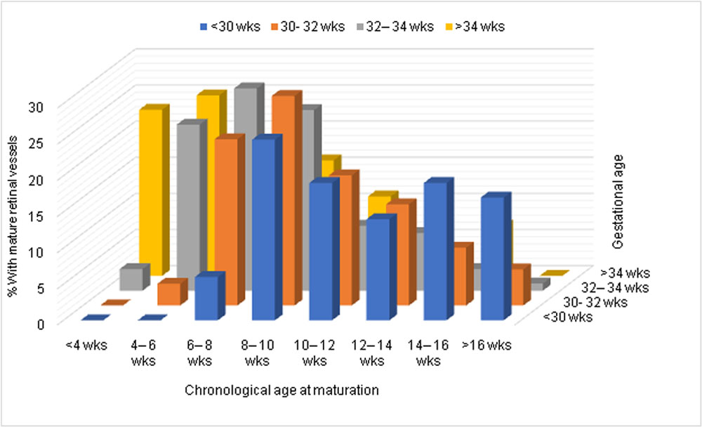
Figure 2. Retinal vessels maturity, by gestational age, in infants with ROP
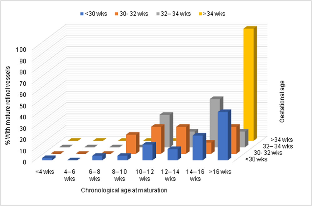
In univariate analysis, infants without ROP, lower GA (<30 weeks) was associated with delayed retinal vessel maturation beyond 12 weeks of age compared to higher GA (>32 weeks)(50%vs 20.7%; risk ratio 2.5, 95% confidence interval (CI) 1.6-3.9; p < 0.001). However in infants with ROP, there was no difference (75.5% vs 71.4%).Infants receiving supplemental oxygen showed very strong evidence of delayed retinal vessel maturation (risk ratio 1.9 (95%CI 1.3- 2.6, p <0.001) (Table 2).
Table 2. Postmenstrual age at retinal vessel maturation by supplemental oxygen administration.
| PMA at maturation | No oxygen | Oxygen | Total | |||
| N | % | N | % | N | % | |
| 44 weeks or less | 145 | 80.1% | 129 | 62.3% | 274 | 72.4% |
| 44.1 weeks or more | 36 | 19.9% | 78 | 37.7% | 114 | 27.6% |
| Total | 181 | 100% | 207 | 100% | 388 | 100% |
P<0.001
Multiple logistic regression analysis was performed to account for effect of other risk factors on retinal maturation. Presence of ROP, lower GA and history of supplemental O2 were all independent risk factors (p <0.001) for delayed retinal vessel maturation None of the other risk factors showed any significant association.
Discussion:
This study demonstrates natural history of retinal maturation in preterm infants with respect to development of ROP. Two previous studies(5,6) had reported status of retinal vascularization in relation with ‘GA’ of a newborn irrespective of whether the child was preterm or full term. In those studies, infants with immature retinal vessels were not followed up to see at what age retinal vessels matured in them. Present study reports ‘PMA’ at which retinal vascular maturation occurs in preterm infants with and without ROP. All the infants enrolled in this study had immature retina at first screening and completed follow up till retinal vascular maturation.
Difference between mean GA and BW of infants with and without ROP in this study re-emphasizes a well-known fact that these two are risk factors for ROP.Among those who developed ROP, it was seen before 42 weeks of PMA in more than 95% infants. In them, retinal vessels maturation was delayed in over half compared to just a quarter of those without ROP. Infants with ROP were 2.3 times more likely to have delayed maturation compared to those without ROP. In phase 1 of ROP, growth of retinal vessels is inhibited.(24) This is often followed by a phase of vasoproliferation. Perhaps time taken by the vasoproliferation to regress before allowing natural growth of the retinal vessels,
adds to the delay in retinal vessel maturation.
Among the infants without ROP, those born before 30 weeks of GA were 2.5 times more likely to have retinal maturation beyond 12 weeks of age compared to those with higher GA. This implies that in infants without ROP, lower the GA more delayed is the retinal vessels maturation. There is a possibility that these extremely premature infants develop what can be called as ‘subclinical stage 1 ROP’ where vessel growth is being inhibited as in phase 1 of the disease but the process somehow gets aborted before a demarcation line develops leading to delay in retinal vessel maturation. In those with ROP retinal maturation did get delayed in more than 70% infants irrespective of GA. Similarly, infants receiving supplemental O2 were twice as likely to have delayed retinal maturation compared to those not needing O2. Deleterious effects of unrestricted O2 on pulmonary and ocular outcomes (ROP) have been established in several previous studies.(25,26) Excessive supplemental O2 is known to cause vasoconstriction in vulnerable retinal tissue of a newborn. This could either lead to development of ROP or delayed resumption of normal growth of retinal vessels. Multivariable analysis showed that lower GA, presence of ROP and supplemental O2 are all independent risk factors for delay in retinal vessel maturation in preterm infants. In India, lack of uniformity in the quality of neonatal care (including unmonitored O2 supplementation) is a major risk factor for ROP.(9,27) Hence this subgroup of preterm infants is at definite risk of delayed retinal vessel maturation.
Delayed maturation of retina in preterm infants does have an implication on the protocol to be followed in any ROP program and timing for ‘discharge’ from screening follow up. In the present study significant number of infants showed retinal maturation later than 44 weeks of PMA and a minority (4.2%) even beyond 50 weeks. One in ten infants with ROP showed delayed maturation beyond 50 weeks of PMA. This implies that burden of follow up visits on parents as well as the health system could be significant. In countries such as India, compliance to follow up is a major challenge. Customized follow up protocol especially for the ‘high risk’ group i. e.infants with lower GA, ROP and history of supplemental O2 could help in identifying infants who need prolonged follow up.Based on the observations of this study it is recommended that all preterm infants enrolled in a ROP screening program be followed up at least until 44 weeks of PMA or maturation of retina whichever occurs earlier. Infants belonging to aforementioned high risk group need follow up until 50 weeks of PMA or maturation of retina whichever occurs earlier. However, none of the infants showed development of ROP after 44 weeks of PMA in this study. Hence it may be safe to discharge an infant from ROP screening follow up if: 1.PMA is 44 weeks in the absence of ROP or 2. There is definite regression of ROP by 44 weeks of PMA irrespective of retinal maturity.
There are certain limitations to this study. Comparison with natural history of retinal maturation in ‘full term’ infants could have provided a strong normative database. Details of supplemental oxygenadministration, such as when oxygen was first administered, duration of oxygen therapy, target range and thefrequency of hyeroxic episodes, were not available for many infants which prevented more in depth analysis. There is a possibility that all the risk factors were not mentioned in the discharge file. This may mean incomplete data on confounders. Possibility of miscalculation of Ballard score to determine GA may mean inaccurate PMA calculation. Maternal risk factors (such as nutrition, anemia) could not be collected. These have shown association with retinal vascularization in a previous study from India.(5)
In conclusion, over two third of preterm infantsattain retinal maturation by 44 weeks of PMA. Those with lower GA, ROP orreceiving supplemental oxygen do show delayed retinal maturation and need follow up at least until 50 weeks of PMA. It is safe to discharge a preterm infant from screening follow up if he/ she has not developed ROP by 44 weeks of PMA.
References:
- Monte DM. The eye in childhood. Am Fam Physician. 1999;60(3):907–18.
- Chan-Ling, T., Gole, G., Quinn, G., Adamson, S., Darlow B. Pathophysiology, screening and treatment of ROP: A multi-disciplinary perspective. Prog Retin Eye Res. 2018;(62):77–119.
- Hughes S, Yang H, Chan-Ling T. Vascularization of the human fetal retina: Roles of vasculogenesis and angiogenesis. Investig Ophthalmol Vis Sci. 2000;41(5):1217–28.
- Gerhardt, H., Golding, M., Fruttiger, M., Ruhrberg, C., Lundkvist, A., Abramsson A, Jeltsch, M., Mitchell, C., Alitalo, K., Shima, D., Betsholtz C. VEGF guides angiogenic sprouting utilizing endothelial tip cell filopodia. J Cell Biol. 2003;161:1163e1177.
- Jalali S, C M, Reddy GP, Nutheti R. Pilot Study on In Vivo Evaluation of Retinal Vascular Maturity in Newborn Infants in the Context of Retinopathy of Prematurity. Am J Ophthalmol [Internet]. 2006;142(1):181–3.
- Roth AM. Retinal vascular development in premature infants. Am J Ophthalmol [Internet]. 1977;84(5):636–40.
- Chan-Ling, T., Gock, B. SJ. The effect of oxygen on vasofromative cell division. Evidence that “physiological hypoxia” is the stimulus for normal retinal vasculogenesis. Invest Ophthalmol Vis Sci. 1995;(36):1201e1214.
- Askie LM, Henderson-Smart DJ, Irwig L, Simpson JM. Oxygen-saturation targets and outcomes in extremely preterm infants. N Engl J Med. 2003;349(10):959–67.
- Clare Gilbert Luz Gordillo,Graham Quinn, Renato Semiglia, Patricia Visintin, on behalf of the International NO-ROP Group AF. Characteristics of babies with severe retinopathy of prematurity in countries with low, moderate and high levels of development: implications for screening programmes. Pediatrics. 2005;115(No 5):518–25.
- Gilbert C. Retinopathy of prematurity: a global perspective of the epidemics, population of babies at risk and implications for control. Early Hum Dev [Internet]. 2008;84(2):77–82.
- Fortes Filho JB, Eckert GU, Procianoy L, Barros CK, Procianoy RS. Incidence and risk factors for retinopathy of prematurity in very low and in extremely low birth weight infants in a unit-based approach in southern Brazil. Eye [Internet]. 2009;23(1):25–30.
- Wang L-W, Lin Y-C, Wang S-T, Huang C-C. Identifying Risk Factors Shared by Bronchopulmonary Dysplasia, Severe Retinopathy, and Cystic Periventricular Leukomalacia in Very Preterm Infants for Targeted Intervention. Neonatology. 2018 Apr;114(1):17–24.
- Chaudhari S, Patwardhan V, Vaidya U, Kadam S, Kamat A. Retinopathy of prematurity in a tertiary care center–incidence, risk factors and outcome. Indian Pediatr. 2009;46(3):219.
- Rekha W, Battu RR. Retinopathy of prematurity: incidence and risk factors. Indian Pediatr. 1996;33:999–1004.
- Zaharie GC, Talu S, Blaga L, Panga G, Popa M. RISK FACTORS FOR RETINOPATHY OF PREMATURITY. Arch Dis Child. 2008;93(Suppl 2):ps378-ps378.
- Rao KA, Purkayastha J, Hazarika M, Chaitra R, Adith KM. Analysis of prenatal and postnatal risk factors of retinopathy of prematurity in a tertiary care hospital in South India. Indian J Ophthalmol [Internet]. 2013;61(11):640–4.
- Simmons AB, Fuerst PG. Analysis of Retinal Vascular Plexuses and Interplexus Connections. Methods Mol Biol. 2018;1753:317–30.
- Mrinalini Hoon, Haruhisa Okawa, Luca Della Santina and ROLW. Functional Architecture of the Retina: Development and Disease. Prog Retin Eye Res. 2014;42:44–84.
- Hendrickson A. Development of Retinal Layers in Prenatal Human Retina. Am J Ophthalmol. 2016;161:29–35.
- Ministry of Health and Family welfare Govt. of India. Guidelines for universal eye screening in newborns including retinopathy of prematurity [Internet]. 2017. Available from: https://nhm.gov.in/images/pdf/programmes/RBSK/Resource_Documents/Revised_ROP_Guidelines-Web_Optimized.pdf
- National Neonatology Forum of India. Evidence Based Clinical Practice Guidelines [Internet]. New Delhi; 2010. Available from: https://www.nnfi.org
- Ballard JL, Khoury JC, Wedig K, Wang L, Eilers-Walsman BL, Lipp R. New Ballard Score, expanded to include extremely premature infants. J Pediatr. 1991 Sep;119(3):417–23.
- Lee AC, Panchal P, Folger L, Whelan H, Whelan R, Rosner B, et al. Diagnostic Accuracy of Neonatal Assessment for Gestational Age Determination: A Systematic Review. Pediatrics [Internet]. 2017;140(6):e20171423.
- Ashton N, Ward B, Serpell G. Effect of Oxygen on Developing Retinal Vessels with Particular Reference to the Problem of Retrolental Fibroplasia. Br J Ophthalmol [Internet]. 1954 Jul;38(7):397–432.
- Askie LM, Henderson-Smart DJ, Ko H. Restricted versus liberal oxygen exposure for preventing morbidity and mortality in preterm or low birth weight infants. Cochrane Database Syst Rev [Internet]. 2009/01/23. 2009;(1):Cd001077.
- Wright KW, Sami D, Thompson L, Ramanathan R, Joseph R, Farzavandi S. A physiologic reduced oxygen protocol decreases the incidence of threshold retinopathy of prematurity. Trans Am Ophthalmol Soc [Internet]. 2006;104:78–84.
- Blencowe H Gilbert C. MS. Update on Blindness Due to Retinopathy of Prematurity Globally and in India. Indian Pediatr. 2016;Volume 53,(November 15):89–92.
Leave a Comment