

Dr.prabhakar g v, Dr.Jagadeesh Kumar Reddy K,Dr.Neeraj Shah,Dr.K S SIDDHARTHAN
Abstract :
AIM:
To evaluate the efficacy of 0.01% atropine to control myopia.
METHODS:
In this retrospective study,300 eyes of 150 patients with documented progression of >/= 1D/year were cyclopegicrefraction,keratometry,mesopic and photopic pupil size were recorded every 6 months.At 2 years data was evaluated and Rate of progression of myopia(change of diopteric power per year)before and after starting on 0.01% atropine was calculated.
RESULTS:
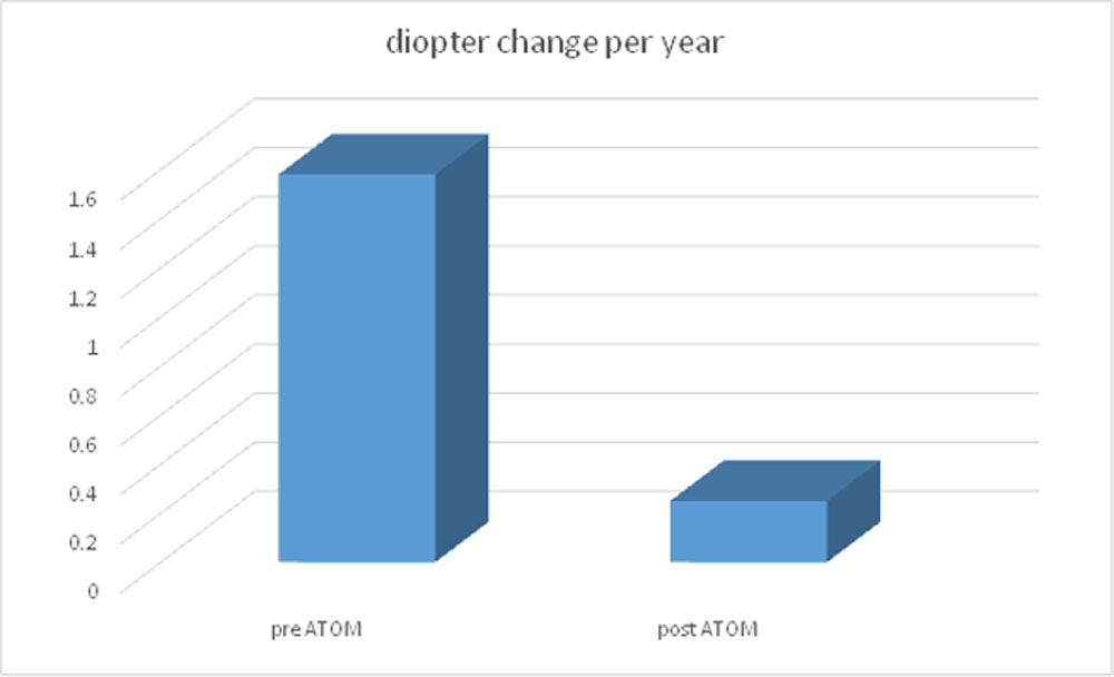
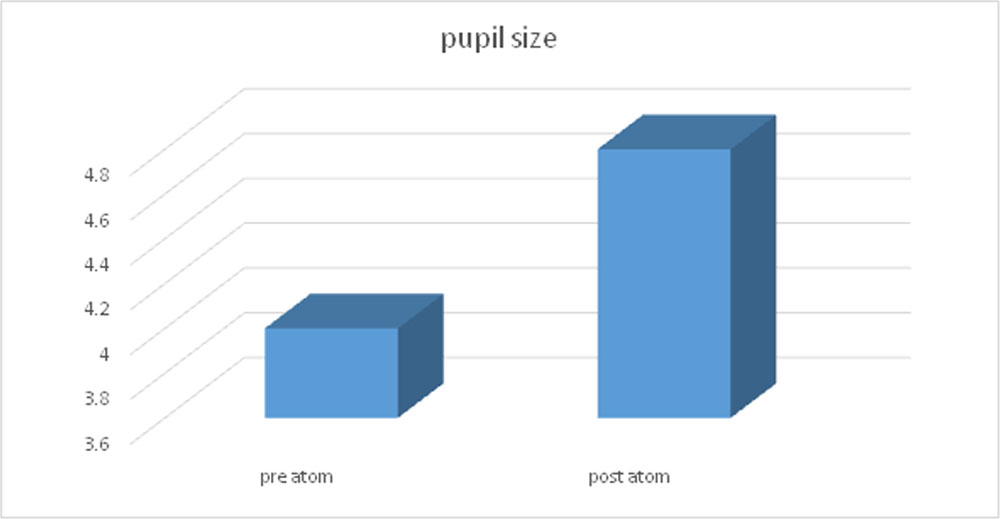
Rate of myopia progression was significantly controlled after starting on 0.01% atropine. Change of diopteric power per year before and after starting on 0.01% atropine was 1.58 D+/-0.36D and 0.25D+/-0.25 / year respectively(p<0.05).The change in SE vs AL was at the rate of 1D/0.33mm.Average change of pupil size was 0.8 mm with no significant change in NPA and NPC.This study shows that 0.01% atropine is effective in controlling progression of myopia with out any significant effect on accomodation.
Introduction :
Myopia is a common and yet perplexing ocular disorder. Once viewed as a benign refractive condition, today myopia, even at low levels, is associated with increased risk for numerous ocular diseases.1Researchers have reported on the myopia epidemic, which is occurring worldwide.2 Although the exact etiology of myopia remains elusive, it appears to have both genetic and environmental components,3making prevention and treatment both challenging and individualized.Myopia is the most common ocular disorder worldwide.4 The prevalence of myopia in the United States has increased from 25% to 44% between 1972 and 2004.5–7 In urban communities in Asia, the prevalence is greater than 80%.8,9 The prevalence is much lower in underdeveloped areas in the world such as Sherpa in Nepal.10
The economic burden of eye diseases is approximately $139 billion in the United States, with nearly $16 billion spent on myopia correction alone.5,6,11 Myopia represents a major risk factor for a number of other ocular pathologies such as cataract, glaucoma, retinal detachment, and myopic maculopathy, which is comparable to the risks associated with hypertension for stroke and myocardial infarction.1,12 Taking into account pathological complications of myopia and other serious pathologies associated with the disease, myopia not only negatively affects self-perception, job/activity choices, and ocular health,13–15 but also represents one of the leading causes of blindness in the world.16 The yearly incidence of retinal detachments is 0.015% in patients with less than 4.74 diopters (D) myopia and it increases to 0.07% in myopia greater than 5 D and 3.2% myopia greater than 6 D.17,18 Myopic patients also have great risk of developing macular choroidal neovascularization, that is, 2X for patients with 1 D to 2 D of myopia; 4X with 3 D to 4 D of myopia; and 9X for 5 to 6 D of myopia.19 It is estimated that 4.8 billion people (one half of the world’s population) will be affected by myopia by 2050.20 A recent study reported that 10% of Asian high school students have high myopia, which increases the risk for future retinal disease.21
Chua et al.22 (ATOM1) studied the effect of 1% atropine in a group of 400 children (13.5% dropout rate) where one group received atropine, whereas the other group received a placebo. Only one eye of each child was chosen for treatment. The mean progression in the control eye after 2 years was 0.6 D/year and in the atropine-treated eye was 0.14 D/year. This represents a 77% reduction in the progression of myopia. Furthermore, the AL measurements in the eyes, which received atropine, remained essentially unchanged (0.02 mm over 2 years). There were no serious adverse events with the atropine being well tolerated.
The ATOM 2 study23 evaluated various concentrations of atropine, including the one below that threshold, that is, 0.5%, 0.1%, and 0.01%. After 2 years, researchers found that all 3 concentrations slowed the progression of myopia.
Methods :Children aged 6 to 12 years with myopic refraction of at least 2.0 D in both eyes, astigmatism of less than 1.5 D, and documented myopic progression of at least 0.5 D in the past year were enrolled in a double-masked, single-center clinical trial. Excluded were those with ocular pathology (e.g., amblyopia, strabismus), previous use of atropine or pirenzepine, an allergy to atropine, or systemic ill health (e.g., cardiac or respiratory illness). Written informed consent was obtained from parents or guardians, and verbal assent was obtained from children.
The study was conducted according to the tenets of the Declaration of Helsinki, with ethics approval was obtainede. Participants received 0.01% atropine once nightly in both eyes. After assessment at the time of recruitment , children were reassessed 2 weeks after starting atropine then at 6, 12, 18 and 24 months. At each visit, distance best-corrected visual acuity (BCVA) logarithm of the minimum angle of resolution (logMAR) was assessed by an optometrist using the Early Treatment Diabetic Retinopathy study chart. Near visual acuity was assessed using best-corrected distance spectacle correction with a reduced logMAR reading chart placed at 40 cm under well-lit conditions. The near point of accommodation was measured using a Royal Air Force near point rule using bestcorrected distance spectacle correction. Children were instructed to move the target inward until the N5 print became slightly blurred and then outward until it just became clear. Accommodation amplitude was calculated as the inverse of near point of accommodation. Mesopic pupil size was measured with the pentacam.
Photopic pupil size was measured using the slitlamp, at least 5 pupil size readings were recorded and averaged. Cycloplegicautorefraction was determined 30 minutes after 3 drops of cyclopentolate 1% were administered at 5 minutes apart using a retinoscope. Five readings, all of which had to be less than 0.25 D apart, were obtained and averaged. Spherical equivalent was calculated as sphere plus half cylinder power. The Zeiss IOLMaster (Carl Zeiss Meditec Inc., Dublin, CA), was used to measure the ocular axial length. Fivereadings, with a maximum-minimum deviation of 0.05 mm or less, were taken and averaged. The primary end point was myopia progression over 2 yearsLevel of myopia progression in each eye was further categorized as mild (<0.5 D), moderate (0.5– 0.99 D), or severe (>1.0 D). Secondary end points included myopia progression at 1 year, change in axial length at 1 and 2 years, and side effect parameters, such as changes in accommodation amplitude, mesopic and photopic pupil size, and distance and near BCVA. Myopia and axial changes, accommodation, pupil size, and visual acuity were monitored from the first visit. During each visit, children and parents were given an openended opportunity to report any medical illness or side effects. They were also specifically asked about symptoms related to allergy, blurred near vision, glare, or visual loss, and if children had been ill or hospitalized since the last visit. Any adverse events, regardless of whether they appeared relevant to atropine use, were documented.
Results :
Rate of myopia progression was significantly controlled after starting on 0.01% atropine. Change of diopteric power per year before and after starting on 0.01% atropine was 1.58 D+/-0.36D and 0.25D+/-0.25 / year respectively(p<0.05).The change in SE vs AL was at the rate of 1D/0.33mm.Average change of pupil size was 0.8 mm with no significant change in NPA and NPC.This study shows that 0.01% atropine is effective in controlling progression of myopia with out any significant effect on accomodation. were included.
Discussion :
Atropine is a nonspecific muscarinic antagonist. It is uncertain how atropine acts to inhibit myopia progression24Initially, inhibition of accommodation was thought to be important, but subsequent studies have shown that atropine also inhibits myopia in animals (e.g., in chickens) that have no accommodative facility. One theory is that atropine and other muscarinic antagonists may have biochemical effects on the retina or sclera, which in turn affect remodeling of the sclera. Another theory suggests that increased ultraviolet exposure (secondary to pupil dilation) may increase collagen cross-linking within the sclera, thereby limiting scleral growth 25
In the ATOM1 study, 400 children aged 6 –12 years with spherical equivalents of 1.00 and 6.00 D were randomly assigned to atropine 1% and placebo medication in 1 eye.16 These children were slightly younger and had lower spherical equivalents and smaller axial lengths (24.8 vs. 25.2 mm) than those in the ATOM2 group. Axial lengths were also measured differently between studies, with the A-scan ultrasonography used in ATOM1 and the IOLMaster used in ATOM2. At the end of 2 years, the mean myopia and axial length progression in the ATOM1 study were.92 D and.35 mm, respectively, in the atropine 1% eyes compared with .69 D and.38 mm, respectively, in the placebo eyes. The progression of myopia in the ATOM2 subjects lies in a dose-related manner between these 2 extremes .Such a dose-related effect on myopia progression was also noted in other studies.
In ATOM2, the progression of myopia on atropine 0.5% was.47 D over 1 year and.60 D over 2 years. This was similar to the progression noted in children receiving atropine 1% in the ATOM1 study and within the ranges noted in studies using atropine 0.5%.
Our study showed similar results in controlling myopia progression.
In conclusion, The lowest concentration of 0.01% atropine thus seems to retain efficacy and is a viable concentration for reducing myopia progression in children, while attaining a clinically significant improved safety profile in terms of accommodation, pupil size, and near visual acuity, and subsequently reduced adverse impact on visual function on indian population.
References :
- Flitcroft DI. The complex interactions of retinal, optical and environmental factors in myopia aetiology. ProgRetin Eye Res 2012;31:622–660. [PubMed]
- Holden BA. The Charles F. Prentice award Lecture 2014: A 50-year research journey: Giants and Great Collaborators. Optom Vis Sci 2015;92:741–749. [PubMed]
- Tkatchenko AV, Tkatchenko TV, Guggenheim JA, et al. APLP2 regulates refractive error and myopia development in mice and humans. PLoS Genet 2015;11:e1005432. [PMC free article] [PubMed]
- Pararajasegaram R. VISION 2020-the right to sight: From strategies to action. Am J Ophthalmol1999;128:359–360. [PubMed]
- Kempen JH, Mitchell P, Lee KE, et al. The prevalence of refractive errors among adults in the United States, Western Europe, and Australia. Arch Ophthalmol 2004;122:495–505. [PubMed]
- Javitt JC, Chiang YP. The socioeconomic aspects of laser refractive surgery. Arch Ophthalmol1994;112:1526–1530. [PubMed]
- Vitale S, Sperduto RD, Ferris FL., III Increased prevalence of myopia in the United States between 1971-1972 and 1999-2004. Arch Ophthalmol 2009;127:1632–1639. [PubMed]
- Lin LL, Shih YF, Hsiao CK, et al. Prevalence of myopia in Taiwanese schoolchildren: 1983 to 2000. Ann Acad Med Singapore 2004;33:27–33. [PubMed]
- Lam CS, Goldschmidt E, Edwards MH. Prevalence of myopia in local and international schools in Hong Kong. Optom Vis Sci 2004;81:317–322. [PubMed]
- Niroula DR, Saha CG. Study on the refractive errors of school going children of Pokhara city in Nepal. Kathmandu Univ Med J (KUMJ) 2009;7:67–72. [PubMed]
- Vitale S, Cotch MF, Sperduto R, et al. Costs of refractive correction of distance vision impairment in the United States, 1999-2002. Ophthalmology 2006;113:2163–2170. [PubMed]
- Saw SM, Gazzard G, Shih-Yen EC, et al. Myopia and associated pathological complications. Ophthalmic Physiol Opt 2005;25:381–391. [PubMed]
- Pesudovs K, Garamendi E, Elliott DB. A quality of life comparison of people wearing spectacles or contact lenses or having undergone refractive surgery. J Refract Surg 2006;22:19–27. [PubMed]
- Rose K, Harper R, Tromans C, et al. Quality of life in myopia. Br J Ophthalmol 2000;84:1031–1034. [PMC free article] [PubMed]
- Takashima T, Yokoyama T, Futagami S, et al. The quality of life in patients with pathologic myopia. Jpn J Ophthalmol 2001;45:84–92. [PubMed]
- Holden B, Sankaridurg P, Smith E, et al. Myopia, an underrated global challenge to vision: Where the current data takes us on myopia control. Eye (Lond) 2014;28:142–146. [PMC free article] [PubMed]
- Arevalo JF, Ramirez E, Suarez E, et al. Rhegmatogenous retinal detachment after laser-assisted in situ keratomileusis (LASIK) for the correction of myopia. Retina 2000;20:338–341. [PubMed]
- Arevalo JF, Azar-Arevalo O. Retinal detachment in myopic eyes after laser in situ keratomileusis. Am J Ophthalmol 2000;129:825–826. [PubMed]
- Steidl SM, Pruett RC. Macular complications associated with posterior staphyloma. Am J Ophthalmol1997;123:181–187. [PubMed]
- Holden BA, Fricke TR, Wilson DA, et al. Global prevalence of myopia and high myopia and temporal trends from 2000 through 2050. Ophthalmology 2016;123:1036–1042. [PubMed]
- Wu PC, Tsai CL, Gordon GM, et al. Chondrogenesis in scleral stem/progenitor cells and its association with form-deprived myopia in mice. Mol Vis 2015;21:138–147. [PMC free article] [PubMed]
- Chua WH, Balakrishnan V, Chan YH, et al. Atropine for the treatment of childhood myopia. Ophthalmology2006;113:2285–2291.
- Chia A, Chua WH, Cheung YB, et al. Atropine for the treatment of childhood myopia: Safety and efficacy of 0.5%, 0.1%, and 0.01% doses (atropine for the treatment of myopia 2). Ophthalmology2012;119:347–354
- Duncan G, Collison DJ. Role of the non-neuronal cholinergic system in the eye: a review. Life Sci 2003;72:2013–9.
- Prepas SB. Light, literacy and the absence of ultraviolet radiation in the development of myopia. Med Hypotheses 2008; 70:635–7
Leave a Comment