

Dr.Sanjeev Kumar Mittal, Dr.Prateek Nishant,Dr. ANUPAM,Dr.Vinita Gupta
Abstract:
We herein describe cavernous malformation of the left orbit and maxillary region presenting with morning eyelid swelling, pain, uneasiness, vomiting and tremors in a young woman. Examination showed a 2x3mm bluish swelling over skin of upper eyelid, a 6×1.5mm horizontal bluish-red soft compressible lesion in lower fornicial conjunctiva, congested soft palate and bright red streaks on uvula. Ultrasound Doppler, CECT, digital subtraction angiography and CEMRI revealed vascular lesion in intra- and extraconal compartments, loss of plane with inferior rectus muscle laterally, and similar lesions in cheek, which faintly enhanced on arterial phase and showed thin draining veins. Raising head-end of the bed by 30 degrees was palliative while CO2 laser resection was curative. The case is unique as such presentation has not been described in literature earlier. Meticulous imaging demonstrated multifocality. Head-raising alleviated symptoms while waiting for surgery.
Key-words: Atypical Cavernous Malformation, Orbit, Palate, Uvula, CO2 laser
Key Messages : Meticulous imaging may demonstrate multifocality of vascular malformations, delineate the extent and characterise the feeding/draining vessels of these lesions.
Vascular abnormalities such as venous malformations may be localised solely within the orbit, or part of a spectrum of vascular anomaly that also affects the periorbital skin, face, central nervous system, paranasal sinuses, or a combination of these sites.1 Highlighting the need for comprehensive radioimaging to demonstrate multifocality of these lesions, we herein report a case of cavernous malformation of the left orbit and maxillary region with a unique presentation of morning eyelid swelling, pain, uneasiness, vomiting and tremors.Introduction:
Case History :A 20-years-old lady from Roorkee, Uttarakhand presented with complaints of diffuse bluish nonpainful nontender swelling of both eyelids of left eye on waking up every morning for the past ten years which had progressively increased in size, and had been noticeable for past 4 years as a spontaneously-developing swelling when assuming recumbent posture for a prolonged period, resolving on staying erect. Occasionally, it became large as to prevent opening of the eyelid. For the past two months, it was associated with eye pain, heaviness, and feeling of uneasiness, with occasional episodes of associated vomiting and tremors at times when the swelling was exceptionally large. She was also carrying a photograph of the swelling (Figure 1A).
There was no history of blurred or distorted vision, double vision, deviation or outward protrusion of the eyeball, limitation or pain on eye movements, redness, acute severe eye pain, colour vision problems or visual field defects, thyroid disorders, tuberculosis, seizures, malignancy, trauma or surgery.
On examination, both eyes had 6/6 uncorrected Snellen’s visual acuity. Orbital margins were bilaterally well-defined. There was no proptosis. Infraorbital sensation was symmetrical. A 2x3mm bluish soft irregular slightly elevated nonpeduculated swelling was seen over left upper eyelid, with a linear superomedial extension (Figure 1B). Hirschberg test was unremarkable (Figure 1C). Ocular movements were full and free. An irregular bluish-red soft compressible nonpulsatile nonreducible lesion about 6×1.5mm was found in the left lower palpebral and fornicial conjunctiva (Figure 1D). Provocative tests like Valsalva manoeuvre and head-lowering increased the size of the swelling. There was no pulsation, resistance or pain on palpation of globes. There were no bruits or venous hums. The pupils were normal-sized and normally reacting. Rest of the anterior and posterior segment examination was unremarkable. There was no field defect in either eye. Otorhinolaryngologic examination revealed red-blue discolouration of the oral mucosa beneath philtrum (Figure 2A), a bluish streak on soft palate and congested uvula with bright red streaks (Figure 2B).
With a provisional diagnosis of atypical cavernous malformation of left orbit and maxillary region and differentials encompassing other vascular malformations, we did B-scan USG for the left eye which was unremarkable (Figure 3). Venous Doppler showed a heterogenously hypoechoic lesion 0.9×0.5cm within the soft-tissue of left maxillary region near nasal bone showing venous waveform. Another similar lesion 1.8×0.7cm in the soft-tissue plane of left cheek had a predominantly venous waveform but a prominent feeding artery.
With normal kidney function tests, we did multiplanar MRI with gadolinium contrast. Precontrast T2-weighted images showed a lesion with irregular margins in extraconal compartment inferiorly, with loss of plane with inferior rectus muscle laterally (Figure 4A). Another small lesion (0.4×0.2cm) was found medial to it and a similar lesion was found on left buccal mucosa (1.5×0.8cm). T1-weighted images showed hypointense lesions with postcontrast enhancement (Figure 4B). Multidetector-CT with four-vessel angiogram and digital subtraction analysis (MDCT-DSA) showed 1.3×0.8cm soft-tissue lesion in inferior intraconal portion of left orbit, faintly enhancing on arterial phase and showing thin draining veins (Figure 4C).
In consultation with departments of plastic surgery, neurosurgery and otorhinolaryngology, we advised the patient to raise the head end of the bed by 30° (2 pillows) and this alleviated the symptoms completely. She underwent CO2 laser resection of the lesion later, and has had an uneventful postoperative course.
Provide appropriate messages of about 35-50 words to be printed in centre box
Please include why this case is unique. If it is rare, how rare, how many cases have been reported.
Include the tables/charts at appropriate places in the text it self. Do not include images in the text. Mark the point of insertion of images (e.g. Figure 1) along with the legends. Send the images separately as jpeg files (not larger than 100 kb each)
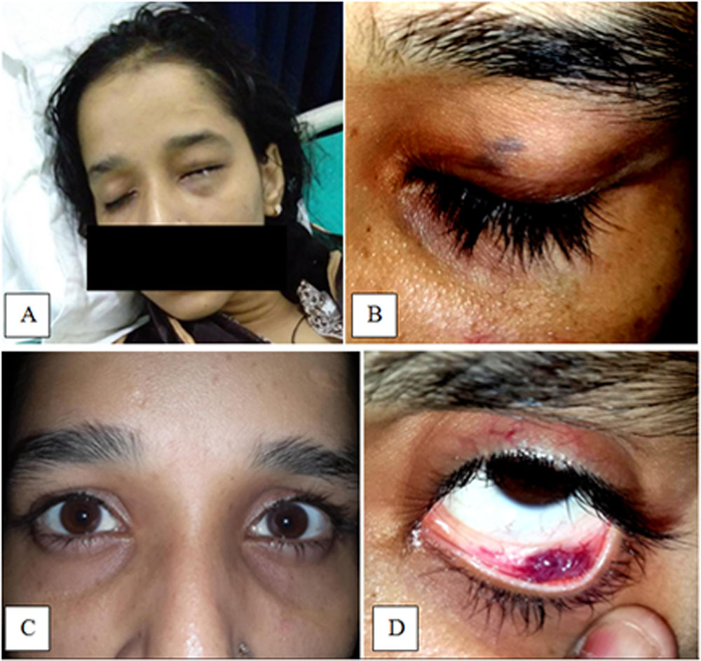
Figure 1: (A) Swelling of left eyelids noticed during morning hours, (B) 2mm x 3mm bluish swelling on left upper eyelid, (C) Normal Hirschberg test, (D) Left eye conjunctival lesion
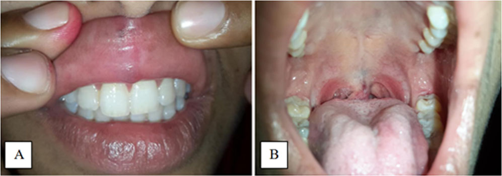
Figure 2. (A) red-blue discolouration of the oral mucosa beneath philtrum, (B) bluish streak on soft palate and congested uvula with bright red streaks
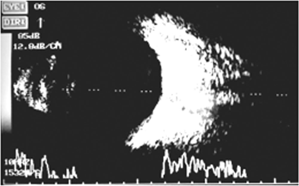
Figure 3: USG B-Scan of left eye with a 10MHz probe at 95dB gain, showing no vitreous echoes, normal position of retina and choroid, and normal orbital fat echoes
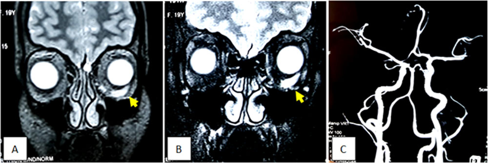
Figure 4. (A) Precontrast T2-weighted image showing lesion in left orbit, (B) Postcontrast T1-weighted image showing enhancement of the lesion with contrast, (C) MDCT-DSA showing thin arterial feeder to the lesion
Discussion:
The location, distribution and extent of vascular malformations are affected by local physiochemical or neural influences.2,3 Cavernous malformations in the orbital region occur most often in women (60-70%) between 18-72 years of age. They slowly enlarge, however, observed association of proptosis during puberty or pregnancy implies a role of hormone- or cytokine-mediated angiogenic factors.4-6 Our case was a young woman, whose lesion had enlarged during her adolescence, and distended with posture and provocative manoeuvres supporting the diagnosis of cavernous malformation. 2
Cavernous malformations often present with retrobulbar/periorbital pain, headache, optic nerve dysfunction, vision disturbances, hypermetropia, diplopia and gaze-evoked amaurosis. Signs may include proptosis, RAPD, colour and field defects, papilloedema, choroidal folds, duction deficits, and strabismus.7 Our case had unique presentation with eye pain, feeling of heaviness and uneasiness, and occasional vomiting and tremors, which has not been described in literature before.
Cavernous malformations usually occur in inferolateral and inferomedial aspects of the intraconal space, and are rarely intramuscular.2,7 USG Doppler shows venous waveform with low amplitude, slow rise and fall, along with feeding and draining vessels. Contrast-MRI delineates the lesion isointense to muscle on T1- and uniformly hyperintense on T2-weighted images. Early (35 seconds) and late films (>6 minutes) of MDCT-DSA make it possible to further delineate the lesion. Rootman et al. recommend arterial and Valsalva-augmented venous-phase CT-angiograms to evaluate distensible vascular lesions. To obtain a dynamic picture of filling and drainage with flow route, voids and differential perfusion, examination with contrast under fluoroscope is now being recommended.2,7,8
Usual indications for surgery are pain, cosmesis, increase in size, ptosis, optic nerve compression, amblyopia, haemorrhage following trauma, and acute thrombosis.3 Ethanol sclerotherapy may cause reduced infraorbital sensation and necrosis in the injected area; thus it is recommended only for superficial well-circumscribed lesions. Surgical debulking alone or as adjunct may cause ectropion of lower lid, iatrogenic strabismus or dystopia or reduction of visual acuity. CO2 laser resection is recommended for superficial diffuse and deep lesions. Deeper lesions require embolisation using coils or cyanoacrylate tissue glue.2,9,10 Head-raising alleviated all symptoms of our patient while awaiting surgery. She later underwent CO2 laser resection and was cured.
In summary, multidisciplinary approach with meticulous imaging is required in all cases of vascular malformations for optimum treatment outcomes.
References :
- Bhat V, Salins PC, Bhat V. Imaging Spectrum of Hemangioma and Vascular Malformations of the Head and Neck in Children and Adolescents. J Clin Imaging Sci. 2014;4:31.
- Osaki TH, Fay A, Waner M. Vascular malformations. In: Fay A, Dolman PJ, eds. Diseases and Disorders of the Orbit and Ocular Adnexa. Edinburgh: Elsevier; 2017. p. 454-61
- Lacey B, Rootman J, Marotta TR. Distensible Venous Malformations of the Orbit: Clinical and Hemodynamic Features and a New Technique of Management. Ophthalmology 1999;106:1197–209.
- Garzon MC, Huang JT, Enjolras O, Frieden IJ. Vascular malformations: Part I. J Am Acad Dermatol. 2007 Mar;56(3):353-70; quiz 371-374.
- Ansari SA, Mafee MF. Orbital cavernous hemangioma: role of imaging. Neuroimaging Clin N Am 2005;15:137–58.
- Zauberman H, Feinsod M. Orbital hemangioma growth during pregnancy. Acta Ophthalmol (Copenh) 1970;48:929–33.
- Rootman DB, Heran MKS, Rootman J, White VA, Leumsamran P, Yucel YH. Cavernous venous malformations of the orbit (so-called cavernous haemangioma): a comprehensive evaluation of their clinical, imaging and histologic nature Br J Ophthalmol 2014;98:880-8.
- Callahan BA, Meyers PM, Garrity JA, Son JH, Petris C, Kazim M, Low-Flow Arterialized Venous Malformations of the Orbit. Ophthal Plast Reconstr Surg 2017;33(4):256-60.
- Arat OY, Mawad ME, Boniuk M. Orbital Venous Malformations Current Multidisciplinary Treatment Approach. Arch Ophthalmol 2004 Aug; 122:1151-8.
- Bonolton LA, Chan K, Steiner F, FitzJohn T, Tan ST. Management of Orbital and Periorbital Venous Malformation. Frontiers J Surg 2017;4:27.
Acknowledgement: Dr Ajai Agrawal, Additional Professor, Department of Ophthalmology, All India Institute of Medical Sciences, Rishikesh for his valuable clinical inputs.
Leave a Comment