

Dr.Santanu Das, D20971, Kiran.Kumar L, Dr.Raghuraj S Hegde
ABSTRACT:
CSF leak during orbital exenteration is rare but can be a life threatening complication if not managed properly and prompt. A good anatomical knowledge as well as a thorough preoperative assessment is important to identify areas where CSF leak can be encountered. Reports of CSF leak during exenteration, incidence and management are few in literature. Management of such cases should be prompt. Various techniques like primary suturing of the dural defect with 6-0 nylon, placing an overlay graft, usage of local pericranial flaps, Calvarial grafts and fascia lata have been advocated to seal the leak intraoperatively. Use of bone wax, glue has also been used to seal small leaks. Postoperatively the patient can be treated conservatively with antibiotics, Acetazolamide and bed rest with head end elevation. If all these techniques fail then only we can go for surgery either endoscopically or intracranially. In this review article we aim highlight the pathophysiology, modes of preventing CSF leak intraoperatively and how to manage this complication as well as the newer techniques available to treat CSF rhinorrhoea after doing a thorough literature search in pubmed. We will also highlight our experience of treating 2 such cases of CSF leak we encountered during orbital exenteration in our hospital and how we managed them both intraoperatively as well as in the postoperative period.
Keywords- orbital exenteration, complication, management, CSF leak,
INTRODUCTION:
Orbital exenteration is a radical and disfiguring procedure which was first described by George Bartisch in 1583.[1,3,12] It is a rare process, Mohr et al reported 77 cases of orbital exenteration over a 20 year period whereas Rathbun et al reported 48 cases over 30 years.[1,27,28] It includes removal of all the orbital contents including globe, orbital fat, conjunctival sac and part or whole of the eyelids.[1] It is of 3 types:
- TOTAL: Involves removal of all the contents of the orbit with or without removal of the eyelids.[2,33]
- SUBTOTAL: Removal of the orbital contents are partial but with removal of the ocular bulb.[2,33]
- EXTENDED: When the adjoining bones and paranasal sinuses are also removed.[2,33]
This procedure is majorly reserved for potentially life threatening malignancies or relentlessly progressive conditions not responding to other modes of treatment.[1,2,3,29,33] Less often it can also aid in palliation of severe deformity, pain.[1,14] Bartley et al reported 100 out of their 102 cases needed exenteration because of malignant disease similar to Levin et al who reported 63 of their 69 cases needed exenteration owing to malignancies.[9,15]Modifications like eyelid sparing, retaining the conjunctiva and the periorbital tissue have been tried to aid in facial rehabilitation.[1]
CSF leak in general can be classified as traumatic or non-traumatic.[5] Traumatic leaks can be further sub classified as surgical and non-surgical.[5] CSF leak although a rare complication while doing orbital exenteration can be potentially life threatening as it can lead to meningitis, delayed cerebral abscess, seizures, CSF hypotension with position dependent headache, occult haemorrhage and sometimes even death.[5,6] Therefore oculoplastic surgeons should be competent in diagnosing CSF leak as early as possible. Since the orbital apex and skull base is involved while doing exenteration of aggressive lesions the risk of CSF leak increases as any damage creates a conduit between the paranasal sinuses and the intracranial space.[7,8] In literature, reported rate of CSF leak during exenteration ranges from 1.6%-16.7%.[5,7] The first CSF leak manifesting as rhinorrhoea was described by GALEN.[5] CSF leak manifesting as otorrhoea was first described by ESCAT in 1897.[5] How to repair CSF leak was first described by Dandy through an intracranial approach whereas an extracranial approach was suggested by Doldman in 1948.[5] Endoscopic repair was first introduced by Wigand in 1981.[5] Exenteration studies are scarce in literature as well as studied on complications like CSF rhinorrhoea during orbital exenteration, therefore this review aims at summarizing the current literature on incidence of CSF leak during orbital exenteration, pathogenesis, methods to avoid such complications and management of CSF leak. This review will also highlight our experience in handling 2 cases of CSF rhinorrhoea during orbital exenteration in our hospital.
INCIDENCE OF CSF LEAK DURING EXENTERATION:
CSF leak into the orbit is rare and the true incidence if difficult to determine.[5] The majority of leaks as stated in literature are due to accidental trauma followed by iatrogenic leak from surgical procedures.[5]CSF leak can also be a risk factor when operating lesion involving the orbital roof and posterior lateral wall.[6]The following table consists of incidence of CSF rhinorrhoea during orbital exenteration reported by various authors over the years and how they went about treating this complication.
TABLE-1
De Conciliis et al also found that dural exposure occurred in 20.5% of exenterations and 30.8% of enlarged exenterations which included removal of one or more orbital walls.[4,6]
PATHOPHYSIOLOGY OF CSF LEAKS: CSF leaks happen when there is a break in the normal anatomical barriers. For a CSF leak to occur there must be a defect in the dura, arachnoid and the bone so that there is a communication between two spaces. Secondly the pressure gradient in the subarachnoid space should be higher for the CSF to flow out.[5] Extended exenteration as it is a more extensive surgery is associated with more incidences of CSF leak as the dura will be exposed during this surgery.[4,5] Even CSF leaks have been reported secondary to application of monopolar cautery to the orbital roof. When there is communication between the subarchnoid space and atmosphere for example paranasal sinuses, a positive intracranial pressure (ICP) allows CSF to come out whereas in orbit the ICP must overcome the pressure of the orbital tissues for CSF to come out.[5] Septoplasty and FESS have high risks of CSF leak and the most common site being the ethmoid and cribriform plate because procedures in this area are associated with high risk of damaging the skull base as well as allowing intracranial communication with the sinuses.[5] Since the sinuses are at atmospheric pressure that is at 0 mm hg and any positive intracranial pressure will result in rhinorrhoea.[5]
Orbital decompression like orbital exenteration is associated with high risk of CSF leak as bones from high risk areas have to be removed like lamina papyracea and sphenoid trigone.[5,17]
CLINICAL FEATURES AND SEQUELAE OF CSF LEAK:
In reviewed literature the description about the timing of CSF leak in studies conducted by Bartley et al and Kuo et al were not available. Taking into consideration the other studies CSF leak during orbital exenteration occurred mainly during the intraoperative period (91.6%) while a few occurred in the immediate post operative day 1(8.3%).
CSF leaks can present in a variety of ways the most common being rhinorrhoea which can be clean or blood tinged.[5,13] The leak can be continuous or intermittent (reservoir sign) if the fluid accumulates in the sinuses and drains out through the nose when the head is in the dependent position. Fluid accumulation in the sinuses is associated with headache and once the fluid comes out the headache is relieved. If the cribriform plate is damaged there might be subsequent damage to the olfactory nerve producing hyposmia or anosmia.[5,6]When the fluid gets collected in the orbits patients might have proptosis eyelid swelling and diplopia which is known as orbitorrhoea which is not very common.[35]
Since there is a communication between the subarachnoid space and the external environment like the paranasal sinuses or external auditory canal any passage of pathogens through this pathway will give rise to symptoms of intracranial infection like fever, headache and other meningeal signs like neck rigidity and projectile vomiting. These are serious clinical features and one has to intervene promptly to prevent complications like seizures, cerebral abscess or even death. In cases of persistent leakage there might be CSF hypotension syndrome characterized by headache worse during standing than during lying down.[6]
OUR EXPERIENCE:
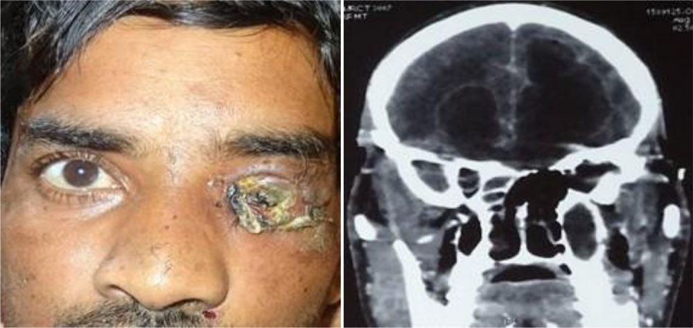
Case 1– A 35year old male came to our OPD with history of fall under influence of alcohol 1 month back with injury to left eye, followed by diminision of vision in the left eye for the same duration.
He also complained of nasal discharge since 15 days and fever associated with headache since 15 days. He was a known diabetic, alcoholic, and a smoker. On further examination he had nasal discharge, left sided lower motor neuron type of facial palsy and random blood sugar was 370mg/dl. In the right eye vision was 6/6 recorded using Snellens chart and the anterior and posterior segments were within normal limits on slit lamp examination. In the left eye there was no perception of light, blackish discolouration of eyelids with gangrenous and disorganised globe consisting of foul smelling and purulent discharge. The posterior segment could not be visualized.
CT scan of orbits and brain 1mm cuts bone windows and soft tissue windows coronal axial and saggital views was done, pus culture and sensitivity showed the growth of E.coli. Routine blood investigations were done and medical management was started with broad spectrum s systemic antibiotics and systemic antifungals with subcutaneous insulin. CT SCAN of orbit and brain shows hypodense areas suggestive of either a brain abscess or a fungal granuloma in the bilateral frontal region of brain. Patient was drowsy with altered sensorium and electrolyte imbalance. A diagnosis of left rhino orbito cerebral mucormycosis was done and the patient was taken up for left sided orbital exenteration with craniotomy to evacuate the accumulated pus after taking consent under general anaesthesia.
FIGURE-1A,B
During Left sided orbital exenteration under general anaesthesia, intraoperative CSF leak was observed from the optic nerve stump and was sealed with periorbital galeal flap.
Necrotic mass from sphenoid maxillary and ethmoid sinuses were removed and sent for histopathological examination which came out to be mucormycosis. During the post operative period there were no more episodes of CSF leak and we did regular post operative dressing with normal saline, betadine solution and with Amphotericin soaked gauze piece.
CASE 2– A 86 year old male presented to our ophthalmology OPD with a swelling in the lower eyelid of the left eye since 3 years insidious in onset and gradually progressive not associated with pain, discharge or bleeding, but associated with slight diminution of vision in the same eye. He also gave history of carcinoma rectum for which he underwent surgery 5 years back but didn’t take postoperative radiotherapy or chemotherapy.
There was a non tender, firm to hard mass measuring approximately 4*4 cm over the left lower eyelid with a granular appearance on inspection confirmed by palpation. It was not freely mobile and was adhered to the underlying structures. Skin over the swelling was normal with no local rise of temperature.
Vision in right eye was 6/24 recorded using Snellens chart .His eyelids and adnexa were normal. He had senile immature cataract with glaucomatous disc changes in the posterior segment. His left eye was pseudophakic with a visual acuity of 6/12 recorded using Snellens Chart. He had a firm to hard granular mass over the lower eyelid and glaucomatous disc changes in his retina.
FIGURE – 2A,B,C,D
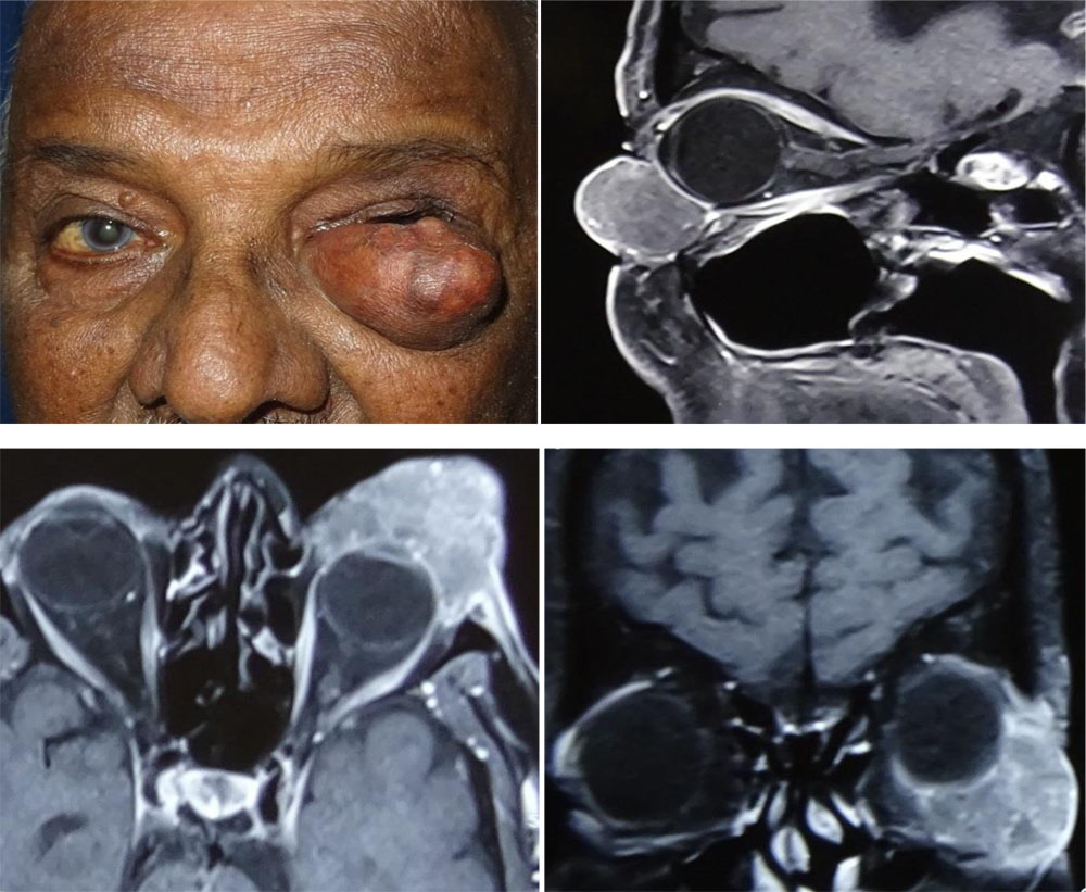
Diagnostic biopsy was done which revealed sebaceous cell carcinoma. MRI was done to find out any intraorbital spread. Since HPE reports revealed the lesion to be sebaceous cell carcinoma and MRI showing orbital spread a lid sparing orbital exenteration was planned inspite of the left eye having visual potential with the consent of the patient and his family. The visual potential in the left eye outweighed the life risk it possessed. Left sided lid sparing orbital exenteration was done. While closing CSF leak was noted, from the stump of the optic nerve. Fascia lata was harvested from the right thigh and the leak was sealed by placing the fascia lata graft over the leak.
FIGURE-3A,B,C,D
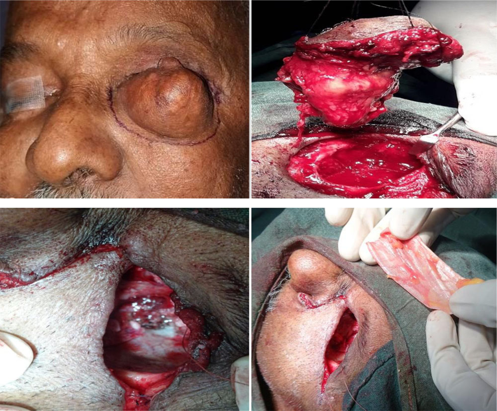
Following surgery in the immediate post op period he was adviced complete bed rest and intravenous broad spectrum systemic antibiotics were administered along with regular dressing of the wound. There were no episodes of CSF rhinorrhoea in the immediate post operative period and he was discharged on the 4th post operative day .When he came for follow after 3 days of discharge he complained of severe headache with CSF rhinorrhoea.
He was readmitted and was treated with systemic antibiotics like Ceftriaxone, Amikacin and Metronidazole and was adviced complete bed rest with head end elevation. He was also started on Acetazolamide tablet 250mg thrice daily along with IV fluids with ringer lactate. Lumbar puncture was done which turned out to be normal to rule out any meningitis. After one week of complete bed rest his headache reduced and there was no CSF rhinorrhoea. He was discharged and was adviced complete bed rest for atleast another 2 weeks. On follow up he didn’t complain of any CSF rhinorrhoea and headache.
DIAGNOSIS OF CSF LEAK:
In practice CSF leaks are generally detected intraoperatively or soon after the surgery is done when leakage of clear fluid is seen.CSF differs from blood when it is placed on a filter paper and produces a halo.[5,6] Measuring glucose content is another method but again it is unreliable because glucose in CSF cannot be distinguished from blood, tears or nasal secretion.[5,6] Beta-2 transferrin assay is a marker protein both sensitive and specific to CSF and can also be done quickly.[5,6] It is the test of choice in almost all the institutions. Majority of the leaks can be localised using non contrast high resolution CT as depicted in the study by Schwaber WI et al.[6,27,44] If not localized then contrast enhanced ct or mri-cisternography can be used.[6,28]
METHODS TO AVOID CSF LEAK DURING SURGERY:
Thorough knowledge of the anatomy and an equally extensive preoperative assessment by looking at CT-Scan or MRI images is essential before embarking on the surgery. Injudicious use of monopolar cautery in areas where the bones are thin may lead to CSF leak and the operating surgeon should be careful while using cautery especially when a lesion is involving the roof.[4,10] Secondly good visualization is also important during surgery especially when the bony anatomy has been distorted by lesions like Osteomas and fibrous dysplasia.[25] During orbital decompression balanced orbital decompression is the preferred technique where the greater wing of sphenoid is removed by mechanical drill as suggested by Graham SM et al and not by high speed cutting burr and the medial orbital wall is removed by an external transcaruncular incision.[18,35] Also there have suggestions by Limawararut et al that the surgeon can use diamond burr when operating near the deep posterior lateral wall as it is less likely to involve the soft tissues.[6] The size of the sphenoid trigone also varies from person to person therefore not only preoperative visualization by CT- scan imaging but also intraoperative image guidance is important to avoid inadvertent damage as suggested by Goldberg et al, Selva D et al and Lefebvre DR et al.[19,20,26,37] Another site where CSF leaks can occur is in the medial wall through the fovea ethmoidalis. Leone suggested that the operating surgeon should use the ethmoidal vessels as a landmark and should not remove the bone beyond these vessels to avoid entering into the anterior cranial fossa.[21] Limawararut et al reported once case of CSF leak while performing anterior orbitotomy for an orbital dermoid involving the roof where they used monopolar cautery to destroy the dermoid lining which they failed to reach with a curettage.[6] So surgeons should avoid overcautery in high risk areas. Good visualization is important during any surgery and the use of a 30-70 degree endoscope may improve visualization in areas like the orbital roof as suggested by Selva D et al. [25]
Selva D, Chen C even suggested the use of stereotactic assisted approach in cases where the normal bony anatomy has been distorted.[26] These are some of the ways by which an operating surgeon can prevent CSF leak during orbital exenteration and other orbital surgeries.
MANAGEMENT:
INTRAOPERATIVE MANAGEMENT:
Reconstruction of the anterior skull base is technically challenging as there is very little tissue available to cover the defects. Regional flaps have been found to be a good option for dural reconstruction, like the pericranial and the galeal flaps as they are easily available and can also be harvested easily. [8] If there is adequate exposure of the dural laceration and can be readily apposed primary repair with 5-0 or 6-0 nylon should be done and care must be taken that it is watertight. Many neurosurgeons also suggest placing an onlay graft mainly harvested from a layer of fat, Temporalis muscle or fascia over the exposed dura to ensure water tightness. [6]
Autologous grafts include: fascia lata, osteomucoperiosteal flaps, mucous membrane and lyophilized heterologous dura. Mucous membranes are associated with high rates of infection whereas although dehydrated lyophilized heterologous dura have been used for decades but their usage is now discouraged because there is risk of Prions disease and other acquired diseases as stated by Limawararut et al in their review article.[4,6]
In cases where dural tear is large or there is limited exposure and primary apposition is difficult onlay grafts are the best choices which are sutured to the surrounding dura. Materials used are autologous fat, mucous membrane, fascia lata, Temporalis fascia and muscle, cartilage from the nasal septum or ear and osteomucoperiosteal flaps.[6,11] specifically in cases of orbital exenteration packing material can be used to hold the fascia against the orbital apex.
Post radiation therapy Galea and Temporalis muscle will be thinned out so their use for reconstruction can be problematic. In such cases Calvarial bone graft can be used. Other uses of biological materials like Calvarial bone graft or a posterior frontal sinus wall graft are reconstruction when the tumour involves the nasal bone or fronto-orbital segment. [8] In the study conducted by Ziv Gill et al they used biological materials and 3D titanium mesh when reconstruction of the medial orbital wall is required whereas they used septal cartilage for reconstruction of small inferior wall defects. For reconstructing defects due to radical maxillectomy the author used lateral thigh free flap or a rectus abdominis free lap to cover such large defects. [8]
Various studies and authors have found that the use of fascia lata is very advantageous as its flexibility allows large defects to be covered including defects of the orbit and paranasal sinuses. Since it is thin and has low mass properties large dural defects can be covered with a single fascia lata. [16] We in our case successfully used fascia lata to stop CSF leak following orbital exenteration which is similar to what Ziv Gill et al did in their study where they used double layer fascia as the standard material for all of their anterior skull base reconstruction. They also used fascia lata for reconstructing large cranial base defects as well used it in patients on radiotherapy where pericranium will be substantially thinned out and raising a flap is difficult. Other advantages of using fascia lata are that not very high technical qualifications are needed like muscle transfer and the procedure is also not time consuming.[16] The authors also conducted a histopathological examination which showed that the graft is uniformly covered by fibrous tissue with invasion of blood vessels. Intraoperatively careful and judicious use of cautery is another way of preventing such complications. [8]
A lot of other materials like tissue adhesives and alloplastic materials have been used in cases of CSF leak. Tissue adhesives like Tisseel fibrin have been used to seal small defects with success. It consists of human fibrinogen. A solid fibrin matrix is created within 5 minutes.[6,19] Cyanoacrylate glue can also be used in closure of CSF leaks as stated by Limawararut et al in their review article.[6,11]
Bone wax can also be used in sealing small defects however Bolger et al reported 3 cases of CSF leak associated with the use of bone wax. They said the reason for this is that it prevents fibrin deposition, spontaneous healing and closure. [6,22] Also there have been reported cases of granuloma with the use of bone wax.[6,23] Other materials that have been successfully used in closing small defects causing CSF leak are Gelfoam, Vicryl mesh and Hydroxiapatite cement that is bone cement.
Recently dural adhesives like Dura seal and Bio glue has been used with success. [38, 44] Dura seal consists of water soluble amine and polyethylene glycol. Bio Glue consists of bovine albumin and glutaraldehyde which produces a mechanical seal within 20 minutes.[38,44] In a case report by Yuen T et al bio glue was seen to persist at the surgical site even 2 years post op.[24]
In the study conducted by Limawararut et al CSF leak during orbital exenteration the optic nerve stump was clamped and covered with gelfoam. Four cases which had CSF leak during orbital decompression 2 were repaired using Temporalis muscle graft and sealed with either fibrin glue or Cyanoacrylate glue. One case was sealed with orbital fat only and one with bone wax. The author also says that during one of the Orbitotomies a 10 mm dural defect was closed with 6-0 nylon followed by an onlay graft was placed and further sealed with Tisseel. [6]
POSTOPERATIVE MANAGEMENT:
Following intraoperative repair of CSF leak it is very important to monitor the patient for continued leakage in the postoperative period as persistent CSF leak in the postoperative period will lead to something known as CSF hypotension syndrome characterized by postural headache, neck tenderness, nausea, vomiting and sometimes blurring of vision and 6th nerve palsy.[6,40,41] CT-scan is needed to rule out intracranial haemorrhage.[6]
Postoperatively the patient should be adviced complete bed rest for atleast 2 weeks with head end elevation at 15-30 degrees so that the venous pressure decreases for the CSF rhinorrhoea to completely subside because with daily activities symptoms might increase. The patient should be instructed to avoid sneezing, coughing and should be administered stool softeners.[6,32] Prophylactic broad spectrum intravenous antibiotics should be started although their use remains controversial. They are thought to decrease the rates of meningitis [32] but Yilmazlar et al reported that antibiotics didn’t affect the incidence of meningitis in such cases.[23] Kerr JT et al reported many surgeons avoid antibiotics to reduce development of resistant organisms.[39] If it doesn’t subside inspite of complete bed rest a spinal catheter can be used to drain 5 to 10 ml CSF per hour. Excessive drainage should be avoided as it might lead to tension pneumocranium and rarely subdural or epidural haematoma.[42] Acetazolamide has been shown to reduce CSF production and is regularly used to treat CSF leaks but one should be careful because it can cause hypokalemia. So regular monitoring of serum electrolytes along with ECG is needed for patients on Acetazolamide.
When all these measures fail, at that time only a surgical intervention is advocated. Endonasal endoscopic repair of leaks at the cribriform plate may be considered [34,39] but persistent or large leaks inaccessible endonasally need a craniotomy and repair.
In both of our cases intraoperative sealing of the leakage with a galeal flap and fascia lata respectively and postoperative bed rest, head end elevation , systemic antibiotics were enough to resolve CSF rhinorrhoea and without any further complications.
CONCLUSION: CSF rhinorrhoea during orbital exenteration is a rare but potentially life threatening complication as it might lead to meningitis, tension pneumocephalus, brain herniation, seizures, cerebral abscess and even death. Once encountered with CSF leak during surgery prompt and immediate covering up the leak with fascia lata or by using regional flaps, alloplastic grafts and glue are useful ways of stopping CSF leak intraoperatively. Postoperatively if there is persistent leak then complete bed rest with head end elevation, avoiding straining, broad spectrum antibiotics along with tablet Acetazolamide administration can completely cure CSF rhinorrhoea without any complications in most of the cases following orbital exenteration or any other orbital surgery.
REFERENCES:
- Orbital Exenteration : A 13 year Manchester experience, I Rahman, AE Cook, B Leatherbarrow, Br J Ophthalmol 2005;89:1335-1340.
- Gabriel de Almeida Ferreira,Natalia Mussi Roberta, Lilian Fernandes de Sousa, Meneghim José Vicente, Tagliarini Mariângela Esther, Alencar Marques, Silvana Artioli Schellini. Orbital Exenteration: a series of cases. Bras.oftalmol. 2016; 75(6):452-5.
- Aryasit et al. BMC Ophthalmology. (2018); 18:186.
- Incidence and treatment of dural exposure and CSF leak during orbital exenteration, Carlo de Conciliis, Giulio Bonavolonta, Ophthalmic Plastic and Reconstructive Surgery 3(2):61-64,1987.
- A. Massoud et al. Cerebrospinal Fluid Leak as a Complication of Oculoplastic Surgery, Seminars in Ophthalmology, 29:5-6, 440-449.
- Limawararut, Vanessa et al. Cerebrospinal Fluid Leaks in Orbital and Lacrimal Surgery. Survey of Ophthalmology.2008; Volume 53: 274 – 284.
- Gill et al, Postoperative Cerebrospinal Fluid Leak After Microvascular Reconstruction of Craniofacial Defects with Orbital Exenteration, Laryngoscope, 127:835–841, 2017.
- Ziv Gil, M.D., Ph.D., Avraham Abergel, M.D., Leonor Leider-Trejo, M.D., Avi Khafif, M.D., Nevo Margalit, M.D., Aharon Amir, M.D., Eyal Gur, M.D., Dan M. Fliss, M.D. A Comprehensive Algorithm for Anterior Skull Base Reconstruction after Oncological Resections, skull base: an interdisciplinary approach. 2007; volume 17:
- Levin PS, Dutton JJ. A 20 year series of orbital exenteration. Am J Ophthalmol 1991; 112:496–501.
- Wulc AE, Adams JL, Dryden RM. Cerebrospinal fluid leakage complicating orbital exenteration. Arch Ophthalmol.1989; 107:827-30.
- Tse DT, Panje WR, Anderson RL. Cyanoacrylate adhesive used to stop CSF leaks during orbital surgery. Arch Ophthalmol.1984; 102:1337-9.
- Bartisch G. Ophthalmodoulcia. Dresden, 1583; 3:208.
- Badilla J, Dolman P. Cerebrospinal fluid leak complicating orbital or Oculoplasty surgery. Arch Ophthalmol. 2007; 125:1631-1634.
- Rose GE, Wright JE. Exenteration for benign orbital disease. Br J Ophthalmol.1994; 78:14–18.
- Bartley GB, Garrity JA, Waller RR, et al. Orbital exenteration at the Mayo Clinic 1967–1986. Ophthalmology, 1989; 96; 468–74.
- Neligan PC, Mulholland S, Irish J, et al. Flap selection in cranial base reconstruction. Plast Reconstr Surg 1996; 98:1159–1166.
- Graham SM, Brown CL, Carter KD, et al. Medial and lateral orbital wall surgery for balanced decompression in thyroid eye disease. Laryngoscope.2003; 113:1206-1209.
- Goldberg RA, Weinberg DA, Shorr N et al. Maximal three wall orbital decompression through a coronal approach. Ophthal Surg Lasers.1997; 28: 832-843.
- Lefebvre DR, Yoon MK. CT based measurements of the sphenoid trigone in different gender and race. ASOPRS. 2013; New Orleans, LA.
- Leone CR, Piest KL, Newman RJ. Medial and lateral wall decompression for thyroid ophthalmopathy. Am J Ophthalmol.1989; 108:160-166.
- Bolger WE, Tadros M, Ellenbogen RG et al. Endoscopic management of cerebrospinal leak associated with the use of bone wax in skull base surgery. Otolaryngol Head Neck Surg. 2005; 132:418-420.
- Katz SE, Rootman J. Adverse effects of bone wax in surgery of the orbit. Ophthal Plast Reconstr Surg.1996; 12:121-6.
- Yilmazlar S, Arslan E, Kocaeli H, et al. Cerebrospinal fluid leakage complicating skull base fractures: analysis of 81 cases. Neurosurg Rev.2006; 29:64-71.
- Yuen T, Kaye AH. Persistence of Bio glue in spinal dural repair. J Clinic Neurosci.2005; 12:100-1.
- Selva D, Lai T, Krishnan S. Orbit frontal cholesterol granuloma: Percutaneous endoscopic assisted- curettage. J Laryngol Otol.2003; 117:892-4.
- Selva D, Chen C, Wormald PJ. Frontoethmoidal osteoma: a stereotactic-assisted sino-orbital approach. Ophthal Plast Reconstr Surg.2003; 19: 237-8.
- Schwaber MK, Netterville JL, Coniglio JU. Complications of skull base surgery. Ear Nose Throat J.1991; 70:648-60.
- Flynn BM, Butler SP, Quinn RJ, et al. Radionuclide cisternography in the diagnosis and management of cerebrospinal fluid leaks: the test of choice. Med J Aust.1987; 146:82-84
- Naquin HA. Exenteration of the orbit. Arch Ophthalmol 1954; 51:850–62.
- Rathbun JE, Beard C, Quickert MH. Evaluation of 48 cases of orbital exenteration. Am J Ophthalmol 1971; 72:191–9.
- Mohr C, Esser J. Orbital exenteration: surgical and reconstructive strategies. Graefes Arch Clin Exp Ophthalmol 1997; 235:288–95.
- Youmans JR. Neurological survey: a comprehensive reference guide to the diagnosis and management of neurosurgical problems. Philadelphia, PA, WB Saunders, 1973, 981-82.
- Hsiu-Fen Lin, MD; Chun-Chung Lui1, MD; Huan-Chen Hsu, MD; Sue-Ann Lin, MD. Orbital Exenteration for Secondary Orbital Tumors: A Series of Seven Cases. Chang Gung Med J. 2002; 25:599-605.
- Schievink WI, Vittorio MM, Atkinson JLD, et al. Surgical treatment of spontaneous spinal cerebrospinal fluid leaks and intracranial hypotension. J Neurosurg. 1998; 88:243-6.
- McCord CD. Current trends in orbital decompression. 1985; 92: 21-33.
- Ide CH, Webb RW. Penetrating transorbital injury with cerebrospinal orbitorrhoea. Am J Ophthalmol.1971; 71:1037-9.
- Goldberg RA, Weinberg DA, Shorr N et al. Orbital Exenteration: results of an individualized approach. Ophthal Plast Reconstr Surg.2003; 19:229-36.
- Kumar A, Maartens NF, Kaye AH. Evaluation of the use of Bio glue in neurosurgical procedures. J Clinic Neurosci.2003; 10:661-4.
- Kerr JT, Chu FW, Bayles SW. Cerebrospinal fluid rhinorrhoea: diagnosis and management. Otolaryngol Clin North Am. 2005; 38:597-611.
- Schievink WI, Meyer FB, Atkinson JL, et al. Spontaneous spinal cerebrospinal fluid leaks and intracranial hypotension. J Neurosurg. 1998; 88:243-6.
- Schievink WI, Maya MM, Riedinger M. Recurrent spontaneous spinal cerebrospinal fluid leaks and intracranial hypotension: a prospective study. J Neurosurg.1996; 84:598-605.
- Buchanan RJ, Brant A, Marshall LF. Traumatic cerebrospinal fluid fistulas, in Winn HR(ed): Youmans Neurological Surgery. Philadelphia, PA, WB Saunders, 2004, p 5629.
- Grotenhuis JA. Costs of postoperative cerebrospinal fluid leakage: 1 year retrospective analysis of 412 consecutive nontrauma cases. Surg Neurol. 2005; 64:490-3.
- Mostafa BE, Khafagi A. Combined HRCT and MRI in the detection of CSF rhinorrhoea. Skull Base 2004; 14:157–62.
Author |
Incidence/reported cases | treatment | outcome | ||
| De Conciliis[4] | 5/39(12.8%) | Dural repair with Temporalis muscle fascia or lyophilized heterologous dura + packing with autogenous fat or Temporalis muscle. | 1 death due to pulmonary complications. | ||
| Bartley[14] | 3/100(3%) | Conservative management | Spontaneous resolution | ||
| Wulc[10] | 3/18(16.7%) | Enlarged bone defect+ primary closure+ gelfoam soaked in thrombin
Enlarged bone defect+ primary closure +drain |
Uncomplicated recovery | ||
| Rahman[1] | 1/64(1.6%) | Local pericranial flap | Uncomplicated recovery | ||
| Limawararut[6] | 1/154(0.6%) | Gelfoam , free flap | Uncomplicated recovery. | ||
| Kuo et al | 6 reported cases | Spontaneous closure | Uncomplicated recovery | ||
| Tse et al[11] | 1case reported during extended exenteration | Surgicel+butyl-2-cyanoacrylate | cerebritis |
Figure-1: (A) left sided mucormycosis; (B) CT scan of brain showing hypodense areas suggestive of either an abscess or fungal granuloma in bilateral frontal lobe region.
Figure-2: (A) left sided lower eyelid sebaceous cell carcinoma; (B), (C), (D) saggital, axial and coronal view showing the size of the tumour with intraorbital spread.
Figure-3: (A) Patient being taken up for left sided lid sparing orbital exenteration; (B) Lid sparing exenteration done and the specimen is sent for histopathological evaluation; (C) during closure csf leak noted from the optic nerve stump; (D) fascia lata harvested from the right thigh being used to seal the leak.
Leave a Comment