

Dr.Shailja Mishra, S21602, Dr.Sukriti Ahuja, Dr.Apjit Kaur
ABSTRACT:
Aim- To analyse and study outcome in 18 cases of Benign cystic orbital lesions treated with foam sclerotherapy.
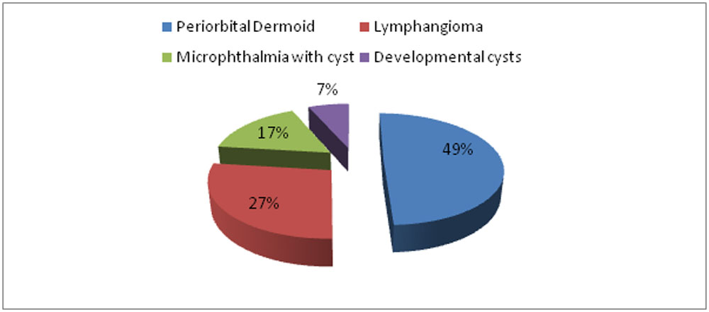
Method-Retrospective study of 18 Benign cystic orbital lesions like Periorbital Dermoids (9), Lymphangioma(5), Microphthalmia with cyst(3)and Developmental cysts(2) who underwent foam sclerotherapy using Sodium Tetradecyl Sulphate (STS). Tessari method was used for preparation of foam. Foam sclerotherapy with STS (30 mg/ml) was injected in a proportion of 10% of total aspirate volume. Patients were followed up on post op Day 1, at 1 month and 3 months. Treatment outcome was assessed on basis of resolution of symptoms and reduction in size of swelling.
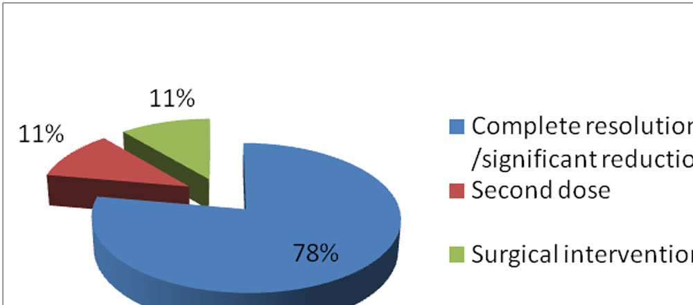
Result- Out of 18 patients ,14 patients had complete resolution of the lesions within 1-3 months . 2 patients showed incomplete response and needed second dose while other 2 needed surgical Intervention.
Conclusion- Foam sclerotherapy gives satisfactory functional and cosmetic outcome in Benign cystic orbital lesions.
KEYWORDS:Foam sclerotherapy, Sodium tetradecylsulphate
INTRODUCTION : Sclerotherapy has been an established modality in the treatment of visceral as well as head and neck cysts. It involves injection of a sclerosing agent intralesional /into an abnormal vasculature resulting in its shrinkage (1,2) . Sclerotherapy is based on the mechanism of endothelial injury
causing inflammation and thrombosis leading to collapse of the abnormal cavity or vessel(1,3).Foaming is a method where the sclerosant is injected as a foam in order to increase its surface area based on the principle of physics (Work done=surface tension x increase in surface area ) . Sodium tetradecyl sulphate
(an anionic surfactant ) is a widely used, safe and effective agent(3,6) .Sclerotherapy can be performed either by direct percutaneous or USG guided technique( 3) . The authors present their work on the extended use of Foam sclerotherapy for treatment of congenital cysts (superficial periorbital dermoid cyst (commonest)(4), microphthalmos/ anophthalmos with cysts and AV malformations) as well as acquired cysts ( orbital Lymphangiomas)(5)
MATERIALS & METHODS
This was a Retrospective study in Tertiary care centre in a study duration of 3 years (2015-2018). 18 patients were included in the study with age group in range of 3 months -50 years
Informed consent taken about the procedure , expected complications , repeating of the injections and other possible treatment modalities Pre and post operative clinical assessment as well as the procedure performed by the single surgeon
Foam preparation & injection
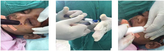
- Under aseptic precautions ,contents of the cyst aspirated through a stab incision with 18G needle
- The Tessari method was used for preparation of foam. STS (30 mg/ml) was injected in a proportion of 10% of the total aspirate volume.
- The affected was bandaged for 1 day and evaluated next day for local side effects (necrosis )or systemic complications
- Antibiotics cover(Amoxicillin ) was given for 5 days
- Follow up was done at 1 and 3 months for cosmetic /functional recovery
RESULTS
18 patients in range of Age =3 months- 50 yrs with ratio of males : females (10:8)
CLINICO RADIOLOGICAL PATIENT DISTRIBUTION OF THE STUDY
Table 1
FINAL OUTCOME ON 3 MONTHLY FOLLOW UP
Table 2
No immediate / late post operative complications noted.
REPRESENTATIVE PHOTO OF THE CASES
CASENO.1
CASE NO.2
CASE NO.3
DISCUSSION
Foam sclerotherapy is a safe and effective alternative to the surgical procedure . In this study we found among 18 patients ,9 ( 78 %)had significant improvement in terms of cosmetic and functional gain after a single dose of injection while 2 patients(11%) needed a second dose and 2 needed surgical intervention(11%) on 3 monthly follow up. The young individuals presented mainly for cosmesis rather than functional disability .Among the patients undergoing surgery one of them had significant reduction at 1 month follow up ,but had increase in the size of the swelling following trauma ,hence the patient was taken for surgery. The other patient had improvement but needed complete resolution of swelling for cosmesis.
Hence , foam sclerotherapy with STS led to the improvement in the quality of life without complications.
CONCLUSION
Foam sclerotherapy either direct percutaneous /USG guided is an effective and easy to perform procedure for orbital benign cystic lesions with satisfactory outcome.
REFERENCES
1. Naik MN, Batra J, Nair AG, Ali MJ, Kaliki S, Mishra DK. Foam sclerotherapy forperiorbital dermoid cysts. Ophthalmic Plast Reconstr Surg. 2014May-Jun;30(3):267-70.
2. Apjit Kaur Chhabra , Sonal Bangwal, Nibha Mishra. A Study on Cyst Volume Dependent Dose of 3% Sodium Tetradecyl Sulphate in Sclerotherapy of Orbitopalpebral Cyst Associated with Severe Microphthalmos/Anophthalmos.Open Science Journal of Clinical Medicine 2015; 3(5): 173-176
3. Smriti Nagpal , Ruchi Goel , Sushil Kumar, Sonam Garg.Sclerosing Agents in Ophthalmology.Del J Ophthalmol 2012;23(3):219-224.
4. Günalp I, Gündüz K. Cystic lesions of the orbit. Int Ophthalmol.1996-1997;20(5):273-7
5. Wojno TH. Sotradecol (sodium tetradecyl sulfate) injection of orbital lymphangioma. Ophthalmic Plast Reconstr Surg. 1999 Nov;15(6):432-7
6.Berenguer B, Burrows PE, Zurakowski D, Mulliken JB. Sclerotherapy of craniofacial venous malformations: complications and results. Plast Reconstr Surg. 1999 Jul;104(1):1-11
Table 1
LEGENDS TO FIGURES:
FOAM PREPARATION AND INJECTION
Fig 1 : Aspiration of content of the cyst/lesion
Fig 2 : Foam preparation from STS
Fig 3: Foam injection
Case 1-
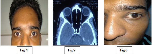
Fig 4 : Pre op image of Superomedial orbital mass (L/S) cause ? Dermoid
Fig 5 : CT Scan image at the time of presentation
Fig 6 : Post op image
Case 2-
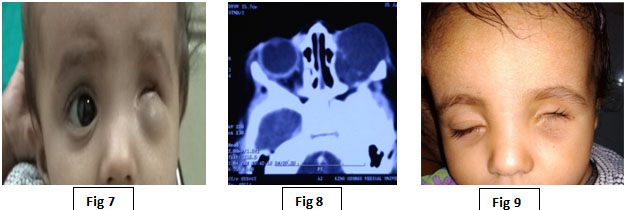
Fig 7 : Pre op image of Microphthalmos with orbitopalpebral cyst(L/S)
Fig 8 : CT Scan image at the time of presentation
Fig 9 : Post op image
Case 3-
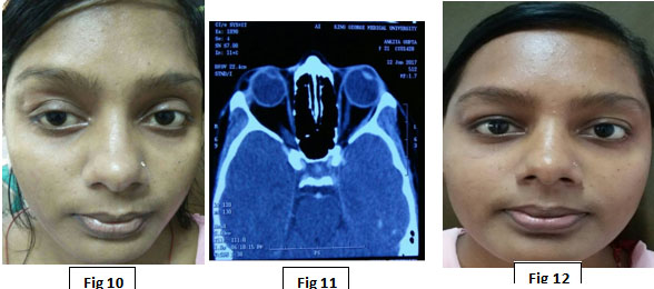
Fig 10 : Pre op image of Left superior orbital mass cause ? dermoid cyst
Fig 11 : CT Scan image at the time of presentation
Fig 12 : Post op image
Leave a Comment