

Dr. Sourav Damodaran, D18998, Dr.Gaurav Garg, Dr.Shreyas Temkar, Dr.Mandeep Singh Bajaj
Introduction
Retinoblastoma (RB) is the most common intraocular malignancy in children [1,2]. Recent advances in management has led to improved survival of children affected with RB with a 5-year cumulative survival rate of greater than 90% [3,4]. As the rate of globe salvage in retinoblastoma cases has increased, the focus has shifted towards tumour monitoring using various imaging modalities [5].Various authors have described the utility of optical coherence tomography (OCT) in the management of different pediatric vitreo-retinal disorders. Hand held spectral-domain OCT (SD-OCT) has been shown to be an useful tool in conditions like shaken baby syndrome, retinopathy of prematurity and ocular albinism [6-8]. OCT is also being increasingly utilised in imaging patients with RB to look for tumour characteristics, monitor therapeutic response, identify recurrence and to look for ophthalmoscopically invisible lesions [9-12]. Majority of these studies have been done using hand held SD-OCT under general anaesthesia. However, SD-OCT has limitations and is not useful for imaging large lesions due to limited depth of penetration [13]. The development of SS-OCT has resulted in rapid image acquisition and improved axial resolution (to the order of 1μm). SS-OCT is an extremely useful tool for imaging choroid and it can even be used for imaging in cases with hazy media. In this study we assess the utility of SS-OCT in cases of RB and describe the OCT patterns of regressed macular RB.
Method
This was a cross-sectional observational study conducted at the Ocular Oncology and Retina Services of Dr Rajendra Prasad Centre for Ophthalmic Sciences, All India Institute of Medical Sciences, New Delhi. The study adhered to the tenets of Declaration of Helsinki. Institute ethics committee clearance was taken prior to the beginning of study. The definition of macular retinoblastoma was based on the definition given by Shields et al [14]. It was defined as tumor, any part of which is within 2DD (3mm) of the centre of macula. Regression was based on the clinical appearance of the tumor after successful response to various modalities of therapy. Only tumours of macular region were taken to facilitate ease of scanning and to avoid artifacts. Children with regressed macular retinoblastoma in one or both eyes (but presence of good fixation in either of the eyes) were included in the study. Children who were not able to cooperate for the procedure were excluded. Detailed consent was obtained as per the institute protocol. Demographic data like current age, age at diagnosis, gender and laterality were noted. Previous treatment history was also noted with respect to the modalities of treatment (chemotherapy and local therapy) and time to regression. The clinical regression patterns were classified as type 0 (no visible residua), type 1 (fully or almost fully calcific residua), type 2 (fleshy tissue with little or no calcification), type 3 (mixed calcific and fleshy), and type 4 (atrophic chorioretinal flat scar) [15]. Fundus photography was done using Retcam 3 (Clarity medical systems, USA) during examination under anaesthesia or on regular fundus cameras (Triton DRI- OCT-fundus camera). Swept source OCT was done on Triton DRI-OCT (Topcon Corporation, Tokyo, Japan). Children were explained regarding the procedure and their need for proper fixation during the whole scanning process. OCT protocol included radial scans centered on the lesions (6mm or 12mm, 12 scans, 1 clock hour apart). Only high resolution scans with good signal strength were selected. Successful OCT scans could be obtained in 13 patients and were included in the analysis.
Results
Demographics
13 patients who met the above inclusion criteria were recruited in the study. Mean age of the tumour diagnosis was 3.16 ± 1.40 years and mean age at inclusion into the study was 6.15 ± 1.95 years. Of the total 13 children recruited, 8 cases (58%) were males and 5 cases (42%) were females. Most common presentation of the tumour was leucocoria (9 cases) followed by strabismus (4 cases).
Tumour Characteristics
Out of the 13 cases, 7 (53%) had bilateral tumours and 6 (47%) had unilateral tumours. Of the bilateral cases, 3 cases had regressed extramacular lesions in the fellow eye and 4 eyes had been enucleated for advanced RB in the course of treatment. Among the 13 eyes of regressed macular RB studied, 7 cases (53%) were group B tumours, 4 cases (30%) were group C tumours and 2 cases (17%) were group D tumours. Tumour involvement of the fovea was seen in 7 eyes. All cases had received triple regimen chemotherapy (Vincristine, Etopside, Carboplatin). No other modality of treatment was used for these macular lesions. Regression pattern of fovea involving tumours was type 1 (4 eyes), type 3 (2 eyes) and type 2 (1 eye). Regression pattern in non-fovea involving tumours was type 2 (2 eyes), type 3 (2 eyes) and type 4 (2 eyes).
OCT characteristics of tumour
OCT findings of regressed macular retinoblastoma are summarised in table 1. Tumour height was not assessed as the posterior extent of tumor could not be clearly visualised in most of the cases either due to shadowing effect or scar formation. Compared to the surrounding normal retina, the regressed tumour was isodense to hyperdense in all the cases. There was no evidence of neurosensory detachment in any cases. Location of the tumor within the retina showed 3 recognizable patterns:
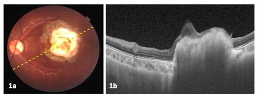
Pattern A (3 eyes) – The tumour predominantly occupied the region of outer and middle retina. In these eyes, the outer retina could not be identified and inner retina was spared which appeared to drape the tumour. Inner retinal layers could be identified only in a few scans and elsewhere the differentiation of layers could not be made out. The tumour had a slight homogenous appearance with smooth surface (figure 1).
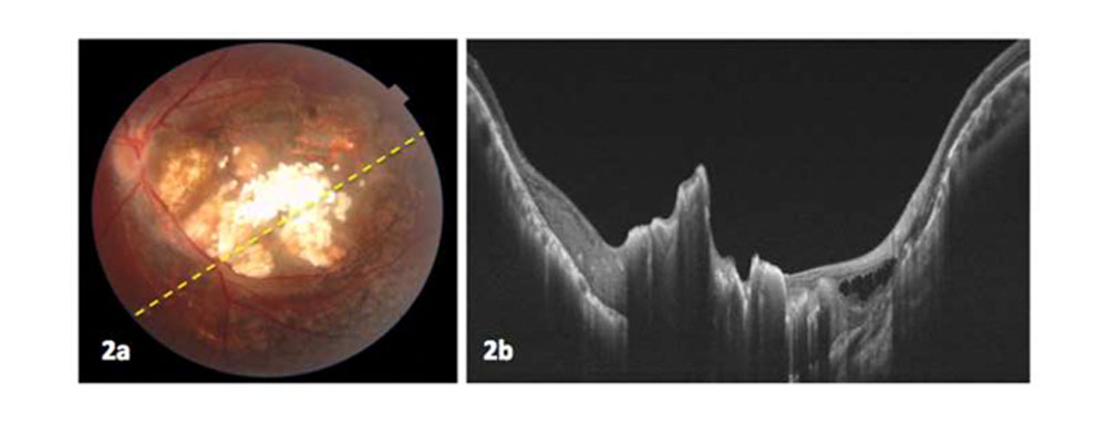
Pattern B (8 eyes) – Mixed involvement (full thickness involvement in centre and outer layers involvement in areas surrounding the central tumour). The tumour predominantly had a heterogenous appearnce with irregular surface (figure 2). Pattern C (2 eyes) – Complete atrophy of retinal tissue forming a depression (figure 3).
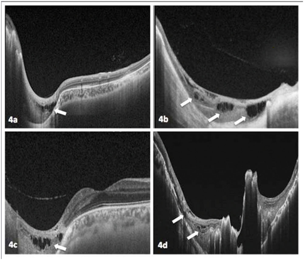
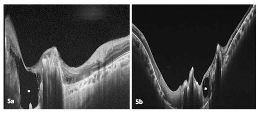
Partial posterior vitreous detachment over the tumour was seen 3 eyes. Foveal dip was clearly identified in all non fovea involving tumors. However, foveal dip could be identified in only 1 eye of fovea involving tumours. Schisis or cavitary changes of variable severity was seen within the substance of tumour in 5 eyes (figure 4 and figure 5).
The choroid couldnot be visualised in scans with dense shadowing from calcification. In scans where there was no shadowing, the choroid showed variable degree of atrophy to complete absence (sclera could be easily visualized in these scans).
Intra-tumour calcification of variable degree was found in 8 eyes. Calcification was invariably found in scans which showed full thickness retinal involvement. Calcification was typically absent or of lesser severity in scans which showed sparing of inner retina over the tumour and in tumours with atrophic (type 4) regression.
There was no evidence of subretinal fluid in any of the scans. Discussion Imaging has become an essential element in the practice of ocular oncology. It plays an important role starting from diagnosis and documentation, decision-making and follow-up. OCT has been shown to be extremely useful for management of posterior segment disorders in children. Shields et al in 2004 had demonstrated the utility of office based time-domain OCT in children as young as 3 to 4 years [16]. However, with the advent of hand held SD-OCT, its utility has been expanded to a number of clinical conditions in children. It has been shown to be useful in abusive head trauma, retinopathy of prematurity, ocular albinism, nystagmus, pediatric optic neuropathies, pediatric glaucoma and pediatric retinal tumours [5-7, 17,18]. In the field of pediatric ocular oncology, hand held OCT has found utility in the management of tumours like combined hamartoma of retina and retinal pigment epithelium, astrocytic hamartoma and retinoblastoma [16, 19, 20]. In cases of retinoblastoma, hand held SD-OCT has been useful in directing diagnosis, taking treatment decisions, monitoring tumour regression and for follow-up [8-12]. It has also been able to detect certain clinically subtle but important findings like detection of ophthalmoscopically invisible tumours, choroidal relapse, optic nerve head infiltration and to know the characteristics of vitreous seeds [8, 21]. Thus OCT has been successfully added as a technological armamentarium in the management of retinoblastoma. However, the use of this technology is sought with certain limitations. Hand held device is not readily available at all centres and has a learning curve with technical difficulties in imaging. Also SD-OCT has limitations in imaging large tumors with limited depth of focus [13]. In our study we selected children who would cooperate for office based examination. All the 13 children were co-operative for the scans. Use of SS-OCT had several advantages in our study. The scanning speed is rapid with upto 10,000 A-scans per second hence overcomes problems related to patient fatigue. It has deep penetration and hence there is clear visualization of the entire tumor from vitroretinal interface to the chorio-scleral junction (except when there was shadowing effect). Also the tracking system incorporated into the system helps to take multiple scans at the area of interest in children who tend to get easily distracted during the scanning session. Of the 13 patients with macular RB, 54% group B tumours, 31% had group C and 15% had group D tumours. This is comparable to that reported by Pica et al (group B – 47%, group C – 40% and group E – 13%) [22]. Involvement of fovea by the tumour is a major determinant of visual acuity as reported by the previous studies [23,24]. In our study, 7 eyes had foveal involvement and 6 eyes did not have tumor involvement of the fovea. However, visual acuity was not documented in this study. Studies have described the features of active RB. The OCT appearance of active RB varies with the size of lesion. Medium sized lesions appear as relatively homogenous hyperdense elevations involving the middle
4
and outer retinal layers, with shadowing of the underlying structures. Small lesions are spherical isodense lesions with a distinctive intraretinal location. The intact inner retina is usually seen to drape the tumour [12,13]. Cao et al have described active RB lesions to have smooth tumour surface with low to intermediate optical density and abrupt transition from normal retina to involved retina. normal, anatomically intact retina appeared draped over the exophytic tumor [25].
With response to treatment, there is a sequential change from a localised, isodense intraretinal mass to a progressively more variably dense, flat, full thickness chorioretinal scar [13]. Cao et al have shown that on OCT, regressed lesions had smooth surface with high optical density with sharp transition from normal retina. The outer retina was thinned or normal and the normal retina was draped over regressed tumor [25]. All these OCT features cannot be generalized to different patterns of regression. We noted that regressed macular RB lesions showed 3 distinct regression patterns on SS-OCT. The cause of this difference in appearance is not exactly known. Previous studies have shown that the variance in clinical regression patterns could be due to difference in initial tumour size, degree of tumour differentiation, mode of therapy and distance from fovea. Similarly in our series, we found that small tumours away from fovea had OCT pattern C and larger tumours had either pattern A or B.
Other features that were documented include partial posterior vitreous detachment noted in 3 eyes and intra-tumour calcification of variable degree in 8 eyes. It is of interest to note that calcification was not detectable even on OCT in rest of the 5 eyes. Also schisis/cavitary changes were noted in 5 eyes. The appearance of schisis in RB is probably due to degeneration of retinal cells with intact Muller cells bridging the variable extent of retina. Cavitary changes which were not seen clinically were detected on OCT in 2 patients. Presence of cavitary changes within the tumour have been speculated to be an indicator of a more differentiated tumor [26].
There are a few limitations in the study. We studied only older children (4 year and above) as the SS-OCT in its current version is only available for office use. Future developments may allow the use of this technology in both operation theatres and in offices. Also this study included only cases of regressed RB. Ideal would be to do a prospective study to include cases of active RB and follow-up them with OCT scans as is done with other disorders of the retina.
SS-OCT was useful to image cases of regressed retinoblastoma including large tumours and provided insight into the understanding of the various OCT based regression patterns of retinoblastoma. Rapid scan acquisition time and tracking facility with SS-OCT system helped in successful imaging even in smaller children.
References
- Shields JA, Shields CL. Intraocular tumours – A text and Atlas. Philadelphia, PA, USA, WB Saunders Company, 1992.
- Dimaras H, Kimani K, Dimba EA, Gronsdahl P, White A, Chan HS, Gallie BL. Retinoblastoma. Lancet. 2012 Apr 14;379(9824):1436-46.
- Tamboli A, Podgor MJ, Horm JW. The incidence of retinoblastoma in the United States: 1974 through 1985. Arch Ophthalmol 1990; 108: 128–132.
- Sanders BM, Draper GJ, Kingston JE. Retinoblastoma in Great Britain 1969–80: incidence, treatment, and survival. Br J Ophthalmol 1988; 72:576–583.
- Stathopoulos C, Gaillard MC, Puccinelli F, Maeder P, Hadjistilianou D, Beck-Popovic M, Munier FL. Successful conservative treatment of massive choroidal relapse in 2 retinoblastoma patients monitored by ultrasound biomicroscopy and/or spectral domain optic coherence tomography. Ophthalmic Genet. 2018 Apr;39(2):242-246.
- Muni RH, Kohly RP, Sohn EH, et al. Hand-held spectral domain optical coherence tomography finding in shaken-baby syndrome. Retina 2010;30:S45–50. 7. Chavala SH, Farsiu S, Maldonado R, et al. Insights into advanced retinopathy of prematurity using handheld spectral domain optical coherence tomography imaging. Ophthalmol 2009;116:2448–56.
- Chong GT, Farsiu S, Freedman SF, et al. Abnormal foveal morphology in ocular albinism imaged with spectral-domain optical coherence tomography. Arch Ophthalmol 2009;127:37–44. 9. Soliman SE, VandenHoven C, MacKeen LD, Héon E, Gallie BL. Optical Coherence Tomography- Guided Decisions in Retinoblastoma Management. Ophthalmology. 2017 Jun;124(6):859-872.
- Lee H, Proudlock FA, Gottlob I. Pediatric Optical Coherence Tomography in Clinical Practice-Recent Progress. Invest Ophthalmol Vis Sci. 2016 Jul 1;57(9):OCT69-79. 11. Seider MI, Grewal DS, Mruthyunjaya P. Portable Optical Coherence Tomography Detection or Confirmation of Ophthalmoscopically Invisible or Indeterminate Active Retinoblastoma. Ophthalmic Surg Lasers Imaging Retina. 2016 Oct 1;47(10):965-968.
- Berry JL, Cobrinik D, Kim JW. Detection and Intraretinal Localization of an ‘Invisible’ Retinoblastoma Using Optical Coherence Tomography. Ocul Oncol Pathol. 2016 Apr;2(3):148-52.
- Rootman DB, Gonzalez E, Mallipatna A, Vandenhoven C, Hampton L, Dimaras H, Chan HS, Gallie BL, Heon E. Hand-held high-resolution spectral domain optical coherence tomography in retinoblastoma: clinical and morphologic considerations. Br J Ophthalmol. 2013 Jan;97(1):59-65.
- Shields CL, Mashayekhi A, Cater J, Shelil A, Ness S, Meadows AT, Shields JA. Macular retinoblastoma managed with chemoreduction: analysis of tumor control with or without adjuvant thermotherapy in 68 tumors. Arch Ophthalmol. 2005 Jun;123(6):765-73.
- Shields CL, Palamar M, Sharma P, et al.. Retinoblastoma regression patterns following chemoreduction and adjuvant therapy in 557 tumours. Arch Ophthalmol 2009;127:282-90.
- Shields CL, Mashayekhi A, Luo CK, Materin MA, Shields JA. Optical coherence tomography in children: analysis of 44 eyes with intraocular tumors and simulating conditions. J Pediatr Ophthalmol Strabismus. 2004 Nov-Dec;41(6):338-44.
- Ghasia FF, Freedman SF, Rajani A, Holgado S, Asrani S, El-Dairi M. Optical coherence tomography in paediatric glaucoma: time domain versus spectral domain. Br J Ophthalmol. 2013 Jul;97(7):837-42. 18. Gospe SM, Bhatti MT, El-Dairi MA. Emerging Applications of Optical Coherence Tomography in Pediatric Optic Neuropathies. Semin Pediatr Neurol. 2017 May;24(2):135-142.
- Mallipatna A, Vinekar A, Jayadev C, Dabir S, Sivakumar M, Krishnan N, Mehta P, Berendschot T, Yadav NK. The use of handheld spectral domain optical coherence tomography in pediatric ophthalmology practice: Our experience of 975 infants and children. Indian J Ophthalmol. 2015 Jul;63(7):586-93.
- Shields CL, Manalac J, Das C, Saktanasate J, Shields JA. Review of spectral domain-enhanced depth imaging optical coherence tomography of tumors of the retina and retinal pigment epithelium in children and adults. Indian J Ophthalmol. 2015 Feb;63(2):128-32.
- Berry JL, Anulao K, Kim JW. Optical Coherence Tomography Imaging of a Large Spherical Seed in Retinoblastoma. Ophthalmology. 2017 Aug;124(8):1208.
- Pica A, Moeckli R, Balmer A, Beck-Popovic M, Chollet-Rivier M, Do HP, Weber DC, Munier FL. Preliminary experience in treatment of papillary and macular retinoblastoma: evaluation of local control and local complications after treatment with linear accelerator-based stereotactic radiotherapy with micromultileaf collimator as second-line or salvage treatment after chemotherapy. Int J Radiat Oncol Biol Phys. 2011 Dec 1;81(5):1380-6.
- Kim JM, Kim JH, Kim SJ, Park KD, Shin HY, Ahn HS, Yu YS. Visual prognosis of retinoblastoma in the posterior pole treated with primary chemotherapy plus local treatments. Korean J Ophthalmol. 2010 Dec;24(6):347-52.
- Weiss AH, Karr DJ, Kalina RE, Lindsley KL, Pendergrass TW. Visual outcomes of macular retinoblastoma after external beam radiation therapy. Ophthalmology. 1994 Jul;101(7):1244-9.
- Cao C, Markovitz M, Ferenczy S, Shields CL. Hand-held spectral-domain optical coherence tomography of small macular retinoblastoma in infants before and after chemotherapy. J Pediatr Ophthalmol Strabismus. 2014 Jul 1;51(4):230-4.
- Mashayekhi A, Shields CL, Eagle RC Jr, Shields JA. Cavitary changes in retinoblastoma: relationship to chemoresistance. Ophthalmology. 2005 Jun;112(6):1145-50.
Legends
Figure legends Figure 1a and 1b: Fundus picture and corresponding OCT image showing Pattern A regression. The tumour predominantly occupies the region of outer and middle retina. The outer retina cannot be identified and inner retina appeared to drape the tumour. Figure 2a and 2b: Fundus picture and corresponding OCT image showing Pattern A regression. There is full thickness involvement in centre and outer layer involvement in areas surrounding the central tumour). Presence of significant calcification and schitic changes can also be noted. The tumour predominantly had a heterogenous appearnce with irregular surface.
Figure 3a and 3b: Fundus picture and corresponding OCT image showing Pattern A regression. There is complete atrophy of retinal tissue at the site of regressed RB lesion forming a depression. Figure 4a-4d: SS-OCT images of various patients (number 1, 3, 10, 12 respectively) showing schisis within the tumour (white arrows).
Figure 5a and 5b: SS-OCT images of patients 3 and 12 showing presence of cavitary changes (white asterisks). Table legends Table 1 showing demographic details, tumour characteristics and swept source-OCT features of patients with macular retinoblastoma (PVD – posterior vitreous detachment, OCT – optical coherence tomography).
colour figure 1 Click here to download colour figure figure 1.tiff
colour figure 2 Click here to download colour figure figure 2.tiff
colour figure 3 Click here to download colour figure figure 3.tiff
colour figure 4 Click here to download colour figure figure 4.tiff
Leave a Comment