

Dr.Payal Shah, S16578, Dr.Rajesh Ramanjulu, Dr.Pradeep Sagar, Dr.Mahesh Shanmugam P
AIM:
To understand the Optical coherence tomography angiography (OCTA) characteristics of intraocular tumours and assess the role of OCTA as a diagnostic adjunct.
METHODS:
It is a retrospective case series of various intraocular tumorsstudied in our hospital.
All patients underwent thorough ophthalmic evaluation with a slit lamp, slit lamp biomicroscopy with 78D lens and indirect ophthalmoscopy with 20D lens, Fundus Fluorescein Angiography with a mydriatic fundus camera (Topcon TRC-50DX, Topcon Corporation, Japan.) and swept source OCTA captured on OCT DRI Triton plus (Topcon Corporation, Japan).
Various vascular (5 cases of retinal capillary hemangioblastomas, 8 solitary and 2 diffuse choroidal hemagiomas), pigmented (5 cases of melanocytomas, 4choroidal melanomas, and 4 choroidal nevi) and glial tumors (1 astrocytic hamartoma, 2 retinal astrocytoma and 5 choroidal osteomas) were studied.
OCT angiography characteristics of different segments was studied. The key to image a tumor is manual segmentation. Every frame was manually segmented to capture the tumor characteristics precisely.
Images with significant motion artifacts and masking due to overlying retinal changes were excluded.
RESULTS:
Retinal capillary hemangioblastomas(RCH): On OCTA, in the superficial and deep retinal layers, a bright well-defined lesion with compact vascularity with minimal to no intervening spaces suggesting highly vascular nature of the tumor is noted. Feeder and drainage vessels can be identified.
Choroidal hemangiomas:Characterized by the presence of irregular network of vessels with variable calibre seen on choroidal layer.In between the vessels we can see intervening signal void areas, which are appreciated more on an inactive tumor compared to an active tumor with associated exudative retinal detachment.Distinct vasculature of tumor can be delineated clearly from the normal choroidal vasculature helping to map the tumor extent.
Choroidal osteomas: Characterised by distinctive dark background with multiple tiny bright punctate spots, which may represent haversian canal. Branching vascular network within the lesion associated with choroidal neovascular membrane was noted in few cases.
Astrocytic hamartomas: Scanty vasculature seen in superficial retinal slab on OCTA.
Retinal astrocytomas: Well defined branching vessel with abundant intervening signal void areas as they are solid tumors in contrast to the vascular tumors.
Optic disc melanocytomas: Signal void areas due to dense pigmentation and minimal vascularity of the tumor.
Choroidal melanomas: Small calibre vessels seen on OCTA within the tumor which are less compact and smaller compared to choroidal hemangiomas.Juxtapapillary melanomas in contrast are densely vascular probably owing to the absence of bruchs membrane and superficial nature of these tumors compared to a choroidal melanoma elsewhere.
Choroidal nevi: May appear dark or bright depending upon overlying RPE changes. In cases of RPE atrophy, bright signals are noted due to unmasking and in cases of RPE hypertrophy, signal void areas are noted due to masking by the overlying RPE.
DISCUSSION:
OCTA is non-invasive compared to FFA. Optical coherence tomography angiography, based on motion contrast imaging, is a promising new imaging modality to understand the intrinsic retinal and choroidal vasculature.1
In cases of RCH, OCTA not only shows the vascular nature of the tumor, but also helps in identification of smaller tumors which may be missed clinically. Also, it helps to identify the draining/ feeder vessels.OCTA also helps in diagnosing exophytic and endophytic juxtapapillaryhemangiblastomas where clinical diagnosis is challenging.2 But the limitation is it can only identify the lesions in posterior pole.OCTA has been shown to be helpful in monitoring response to treatment and identification of harmless nonvascular lesions in VHL disease.3
Clinical diagnosis of Diffuse Choroidal Hemangioma is challenging because of pigmentation of the fundus in Asian population, making it difficult to identify the lesion. In such scenarios, we propose comparing the choroidal vascular pattern of the two eyes using OCTA, aids in the identification of abnormal choroidal vasculature.Characteristic OCTA features of choroidal hemangiomas were the presence of irregularly arranged choroidal vessels in both superficial and deeper choroidal slabs, the presence of club-like appearance in deeper choroidal slab which was more prominently seen in active tumors, and signal void areas in deeper choroidal slabs which was more prominent in dormant tumors.Dark signals in CH on OCTA were reported by Takkar et al.4
In cases of choroidal osteomas, the CNV complex appears as a vascular network in the superficial part of the osteoma and is well identified on OCTA images. Intrinsic vascular network and feeder vessels of the tumor are seen prominently in its early stage. A layer of vessels arranged in a medusa-head appearance represents the vascular network within the tumor.5
OCTA helps in distinguishing neoplastic and non-neoplastic glial cell tumors. Also helps in differentiating optic disc melanocytoma from melanoma.
OCTA can also play a role in assessing the treatment response of tumors in future.
Limitation of OCTA is difficulty to image larger and peripheral tumors.
CONCLUSION:
OCTA is a non-invasive tool and can aid in diagnosing intra-ocular tumors, help in study the vascular nature of the tumor and disease activity and progression.
REFERENCES:
- De Carlo TE, Romano A, Waheed NK, Duker JS. A review of optical coherence tomography angiography (OCTA) Int J Retina Vitreous. 2015;1:5
- Sagar P, Rajesh R, Shanmugam M, Konana VK, Mishra D. Comparison of optical coherence tomography angiography and fundus fluorescein angiography features of retinal capillary hemangioblastoma. Indian J Ophthalmol. 2018;66:872–6.
- Lang SJ, Cakir B, Evers C, Ludwig F, Lange CA, Agostini HT, et al. Value of optical coherence tomography angiography imaging in diagnosis and treatment of hemangioblastomas in von Hippel-Lindau disease. Ophthalmic Surg Lasers Imaging Retina 2016;47:935-46.
- Takkar B, Azad S, Shakrawal J, Gaur N, Venkatesh P. Blood flow pattern in a choroidal hemangioma imaged on swept-source-optical coherence tomography angiography. Indian J Ophthalmol2017;65:1240-2.
- J.T. Szelog, M.A. BoniniFilho, D.R. Lally, et al.Optical coherence tomography angiography for detecting choroidal neovascularization secondary to choroidal osteoma Ophthalmic Surg Lasers Imaging Retina, 47 (2016), pp. 69-72
LEGENDS:
FIGURE 1: Color photo and OCTA of retinal capillary hemangioblastoma showing dense compact vascularity.
FIGURE 2: Color photo of diffuse choroidal hemangiomashowing orangish hue and OCTAshowing spaghetti-like appearance within the tumor
FIGURE 3: OCTA of disc melanoma(right) showing intrinsic tumor vascularity in contrast to disc melanocytoma(left) showing signal void area.
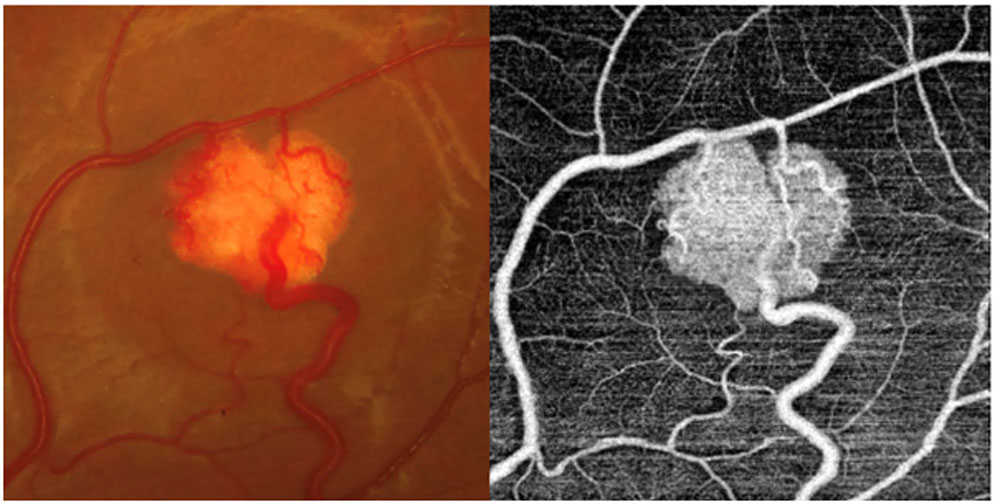
FIGURE 1
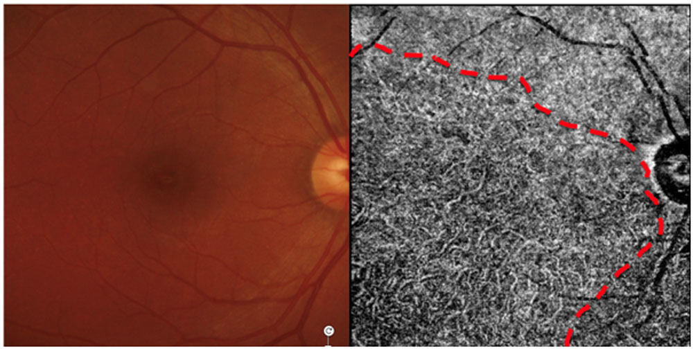
FIGURE 2
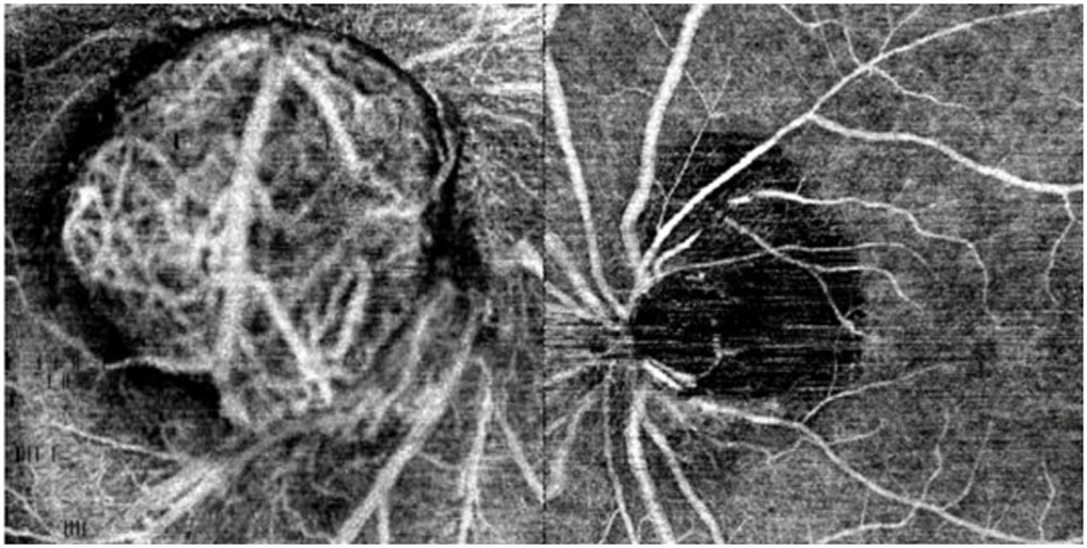
FIGURE 3
Leave a Comment