

Dr.Bhawesh chandra saha, Dr.Rashmi kumari
ABSTRACT
Introduction: Acute dacryocystitis is an uncommon but serious condition in infants and needs immediate treatment. Although, medical management with systemic antibiotics remains the mainstay of initial treatment, there are recent studies justifying simultaneous Naso Lacrimal Duct (NLD) probing with acceptable success rate. Aim: To assess the success rate of probing in the management of infantile acute dacryocystitis and to analyse the factors affecting it. Materials and Methods: A prospective interventional study was designed and infants presenting with acute dacryocystitis with or without complications like dacryocystopyocele/ lacrimal abscess/preseptal cellulitis at a Tertiary Eye Care Centre from May 2015 to April 2016 were enrolled. Demographic details and baseline clinical characteristics were noted. Intravenous antibiotics were started and probing under general anaesthesia was done.
Follow up was done after one month.Success was defined as subsidence of acute attack and resolution of epiphora at final follow up of one month. Results: A total 20 eyes of 18 patients were included with male: female ratio 10:8. Mean age of patients was 6.5 months and the mean duration of symptoms was 5.6 days. Dacryocystitis with lacrimal swelling was present in five eyes, preseptal cellulitis was present in two eyes while the rest 13 eyes presented with simple inflammation over the lacrimal sac. After one month, 85% (17eyes) had complete resolution of symptoms. Recurrence of epiphora was seen in 15% (three eyes) after initial improvement. Conclusion: Medical management with simultaneous probing of nasolacrimal duct has fairly good success rate in infantile dacryocystitis. Presence of dacryopyocele or dacryocystocele can lead to failure of probing owing to presence of intranasal cysts.
Keywords: Epiphora, Intranasal cysts, Orbital cellulitis
INTRODUCTION: Congenital Nasolacrimal Duct Obstruction (CNLDO) is the most common cause of epiphora in infants and children and a common problem encountered in paediatric ophthalmology [1]. Embryologically, lower NLD is last to canalize during development and its failure leads to CNLDO [1,2]. Although actual incidence in infants is 50% but due to spontaneous perforation of membranous valve of Hasner at lower nasolacrimal duct after birth, symptomatic CNLDO are seen in approximately 6-20% full term newborn children [1,3]. The symptoms include classic triad of watering, discharge and matting of eyelashes [3]. Acute dacryocystitis may occur in 2.9% of the cases when pain and redness accompanies the classic triad [1,3]. It can complicate into orbital cellulitis, orbital abscess or cavernous sinus thrombosis which are life threatining [4,5]. In neonates, it differs in several aspects from the chronic low grade dacryocystitis that occurs in typical NLDO in adults. Its presentation is often acute and usually associated with palpable masses that arise from the lacrimal sac [5,6].
Different names have been used to describe these lesions like dacryocystocele, dacryocele, mucocele or amniotocele [6]. Acute dacryocystitis can occur in dacryocele which is a bluish purple dilated lacrimal sac seen in infants with CNDLO [7]. They are typically noticed at or within a few days of birth located medial to the eye beneath the medial canthus [7]. The lesions may resolve spontaneously, but they frequently become infected and progress rapidly to acute dacryocystitis and lacrimal sac abscesses [8]. It is important to carefully follow and treat dacryocele early as it is associated with intranasal cyst in NLD which can cause respiratory problems [7]. Acute dacryocystitis in infants needs immediate treatment because infection has fulminant nature [7,8].
Conservative management consist of systemic antibiotics preferably through intravenous route. Since underlying cause is usually NLD obstruction, probing seems to be the definitive treatment in such cases [5]. Conventionally, it is reserved as secondary treatment for a later date after the subsidence of acute phase [6,7]. There have been very few reports on simultaneous probing in acute dacryocystitis in infants with acceptable success rate [8,9].Therefore, the present study was conducted to assess the success rate of probing in the management of infantile acute dacryocystitis and to unravel any association of the failure rate with its varied clinical presentation.
MATERIALS AND METHODS: This prospective interventional study was designed after getting Ethical Clearance from Institutional Review Committee. Infants presenting with acute dacryocystitis or lacrimal abscess in the Department of ophthalmology, All India Institute of Medical Sciences, Patna, Bihar, India, from May 2014 to April 2016 were enrolled with due consent of their parents. Critically ill infants or those with any nasal pathology or facial asymmetry were excluded from the study. All the enrolled patients were admitted and demographic and clinical characteristics were noted before starting the treatment.
Routine blood investigations along with relevant investigations for fitness for anaesthesia were done. Intravenous antibiotics after paediatric consultation for proper dosing, along with topical antibiotics and nasal decongestants were given for two days. On day 3, all cases underwent probing and syringing under general anaesthesia. In addition, percutaneous incision and drainage of the abscess was done in five eyes with cystic swelling. Systemic antibiotics were continued for five days postoperatively. Topical antibiotic and nasal decongestants were also continued for one week. Success was defined as resolution of acute attack as well as resolution of epiphora at one month.
RESULTS: A total of 20 eyes of 18 patients that were included in the study with complete follow up of one month were analysed. There were 10 male and eight female infants with mean age 6.5 months and range being 4.5-9.5 months [Table/Fig-1].
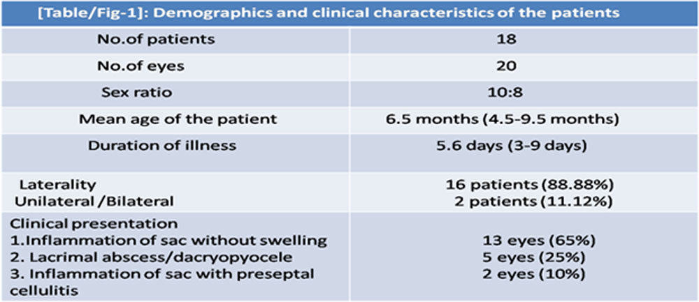
All infants had unilateral presentation except two females with bilateral presentation.History of epiphora with occassional discharge was common to all with acute onset of pain and inflammation over the lacrimal area. Dacryocystitis with lacrimal abscess was present in five eyes, preseptal cellulitis was present in two eyes while the rest 13 eyes presented with simple inflammation over the lacrimal sac. Out of 18 patients, 16 (88.8%)
were under conservative treatment with sac massage. On enquiring about the technique of massage it became obvious that 13 (72.22%) patients were getting it improperly. Prior intervention in form of incision and drainage of abscess was done in three patients (16.67%) including one with preseptal cellulitis. After four weeks, on follow up, 85% (17 eyes) had complete resolution of symptoms while 15% (three eyes) reported recurrence of epiphora after initial resolution [Table/Fig-2].
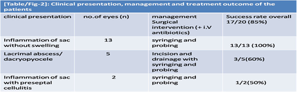
Failed three cases included the two with history of prior incision and drainage. No difference in outcome was observed in patients with bilateral disease.
DISCUSSION: Acute dacryocystitis is an emergency condition in infants due to its fulminant nature that can lead to dreaded complications such Our study noted a success rate of 85% which is quite different from those discussed above probably because of the presenting age of the patients. These studies have compared conventional probing to endoscopic probing in neonates, which is more likely to be associated with dacryocystoceles, reporting a 100% success rate with the later technique as it facilitates simultaneous cyst removal [Table/Fig-3].
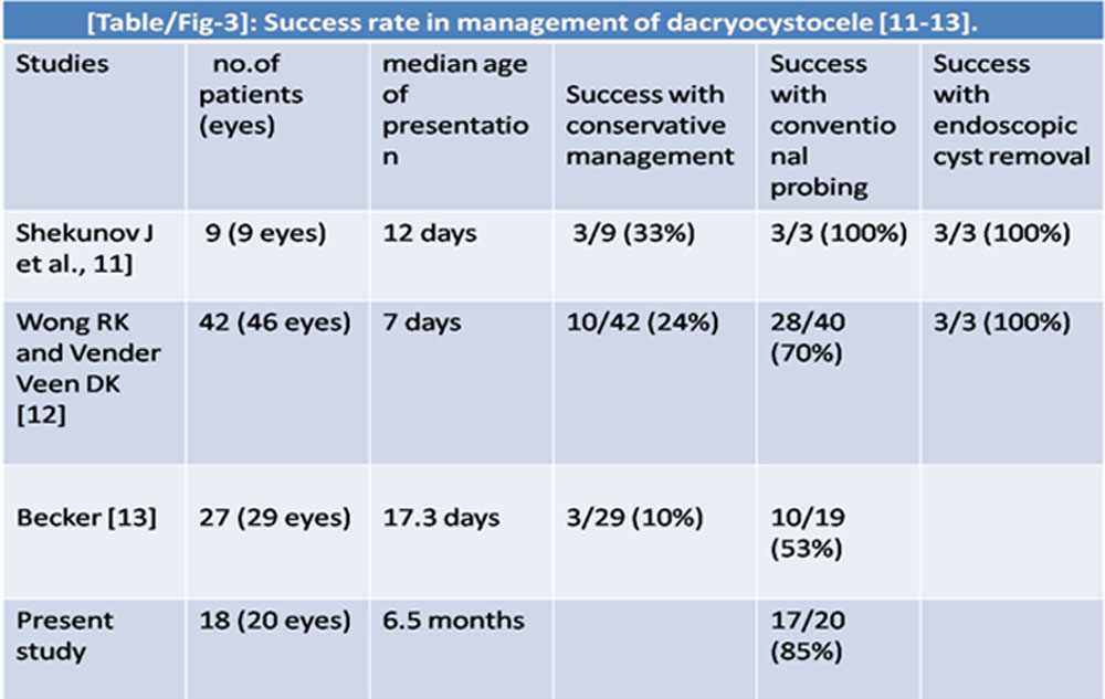
In contrast, present study includes infants with mean age of presentation at 6.5 months decreasing the possibility of dacryocystocele. Out of three eyes with failed conventional probing, two eyes (66.67%) had history of prior surgical intervention for lacrimal swelling while all of them (three eyes) were associated with cysting swelling over the sac area. Probably all failed cases were dacryocystocele which are known to be associated with intranasal cysts in almost all the cases warranting simultaneous treatment if they does not resolve spontaneously and gets infected [11-16]. These need to be removed urgently especially in neonates as they can cause respiratory problems [14,15]. Lueder GT et al., found intranasal lesions at the distal lacrimal duct in 6% of children with NLDO who were older than 18 months at the time of their initial probing and 9% of children who had persistent symptoms of NLDO following previous probing [15].
Another study by Gregg et al., done to investigate the association of neonatal dacryocystoceles and dacryocystitis with NLD cysts, and to report the outcomes of treatment of these disorders concluded that neonatal dacryocystoceles are almost always associated with NLD cysts and the success rate of NLD probing and endoscopic cyst removal in these patients is excellent [16]. In our study, use of endoscopic guided probing in failed cases could have been a better option as it not only identifies these cysts but also facilitates simultaneous management by internal drainage [8]. Since NLD cysts were also present in many young infants with severe sac inflammation even in the absence of any swelling, nasal endoscopy seems to be an important adjunct to the management of all infants with CNLDO [16,17].
LIMITATION: Small sample size and shorter duration of follow up are the limitations of our study but since its a rare condition with inclusion of infants only, even smaller number holds significance with its initial one month of follow up. Moreover any surgery on inflamed tissue is considered difficult.We agree that a study which compares the approach of medical treatment with simultaneous surgical intervention like ours with medical management followed by surgical intervention at second sitting, would give us more information on the apparent success rate.
CONCLUSION: Infantile acute dacryocystitis is rare but serious condition. Early surgical intervention in the form of probing along with intravenous antibiotic has a good success rate.Differentiating lacrimal swelling from infected dacryocystocele is crucial as the later is associated with intranasal cysts which can be a cause of failure. Endoscopic guided probing should be tried in failed cases for simultaneous detection and management of the intranasal cysts
REFERENCES
[1] Yeatts RP. Lacrimal Drainage System Surgery. In: Principles and Practice of Ophthalmology. Albert DM, Jakobiec FA, Azar DT,Gragoudas ES. 2000 WB Saunders Company USA. Vol 4, Ch 268, pp3556-58.
[2] Macewen CJ. Congenital nasolacrimal duct obstruction. Compr Ophthalmol Update. 2006;7:79–87
[3] Kim YS, Moon SC, Yoo KW. Congenital nasolacrimal duct obstruction: Irrigation or probing? Korean J Ophthalmol. 2000;14:90–96
[4] Syed SH, Arif M, Sultan Mahmood M. Syringing and probing results for congenital nasolacrimal duct obstruction. APMC. 2009;3:67–70.
[5] Ali MJ, Joshi SD, Naik MN, Honavar SG. Clinical profile and management outcome of acute dacryocystitis: Two decades of experience in a tertiary eye care center. Semin Ophthalmol. 2015;30:118-23.
[6]Ali MJ. Paediatric acute dacryocystitis. Ophthal Plast Reconstr Surg. 2015;31:341-47.
[7]Thomas SA, Drack AV, Sands RE, Yoon P, Friedman NR, Chan, K. Clinical and histopathologic features of congenital dacryocystoceles associated with intranasal cysts. JAAPOS. 2000;4:46-53.
[8] Ali MJ, Psaltis AJ, Brunworth J, Naik MN, Wormald PJ. Congenital dacryocele with large intranasal cyst: Efficacy of cruciate marsupialization, adjunctive procedures, and outcomes. Ophthal Plast Reconstr Surg. 2014;30:346-51.
[9]Lueder GT. Endoscopic treatment of intranasal abnormalities associated with nasolacrimal duct obstruction. J AAPOS. 2004;8(2):128-32.
[10] Baskin DE, Reddy AK, Chu YI, et. al. The timing of antibiotic administration in the management of infant dacryocystitis. J AAPOS. 2008;12:456–59.
[11]Shekunov J, Griepentrog GJ, Diehl NN, Mohney BG. Prevalence and clinical characteristics of congenital dacryocystocele. J AAPOS. 2010;14(5):417-20.
[12]Wong RK, VanderVeen DK. Presentation and management of congenital dacryocystocele. Paediatrics. 2008;122(5):e1108-12 .
[13]Becker BB. The treatment of congenital dacryocystocele. Am J Ophthalmol. 2006;142(5):835-38.
[14] Ali MJ, Kamal S, Gupta A, Ali MH, Naik MN. Simple vs complex congenital nasolacrimal duct obstructions: Aetiology, Management and outcomes. Int Forum Allergy Rhinol. 2015;5:174-77.
[15]Lueder GT. Paediatric lacrimal disorders. In: Wilson ME, Saunders RA, Trivedi RH, eds. Paediatric Ophthalmology. Current Thought and A Practical Guide. Berlin, Heidelberg: Springer-Verlag; 2009:pp275-85.
[16]Gregg T, Lueder MD. The association of neonatal dacryocystoceles and infantile dacryocystitis with nasolacrimal duct cysts. Trans Am Ophthalmol Soc. 2012;110:74–93.
[17]MacEwen CJ, Young JD, Barras CW, Ram B, White PS. Value of nasal endoscopy and probing in the diagnosis and management of children with congenital epiphora. Br J Ophthalmol. 2001;85:314–18
Leave a Comment