

Dr.D N rakash, Dr.SUJATA,Dr.Raghavendra Ramappa,Dr.AMULYA PADMINI H M
ABSTRACT:
Objective :To study the therapeutic effects of 2% rebamipide ophthalmic suspension in dry eye disease.
Methods:This was a Prospective study of 50 patients diagnosed with dry eye from Jun 2016 to Apl 2017 & were treated with 2% rebamipide suspension.Dry eye-related Symptom Score, anterior segment examination with slit lamp, Tear Film Break-Up Time, Fluorescein Ocular Surface Staining Score(FOS) &Schirmer test were used to collect data from patients at baseline & at 2, 4, 8 & 12 week visits.
Results:Mean dry eye-related symptom score showed a significant improvement from baseline at 2, 4, 8 & 12 weeks (9.80, 7.20, 7.40 & 7.83 points). Median FOS also showed significant improvement from baseline (3.0 points) at 2, 4, 8 & 12 weeks (2.0, 2.0, 1.0 & 1.0 points respectively). TBUT &Schirmer test values also improved but were not significant after treatment.
Conclusion: 2% rebamipide ophthalmic suspension is an effective treatment for dry eye. It improves both ocular &tear film stability.
INTRODUCTION
TFOS DEWS II has redefined dry eye as: “Dry eye is a multifactorial disease of the ocular surface characterized by a loss of homeostasis of the tear film, and accompanied by ocular symptoms, in which tear film instability and hyperosmolarity, ocular surface inflammation and damage, and neurosensory abnormalities play etiological roles”[1].
Dry eye is a very common disorder encountered in daily ophthalmological practice commonly affecting females in menopausal age group and elderly patients. The cause of dry eye could be either aqueous deficiency which encompasses Sjogrens and Non-Sjogrens syndrome or due to excessive evaporation of tears due to defective tear film caused by improper mucin or lipids in tear film2.
Apart from other factors which include aging, female gender, hormonal factors(low androgens), environmental factors (air conditioners, low humidity), lid abnormalities which prevent the spread of an even tear film and of late- the rising trend of refractive surgeries like LASIK. The prevalence is 5- 16% as described in few studies in America and Australia while it is as high as 27-33% in Asian countries. 3 It has a negative impact on the patients quality of life, sometimes with potential sight threatening consequence in long run if left untreated in few cases.4,5
Rebamipide is a quinolinone derivative with mucin secretagogue activity. It has been shown to increase gastric endogenous prostaglandin E2 and I2, promote gastric epithelial mucin, is an oxygen free radical scavenger and has other anti-inflammatory actions as well.6 In addition to its use for the gastrointestinal mucosa, the biological effects of rebamipide 2%, which include cytoprotection, wound healing, and anti-inflammatory properties, are known to be universal for a variety of tissues.After discovering the pharmacological actions of rebamipide 2%, began its development for use as a dry eye medication and rebamipide ophthalmic suspension was recently approved for the treatment of dry eye.
The therapeutic effects of rebamipide ophthalmic suspension are considered to be due to the increase of corneal and conjunctival mucin, and its safety and efficacy have been established in clinical trials and some experimental reports. It also acts by suppressing the production of inflammatory cytokines like IL 8, TNF-α and NFkB.7
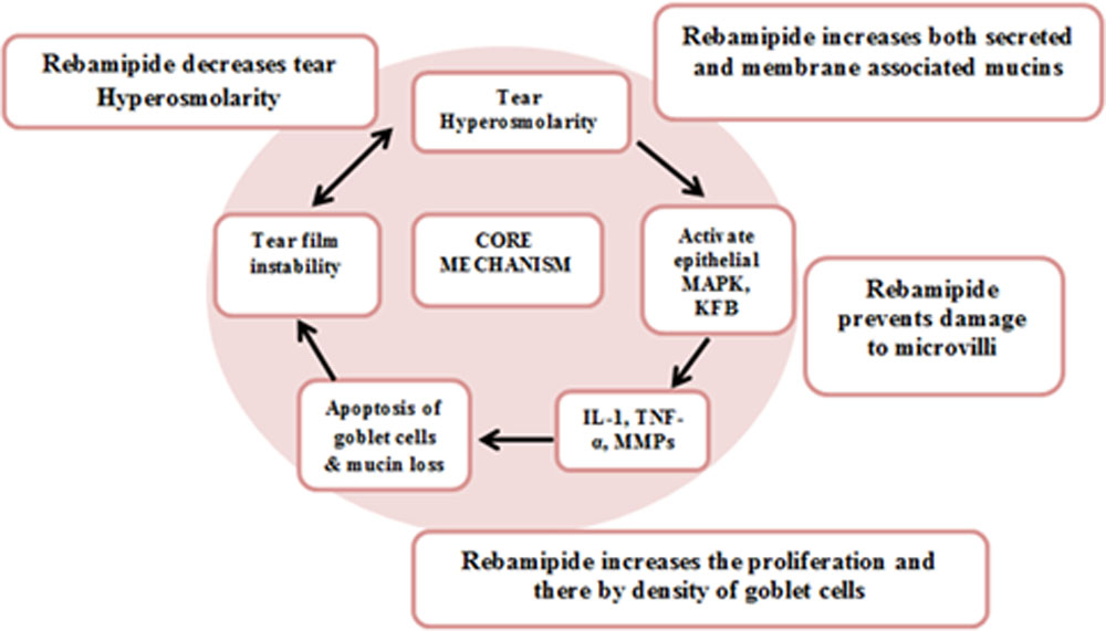
Objectives:To study the therapeutic effects of 2% rebamipide ophthalmic suspension in dry eye disease
MATERIALS AND METHODS
SOURCE OF DATA COLLECTION
Primary source of information with observational methodology was used on the patients diagnosed to have Dry eye disease who attended Outpatient Department in tertiary eye care government hospital.
SAMPLE SIZE : This was a prospective study of 50 patients from June 2016 to April 2017
INCLUSION CRITERIA
- All diagnosed cases of dry eyes for a minimum period of 6 months and willing to participate in the study.
EXCLUSION CRITERIA
- Presence of infective pathologies on the ocular surface and adnexa.
- Patients using other topically instilled ocular medications during study.
- Use of contact lenses.
- History of ocular surgery within 12 months.
METHOD OF STUDY
Patients who attended OPD between from June 2016 to April 2017 and diagnosed to have Dry eye were included in this study.
The diagnosis of dry eye was based on the diagnostic criteria of the Dry Eye Research group8. Patients with three essential problems, which are dry eye-related symptoms, abnormality of tear stability or secretion, and ocular surface damage were regarded as suffering from dry eye. Patients with two out of three problems were regarded as suspected of suffering from dry eye. The tear secretion was regarded as abnormal if the Schirmer I test resulted in equal to or less than 5 mm, and the tear stability is regarded as abnormal if TBUT (tear break up time) values resulted in equal to or less than 5seconds. The ocular surface damage is regarded as existing if the van Bijsterveld system score is equal to more than three points. In the current study, patients with two or three problems were enrolled.
From all patients detailed history was taken and routine ophthalmic examination was carried out. Slit-lamp biomicroscopy, fluorescein ocular surface staining (FOS), TBUT measurement, Schirmer’s test and interviews related to subjective dry eye symptoms were also conducted. All patients were prescribed 2 % rebamipide ophthalmic suspension to be administered four times in a day and followed up. Clinical data will be assessed at the following times: baseline visit and visits at 2 weeks, 4 weeks, 8 weeks, and 12 weeks.
Assessment
The subjective dry eye symptom was evaluated by conducting interviews covering same 11 parameters, which included foreign body sensation (as a feeling that something is in the eye), photophobia, itching, eye pain, dryness (as a dry sensation in the eye), heaviness, blurred vision, eye fatigue, eye discomfort (as a vaguely felling unpleasant sensation), eye discharge and lacrimation, in the same manner as the parameters were used in the previous reports 9,10. Each of the parameters was scored on a 4-point scale from 0 to 3 as follows: 0 = no symptoms, 1 = mild, 2 = moderate, and 3 = severe.
FOS assessment
Fluorescein was instilled by means of a fluorescein strip wetted with a drop of saline. Then the strip was tightly shaken once after saline moistening to remove excess fluorescein solution from the strip. The examiner gently retracts the patient’s lower lid and touch the edge of that with the strip.11 Fluorescein staining was evaluated with a blue free barrier filter according to the van Bijsterveld system, which divided the ocular surface into three zones: nasal bulbar conjunctiva, temporal bulbar conjunctiva, and cornea12. Each zone was evaluated on a scale of 0 to 3, with 0 indicating no staining, 1 indicating few separated spots, 2 indicating many separated spots and 3 indicating confluent staining; the maximum possible score with this system was 9.
Tear film break‑up time(TBUT)
The TBUT was measured by applying a slightly moistened fluorescein strip to the bulbar conjunctiva and asking the patient to blink. The tear film was then scanned under a slit‑lamp with a cobalt filter while the patient refrains from blinking. The time that elapses before the first dry spot appears in the corneal fluorescein layer is the TBUT.It is regarded as abnormal if values resulted in equal to or less than 5 seconds.
Schirmer test
Tear production was measured by the Schirmer test using Whatman 41 filter paper Strip without anesthesia. The strip was placed into the mid and temporal thirds of the lower lid (between the palpebral conjunctiva and the bulbar conjunctiva) without touching the cornea and the wetted length of the strip was read after 5 min.It is regarded as abnormal if wetted length is equal to or less than 5 mm.
RESULTS:
A total of 50 patients diagnosed with dry-eye syndrome were included in this study. The majority of the patients were between 51 to 60 years. The study included 61.3% women and 38.8% men.
Table 1: distribution of patients according to age
| AGE | FREQUENCY |
| <30Y | 1 |
| 31-40Y | 5 |
| 41-50 | 8 |
| 51-60Y | 25 |
| >60Y | 11 |
| TOTAL | 50 |
Subjective dry eye symptom was evaluated by conducting interviews covering same 11 parameters. The most common complaints of the patients were Foreign body sensation and sensation of dryness, followed by itching, discomfort, watering. The means score at baseline was 10.8±2.9. Dry eye symptom score at 2week, 4weeks, 8weeks, 12weeks was 9.8±2.6, 8.2±2.3, 7.4±2.0 and 6.8±1.8.Symptoms of dry eye improved significantly over 3months in majority of the patients following treatment.
FOSS score at baseline 3.0±2.5. Significant decreases in fluorescein uptake were observed in eyes treated with rebamipide. At 2week, 4week, 8week and 12week was 2.0±2.3, 2.0±1.7, 1.0±2.4 and 1.0±2.1 respectively which was statistically significant.
Schirmer’stest before the beginning of the treatment showed a wetting of 6.0±2.0 mm of the paper, and improved after 2week, 4week, 8week and 12week to 6.5±1.5, 6.5±1.5, 7.0±1.5 and 7.5±1.0 but statistically it was not significant
TBUT baseline 6.0±1.4, 6.3±1.2, 6.5±1.5, 7.3±1.8, 7.5±1.3 improved from baseline but statistically not significant
Table 2: Score of parameters at baseline, 2, 4, 8 and 12weeks
| DEWS | FOSS | ST | TBUT | |
| BL | 10.8±2.9 | 3.0±2.5 | 6±2.0 | 6.0±1.4 |
| 2week | 9.8±2.6 | 2.0±2.3 | 6.5±1.5 | 6.3±1.2 |
| 4week | 8.2±2.3 | 2.0±1.7 | 6.5±1.5 | 6.5±1.5 |
| 8week | 7.4±2.0 | 1.0±2.4 | 7.0±1.5 | 7.3±1.8 |
| 12week | 6.8±1.8 | 1.0±2.1 | 7.5±1.0 | 7.5±1.3 |
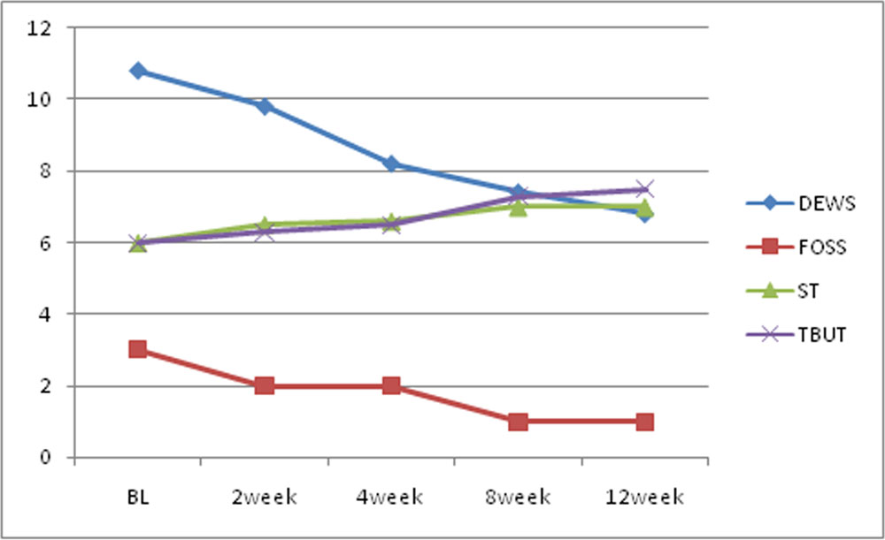
Graph showing change in different parameter from baseline to 12weeks
The most frequently observed adverse event was bitter taste in this study. This was observed in 5 patients. Eye irritation and stinging sensation were observed in one patient and two patients, respectively.
DISCUSSION:
The study population was predominantly female and included majority of patients aged 51-60 years. This population was representative of patients with dry eye in the general population13.
Subjective symptoms like foreign body sensation, feeling of dryness, photophobia showed improvement from baseline to 6 weeks. Study by ueda at el aslo showed similar results.Such improvements in subjective symptoms should contribute to improved quality of life in patients with dry eye6.
FOSS also showed significant improvement from baseline. efficacy results demonstrate that treatment with rebamipide, was associated with improvements in both corneal and conjunctival parameters of a dry eye condition.Kinoshita et al14 reported improvements of the fluorescein corneal staining score, lissamine green conjunctival staining score . These results were also similar to previous trials.14 An increase of mucin-like substances in the cornea and conjunctiva was observed with rebamipide in a preclinical model15.
Rebamipide has been shown to increase the number of periodic acid–Schiff-positive cells (goblet cells) in the conjunctiva16 and the mucin level on the cornea and conjunctiva16,17. Because decreased mucin levels on the surface of the cornea and a decreased density of goblet cells have been observed in patients with dry eye,18 the method of action of rebamipide is expected to be beneficial for this disease.
TBUT also showed improvement from baseline to 12 weeks. It was statistically significant only at 4 weeks but not at 12weeks. Koh et al19 reported that there was significant increases in the mean tear film break-up time, and there were significant upward curves from the baseline in terms of the total corneal higher order aberrations, coma-like aberrations, and spherical-like aberrations after the application of rebamipide. Kinoshita et al14 reported improvements of the tear film break-up time score during their investigation of the rebamipide dose response. Study by Udeet al6 reported that TBUT was significantly improved only at 8 weeks.
ST showed improvement from baseline but was not statistically significant. Kinoshita et al14 also found no significant difference for Schirmer’s test values between the rebamipide and placebo groups. Ude el al6 also found similar result.
The advantage of the rebamipide ophthalmic suspension, in that it does not contain preservatives, which can cause destabilization of the tear film, disruption of the corneal epithelium, a decrease of the density of the goblet cells, conjunctival squamous metaplasia with apoptosis, and damage to the deeper ocular tissues. The most frequently observed adverse event for the rebamipide ophthalmic suspension in the Kinoshita et al20 Phase III study was dysgeusia (bitter taste); however, this was only observed in 9.7% of the 2% rebamipide group.
CONCLUSION
The improvement of ocular surface conditions after its application is remarkable for the patients, especially in severe conditions and well-tolerated profile of rebamipide, makes it a potentially useful treatment option for dry eye.
REFERENCES
1.Bron AJ, de Paiva CS, Chauhan SK, Bonini S, Gabison EE, Jain S et al. TFOS DEWS II pathophysiology report. Ocular Surface 2017, july,15; vol 15(3); 438-510.
2.American Academy of Ophthalmology Cornea/External Disease Panel. Preferred Practice Pattern Guidelines. Dry Eye Syndrome. Limited revision. San Francisco, CA: American Academy of Ophthalmology; 2011. Available at: https://one.aao.org/CE/PracticeGuidelines/PPP.aspx. Accessed July 26, 2012.
3.Gayton JL. Etiology, prevalence, and treatment of dry eye disease.ClinOphthalmol 2009;3:405–12.
4.Friedman NJ. Impact of dry eye disease and treatment on quality of life.CurrOpinOphthalmol 2010;21:310–6.
5.Reddy P, Grad O, Rajagopalan K. The economic burden of dry eye: a conceptual framework and preliminary assessment. Cornea 2004;23(8):751–761.
6.Dhawan M, Hans T, Singh B.Rebamipide 2% Ophthalmic Suspension for the Treatment of Allergic Keratoconjunctivitis: A Hospital Based Study.DJO 2016;26:246-249.
7.Kaori Ueda, WataruMatsumiya*, Keiko Otsuka, Yoshifumi Maeda, Takayuki Nagai and Makoto Nakamura.Effectiveness and relevant factors of 2 % rebamipide ophthalmic suspension treatment in dry eye. BMC Ophthalmology (2015) 15:58.DOI 10.1186/s12886-015-0040-0
7.Dorennavar L, Maurya RP, Singh VP, Singh MK, Sharma K, Sharma R. The role of Rebamipide ophthalmic suspension in management of dry eye disease . Indian Journal of Clinical and Experimental Ophthalmology, October – December 2015;1(4):191-196
8.Shimazaki J. Definition and Diagnosis of Dry Eye 2006. J Eye. 2007;24:181–4.
9.Matsumoto Y, Ohashi Y, Watanabe H, Tsubota K. Efficacy and safety of diquafosol ophthalmic solution in patients with dry eye syndrome: a Japanese phase 2 clinical trial. Ophthalmology. 2012;119: 1954–60.
10.Hikichi T, Yoshida A, Fukui Y, Hamano T, Ri M, Araki K, et al. A multicenter study on definite and probable dry eyes.Jpn J Clin Ophthalmol.1994;48:1621–5.
11.Kanaya Y, Hori Y, Muramatsu R, Deguchi Y, Shiba T, Maeno T. Comparison of Tear Meniscus Height after Different Methods of Fluorescein Staining. J Eye. 2013;30: 1750–3.
12.VanBijsterveld OP. Diagnostic tests in the sicca syndrome.Arch Ophthalmol. 1969;82:10–4.
13.The epidemiology of dry eye disease: report of the Epidemiology Subcommittee of the International Dry Eye WorkShop (2007). Ocul Surf 2007;5:93–107.
14.Kinoshita S,Awamura S,Oshiden K, Nakamichi N, Suzuki H, Yokoi N; Rebamipide Ophthalmic Suspension Phase II Study Group. Rebamipide (OPC-12759) in the treatment of dry eye: a randomized, double-masked, multicenter, placebo-controlled phase II study. Ophthalmology. 2012 Dec;119(12):2471-8.
15.Urashima H, Okamoto T, Takeji Y, et al. Rebamipide increases the amount of mucin-like substances on the conjunctiva and cornea in the N-acetylcysteine-treated in vivo model. Cornea 2004;23:613–9.
16.Urashima H, Takeji Y, Okamoto T, et al. Rebamipide increases mucin-like substance contents and periodic acid Schiff reagent-positive cells density in normal rabbits. J OculPharmacolTher 2012;28:264 –70.
17.Takeji Y, Urashima H, Aoki A, Shinohara H. Rebamipide increases the mucin-like glycoprotein production in corneal epithelial cells. J OculPharmacolTher 2012;28:259 – 63\
18.Rivas L, Oroza MA, Perez-Esteban A, Murube-del-Castillo J. Morphological changes in ocular surface in dry eyes and other disorders by impression cytology. Graefes Arch ClinExpOphthalmol 1992;230:329 –34.
19.Koh S, Inoue Y, Sugmimoto T, Maeda N, Nishida K. Effect of rebamipide ophthalmic suspension on optical quality in the short break-up time type of dry eye. Cornea. 2013;32(9):1219–1223.
20.Kinoshita S, Oshiden K, Awamura S, Suzuki H, Nakamichi N, Yokoi N, Rebamipide Ophthalmic Suspension Phase 3 Study Group A randomized, multicenter phase 3 study comparing 2% rebamipide (OPC-12759) with 0.1% sodium hyaluronate in the treatment of dry eye. Ophthalmology. 2013;120(6):1158–1165.
Leave a Comment