

Dr.Harshavardhan horpade, Dr.Moreker Sunil Ratilal
Introduction: – Dua et al have demonstrated that trabecular mesh work is a direct extension of Dua’s layer. The collagen matrix of the human trabecular meshwork is an extension of the novel pre-Descemet’s layer (Dua’s layer)(1). Deep Sclerectomy has been used for control of intraocular pressure since a decade and more. Deep sclerectomy has definite advantages of non-penetrating methodology like lower risk of endophthalmitis, lower hypotony maculopathy incidences and low incidences of choroidal effusion. Some cases show good results while some fail. The cases which fail need a YAG gonio puncture. There is no way of predicting which cases will succeed and which will fail (2)(3). Guedes et al(4) have investigated the factors affecting the success of this surgery.
George Kistos et al (5) have discussed a modified deep sclerectomy to deal with failure and they postulated an external removal of the inner wall of Schlemm’s canal and the external layers of the trabecular meshwork with external trabeculectomy. Iordanidou et al (6) have shown how the bio mechanics of the cornea changes after deep sclerectomy and this may be responsible for success in stabilising field defects independent of the intra ocular pressure. Dua’s layer has been shown by Dua et al to affect the corneal biomechanics. It now seems that the removal of the Dua’s layer may be central to success of deep sclerectomy due to various reasons, one being imperviousness of this layer (which gets confused with Descemet’s layer) and the other being the fact that the biomechanics of the cornea with removal of this layer. This layer is impervious to water. We hypothesized that if this layer is selectively removed then the aqueous drainage may improve. Electron microscopy is the gold standard to identify Dua’s layer in histology specimen. There are two kinds of electron microscopy, scanning and transmission.
Methods: – Triple blind random comparative case-control electron microscopy study to look for Dua’s layer in excised specimen received after non- penetrating surgery for glaucoma in which deep flap removed from failed cases and successful cases (control) were examined. We did non- penetrating glaucoma surgery on 40 eyes of twenty patients. In one eye trabeculectomy was done and in the other eye a modified deep sclerectomy was done in which selectively Dua’s layer was removed. Conjunctival flap was raised as is done during filtering surgery.
The superficial scleral flap was done with crescent knife. Deep flap was fashioned to include Duas layer and extended into the cornea for 2 mm beyond white line. Deep flap with Dua’s layer was removed and we sent the dissected tissue for Electron microscopy and to look for Dua’s layer in cases which failed and cases of Deep sclerectomy which succeeded. Success qualified as 12 percent reduction of IOP as compared to baseline IOP without drops. Patients in whom target pressure was not reached were given additional pressure lowering agents to bring the IOP to target level. A video audit was done to identify whether in the cases which qualified as success and in whom Dua’s layer was identified in electron microscopy had a egress of trypan blue during surgery and whether in those in which egress did not occur intra operatively, whether they had Dua’s layer seen in electron microscopy.
Results: – Three eyes of patients who qualified as success of deep sclerectomy were selected and the tissue was sent for electron microscopy. Dua’s layer was identified in all the three. Corresponding matched three eyes in which surgery failed were also sent for electron microscopy and Dua’s layer was not identified in these. The electron microscopy was reviewed by two pathologists and two eye surgeons who were blinded to which group the tissue belonged. The investigator and statistician were also blinded
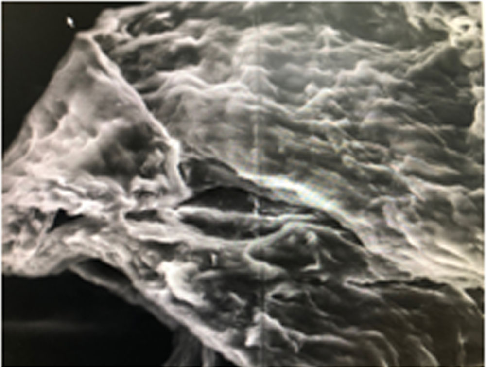
Discussion: We have been doing modified deep sclerectomy since 5 years at our hospitals and go a little ahead anteriorly into the cornea and create a Descemet’s window without perforating that window and sometimes we do not see egress of fluid at the removal of deep scleral flap and deroofing of the schlemm’s canal and the Descemet’s window. We routinely do a video audit of our cases. In our video audit we have noted that in cases where there is no fluid egress the “descemet’s membrane” withstands pressure and the air bubble in the anterior chamber is visible and even on increasing the anterior chamber pressure with a large bubble the “descemets membrane” does not rupture. Dua et al (7) have discussed the properties of this layer and it is said to be impervious to air and known to withstand pressures upto 200kPa.We postulate that
This is a combination of descemet’s layer and Dua’s layer which has not been removed. These are the cases in which the deep sclerectomy fails and needs a gonio puncture. In other cases the “descemet’s membrane” is very thin and shows egress of fluid spontaneously and sometimes ruptures if the anterior chamber pressure is high due to large air bubble, releasing a small air bubble but no prolapse of iris or sometimes leading to iris prolapse without the membrane being touched. In these cases there is good egress of fluid even from the area of schlemm’s canal. These cases of deep sclerectomy are successful. These are the cases where the Dua’s layer has been removed and hence the layer remaining in the eye is purely Descemet’s layer and allows egress of fluid and cannot withstand pressure. We postulate that the thick membrane that withstands pressure and does not allow egress of fluid easily is a sclerosed peripheral part of Dua’s layer which if not removed causes failure of deep sclerectomy necessitating a gonio puncture later. Removing this layer is probably the key to success of Deep Sclerectomy.
Conclusion: – Dua’s layer removal may be vital for success of deep sclerectomy. Glaucoma surgeons may have to be trained in lamellar surgery to make sure that this important layer is removed so as to ensure success in glaucoma surgery. Further evaluation is needed to substantiate this hypothesis though our video audit shows that, cases in which the layer has not been removed, fail and need a gonio puncture and the cases in which the Dua’s layer is removed are successful and do not need a gonio puncture suggesting that removal of Dua’s Layer is essential for success of Deep Sclerectomy, modified or otherwise.
References:-
1)Harminder S Dua, Lana A Faraj,Matthew J Branch,Aaron M Yeung,Mohamed S Elalfy,Dalia G Said,Trevor Gray,and James Lowe.The collagen matrix of the human trabecular meshwork is an extension of the novel pre-Descemet’s layer (Dua’s layer)Br J Ophthalmol 2014 98:691-697;
2)El Sayyad F(1), Helal M, El-Kholify H, Khalil M, El-Maghraby A.Nonpenetrating deep sclerectomy versus trabeculectomy in bilateral primary open-angle glaucoma.Ophthalmology. 2000 Sep;107(9):1671-4.
3)Zsolt Varga and Tarek Shaarawy.Deep Sclerectomy: Safety and Efficacy.Middle East Afr J Ophthalmol. 2009 Jul-Sep; 16(3): 123-126.
4)Guedes RA(1), Guedes VM, Chaoubah A. Factors associated with non-penetrating deep sclerectomy failure in controlling intraocular pressure.Acta Ophthalmol. 2011 Feb;89(1):58-61.
5)George Kitsos,Miltiades Aspiotis, Yannis Alamanos, Konstantinos Psilas. A modified deep sclerectomy with or without external trabeculectomy: a comparative studyClinical Ophthalmology 2010:4 557-564
6)Iordanidou V(1), Hamard P, Gendron G, Labb? A, Raphael M, Baudouin C.Modifications in corneal biomechanics and intraocular pressure after deep sclerectomy.J Glaucoma. 2010 Apr-May;19(4):252-6.
7)Dua HS, Faraj LA, Said DG, Gray T, Lowe J (September 2013). “Human corneal anatomy redefined: a novel pre-Descemet’s layer (Dua’s layer)”. Ophthalmology 120 (9): 1778-85
Leave a Comment