

Dr.Dipankar Das,Dr.Sarma Prafulla,Dr.Harsha Bhattacharjee
Introduction: Glaucoma is an important vision threatening disease that has significant economic burden.1,2,3,4 Optic nerve head is directly susceptible to intraocular pressure (IOP) elevation.1 It is the optic nerve head and nerve fibre layer containing retinal ganglion cells (RGCs) axons that are most evidently linked with glaucomatous visual loss.1,2,3,4,5 Computational cellular and mathematical modelling is a newer method of understanding the human biology and system.1, 8, 9, 10 This branch of science opens our mind towards the ocular research. Till date, there were many drugs that had been used in the medical management of various glaucomatous conditions. Peripapillary uveal and dendritic cells (DCs) can be the potential drug delivery vehicles in anti glaucoma treatment.
Aim: To present cellular modelling in enucleated eyeballs’ optic nerve and its understanding that can open up the idea for future drug delivery vehicle in glaucoma
Methods: Design-A laboratory based study.
Analysis of enucleated eyeballs’ optic nerve in a gross pathology, stained specimen and immunohistochemistry (IHC) that were carried out to know the anatomical pigmented cells arrangements in an around the edges of the optic disc.
Total eight enucleated specimens from seven patients for various other indications were studied including two postmortem eyes from a patient which were taken after proper consent.
Optic nerves were studied by microscopy (Leica S6D and Axioskop 40 with AxioCam MRc, ZEISS, Germany). Routine stains with H&E, diffractive photography, hematoxylin-eosin (H&E) and molecular pathologic study were done in each of the optic nerve cross sections.
Trabecular meshworks (TM) and retinal ganglion cells (RGCs) of these cases were studied for the association.
Results: Five cases had some clue of glaucomatous change. Two eyes of one patient was enucleated after death(postmortem) with proper eye-bank consent for research who had 30 years of glaucoma; One, post trabeculectomised eye with history of trauma and painful blind eye, two other cases removed for painful blind eyes with raised IOPs which include one phthisical eye. Other three eyes were of different indications removed in OT after pre-operative consent. The age of the patient varied from 13 years-79 years; Male [(2) +4]: Female eyes [2]
Pigmented cells from the uveal tissues were spotted near the peripheral edges of the optic nerve. Their morphological sizes, shape, direction of advancement were documented. In all these cases, the TM and RGCs were studied for the similar pigmented cells for the correlation.
Peripapillary cells were pigmented, dendritic shaped, globular, varied from 90 microns to 170 microns. Peripapillary cells were in centrifugal configuration. TM cells were mostly had dendritic pattern. IHC with control were done for BAX, BCL2 and CD 68 for peripapillary cells, TM and RGCs. IHCs were done using kit methods (Standardized) and adequate controls were taken for each sample.
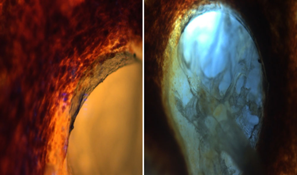
Fig 1, 2: Deep glaucomatous cups in enucleated eyes
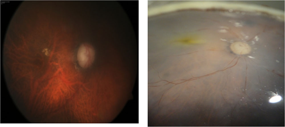
Fig 3, 4: Glaucomatous cup fundus picture of a patient 11 month before his death and right side picture was the studied postmortem enucleated right eye. Other eye had glaucomatous optic atrophy
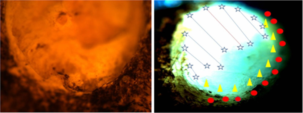
Fig 3, 4: Flat preparation in enucleated eyeball showing peripapillary pigmented cells near neuroretinal rim, Right side picture showing whitish cup and also red zone where peripapillary pigmented cells were traced.
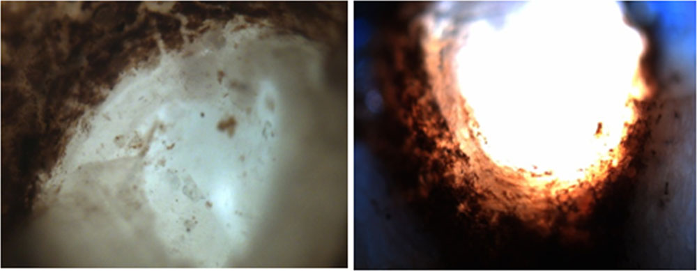
Fig 5, 6: Postmortem glaucomatous optic atrophied in the left eye, also showing pigmented uveal cells at the edges; Right hand picture showing deep cup in juvenile glaucoma with prominence of beta zone and few pigmented cells around the disc margin.
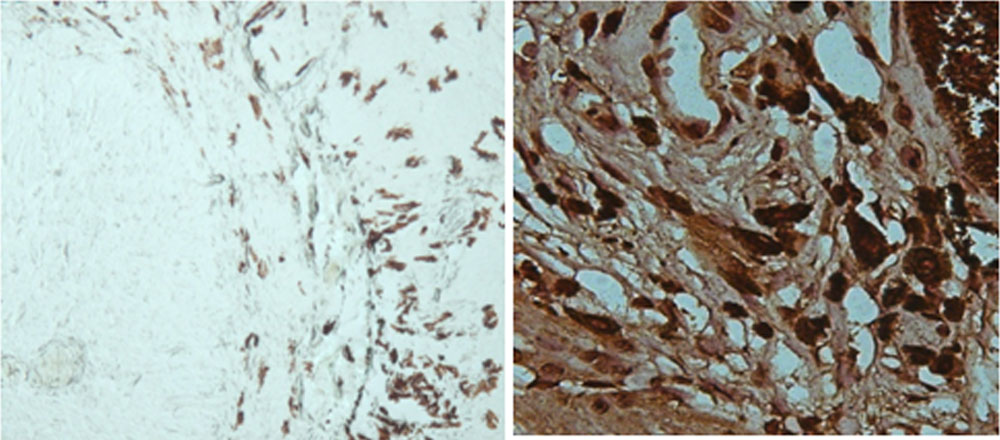
Fig 7, 8: Peripapillary cells stained with CD 68 and right hand picture TM cells stained with CD 68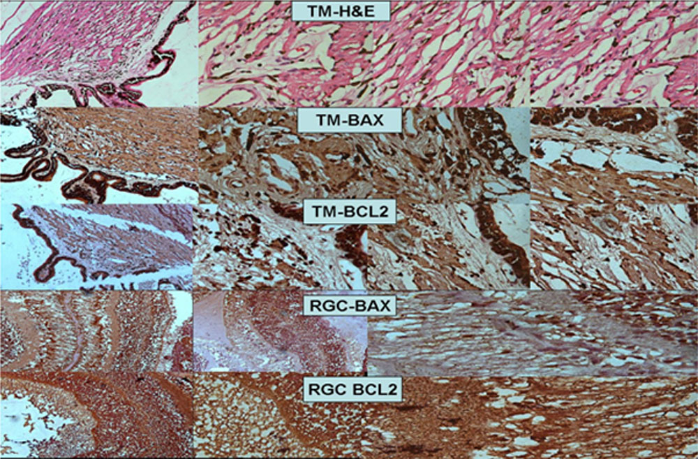
Fig 9: H&E and IHCs on TM and RGCs (BAX, BCL2; Bio-Genex, USA).
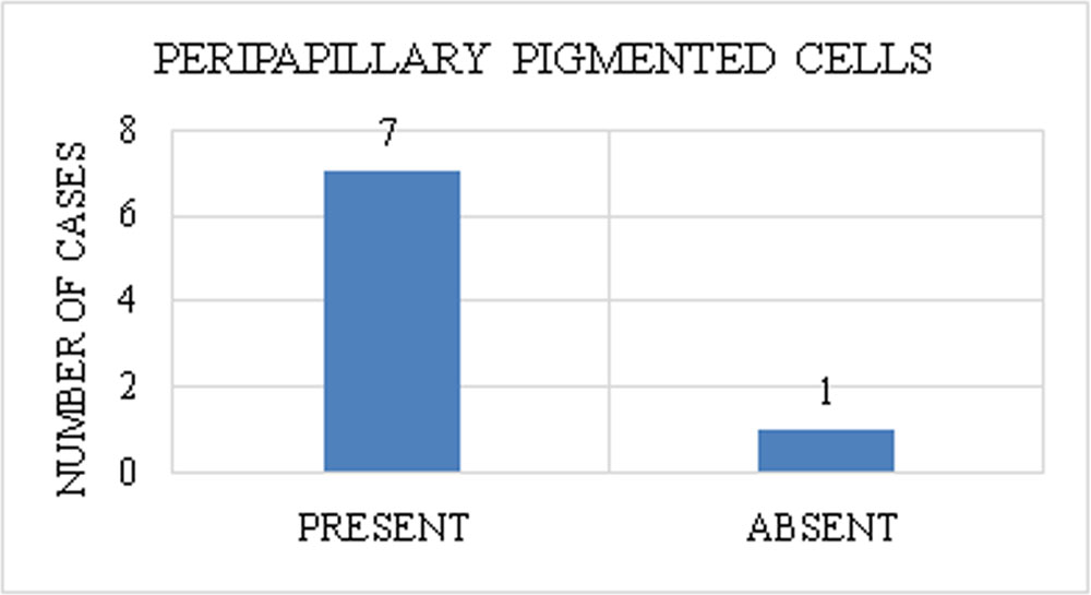
Graph1: Out of 8 enucleated eyeballs, 7 showed peripapillary pigmented cells.
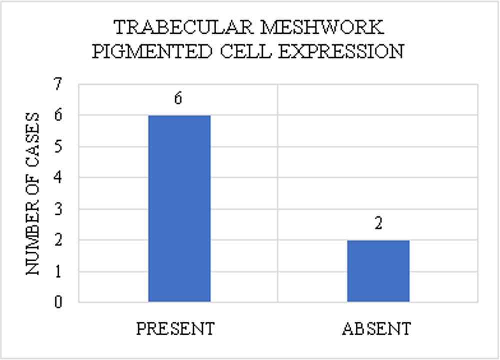
Graph 2: Out of 8 enucleated eyeballs, 6 showed trabecular cells stained with IHC for the pigmented cells particularly, DC.
Discussions:
It is well known that optic nerve head composed of axons and as well as astroglial and collagen support but role of peripapillary pigmented cells were not explored by the experts, till date. Connection of peripapillary pigmented cells, RGCs near the optic nerve head and TM anteriorly may exist. BCL 2, BAX and CD68 (dendritic cells) expression were seen here in glaucomatous eye and the control (postmortem) eye. BCL2, anti-apoptotic protein was involved in neuroprotective effect directly or indirectly related to ocular hypertension in rat model.8 Our study showed BCL2, BAX and CD 68 protein expression in all these tissues. In traumatic glaucomatous optic atrophy models, these molecules showed significant change.8 DC expression (CD68) in RGC, peripapillary optic nerve cells and TM had shown expression in three cases and these cells can be targeted in future drug delivery. We knew that peripapillary pigment change in alpha and beta zone is frequently associated with glaucomatous optic atrophy but they are also seen in other conditions like myopia and aging. 5, 6, 7 Whether reversal of glaucomatous cup can be possible, we do not know exactly? 5,6,7,8,9,10 Pigmented cells in those areas have cellular interconnected link from optic disc to TM for those three molecules studied.
Mastropasqua et al had shown that by confocal imaging and in-vivo dendritic cell expression with staining positive in corneal/limbal tissue in glaucoma patient 9 and we had seen the DC expression in TM, peripapillary cells as well as RGCs, thereby, possible drug delivery can be found out at the targeted site where the vehicle of drug will be those specialized cells. Since there were no drug delivery system in glaucoma that influences those pigmented DCs near the optic disc, RGCs and TM, this study can be an eye opener for new drug delivery vehicle in glaucoma.
Acknowledgements:
Prof. Panna Deka, MD, Co-Pathologist, Sri Sankaradeva Nethralaya (SSN), Guwahati, India
Mr. Apurba Deka, Msc, Technician, Ocular Pathology Lab, SSN, Guwahati, India
Dr. Shahinur Tayab, MS, Associate Consultant, Glaucoma Clinic, SSN
Dr. Saurabh Deshmukh, MBBS, DNB PGT, SSN
References:
- Quigley HA, Brown AE, Morrison JD, Drance SM. The size and shape of the optic disc in normal human eyes. Arch Ophthalmol. 1990;108:51-7.
- Hayreh SS. Anatomy and physiology of the optic nerve head. Trans Am Acad Ophthalmol Otolaryngol. 1974;78:240-54.
- M Ko, D Kim, Y Ahn. Morphological variations of the peripapillary circle of Zinn-Haller by flat section. Br J Ophthalmol. 1999;83:862-6.
- Quigley HA, Green WR. The histology of human glaucoma cupping and optic nerve damage: clinicopathologic correlation in 21 eyes. Ophthalmology. 1979;86:1803-30.
- Danesh-Meyer HV, Levin LA. Neuroprotection: extrapolating from neurologic diseases to the eye. Am J Ophthalmol. 2009;148:186-91.e2.
- Kelley MJ, Rose AY, Keller KE, Hessle H, Samples JR, Acott TS. Stem cells in the trabecular meshwork: present and future promises. Exp Eye Res. 2009;88:747-51.
- Tezel G, Edward DP, Wax MB. Serum autoantibodies to optic nerve head glycosaminoglycans in patients with glaucoma. Arch Ophthalmol. 1999;117:917-24.
- Martin KR, Quigley HA, Zack DJ, Levkovitch-Verbin H, Kielczewski J, Valenta D, et al. Gene therapy with brain-derived neurotrophic factor as a protection: retinal ganglion cells in a rat glaucoma model. Invest Ophthalmol Vis Sci. 2003;44:4357-65.
- Mastropasqua R, Agnifili L, Fasanella V, Lappa A, Brescia L, Lanzini M, et al. In Vivo Distribution of Corneal Epithelial Dendritic Cells in Patients With Glaucoma. Invest Ophthalmol Vis Sci. 2016;57:5996-6002.
- Lo R, Li PY, Saati S, Agrawal RN, Humayun MS, Meng E. A passive MEMS drug delivery pump for treatment of ocular diseases. Biomed Microdevices. 2009;11:959-70.
Leave a Comment