

Dr.Nabanita Barua, Dr.Chitra Sitaraman,Dr.Sonu Goel
Purpose: Slit lamp examination and IOP monitoring have been used as surrogate marker for assessment of bleb functionality for ages. In general, bleb appearance can be classified clinically as diffuse, cystic, encapsulated and flattened. Good intraocular pressure (IOP) control (>21 mm Hg) after filtration surgery was commonly found to be associated with low lying diffuse blebs. OCT is now being used to look for the internal morphology to predict the long term functional viability of the bleb. A prospective and descriptive study analyzing 60 eyes that had earlier undergone trabeculectomy was performed. All blebs were imaged postoperatively with a spectral domain OCT, (RTVue Inc., Fremont, CA). The architecture was described and functional correlation was evaluation.
Method: Bleb assessment of all patients operated for only trabeculectomy or combined procedure were done by slit lamp biomicroscopy and spectral domain OCT. the time span was between 1st post operative day to 1 year. Any patient with re-surgery, previous conjunctival disease or systemic antimetabolite therapy were excluded.
Results: Of the 30 participants, 20 (66.66%) were males and 10 (33.3%) were females . age group 12-62y. Ten eyes (33.3%) had primary open-angle glaucoma, 10 eyes (33.3.0%) had angle closure glaucoma, 3 eyes (.1%) had exfoliation glaucoma, 2 eyes (.06%%) glaucoma secondary to uveitis, 3 eyes (.1%) had neovascular glaucoma and 2 eyes (.06%%) had developmental glaucoma. All procedure were fornix based. The mean IOP was 12.1±5.4mm Hg. The interval between surgery were 1 day-12 months post operative day. The IOP were significantly correlated with the bleb height 650 microns (range 468-1235), wall thickness 125 microns (range 97-257), internal cavity height 847 microns (range 568-1380) . The mean preoperative IOP of the 20 eyes was 28 mmHg (range 18-36 mmHg) and the final average postoperative IOP at the time of RT OCT-measurement was 12.1 mmHg (range 5-30 mmHg). Internal cavity parameters and bleb reflectivity correlated well with the IOP of a functional bleb.
Conclusions: The in vivo architecture of filtering blebs can be objectively described using OCT according to quantitative and qualitative parameters. Internal cavity height and extent, as well as bleb reflectivity measurements are related to the functional aspect of a filtering bleb.
Key Words: Bleb Analysis, Failing Bleb, OCT evaluation of bleb
Manuscript Organization
Introduction:
Since the introduction of trabeculectomy by Cairns in 1968 various modalities have been suggested to assess the functionality of bleb. IOP and slit lamp examination are taken as surrogate marker for viability of bleb. Achieving target pressure without any supplementary topical medication are the ultimate goal.
- UBM gives finer clue about internal milieu of bleb.
- There are various classification system based on the morphological analysis : The Indiana Bleb Appearance Grading Scale (IBAGS) , Moorfield and Kronfeld classification system are all helpful but these are all qualitative and observer dependent.
- With the advent of anterior segment optical coherence tomography (OCT), non-contact, high resolution tomographic cross sectional imaging of anterior segment structures has become possible.
- This documents internal morphology which could be a better predictor of functionality of
Yamamoto et al has described the bleb based on the reflectivity
inside the bleb, blebs were classified into low reflective (type L), high reflective (type H), encapsulated (type E) , flattened (type F).
Good IOP control was found in type L blebs whereas type H, E or F blebs were, in general, associated with fair to poor IOP control.
Savini et al reported the use of the Stratus OCT (Carl Zeiss Meditec, Dublin, California, USA) to image filtering blebs.
Type A had a thick wall and a single large fluid filled space
Type B had a thin wall and multiple large fluid filled spaces
type C had multiple flattened fluid filled spaces.
Materials and Methods
- The study was conducted in accordance with the ethical standards of the 1964 Declaration of Helsinki and approved by the local clinical research ethics committee.
- 30 eyes of 22 patients were included.
- Patients were all subjected to examination with OCT after due consent
- Day of examination varied from immediate second post-operative day to 1 year follow up.
- Patient were evaluated on slit lamp and applanation tension was recorded. (for recent post op NCT was done ) .With RT optovue OCT with anterior segment module bleb photographs were taken. Inclusion criteria: Operated trabeculectomy/combined surgery upto 12 months
- Exclusion criteria: Resurgery
previous failed bleb
Evidence of previous conjunctival disease
Patient on antimetabolite therapy
Measurements:
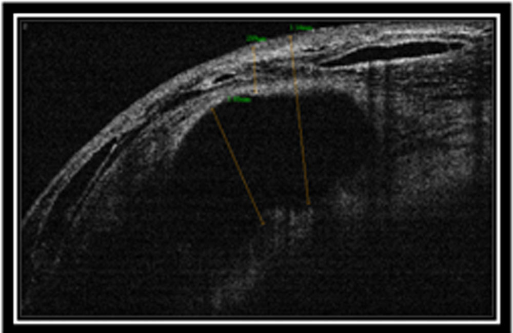
- Bleb wall thickness is defined as the distance between the first reflective signal from the conjunctiva to the top of the subconjunctival fluid space. As bleb wall thickness may vary along the scan, only the maximum distance was measured. (d)
- Bleb height was measured from first signal from conjunctiva to the base of subconjunctival fluid space.(a)
The subconjunctival fluid space was measured as the maximal thickness of the hyporeflective area directly beneath the bleb wall in the vertical cross sectional image. (b)
| Type | Average | SD | ||
| Diffuse elevated | Cystic | |||
| Bleb Wall | 187.7 (150-350) | 73.85 | 340 (120-480) | 99 |
| Subconjunctival fluid | 69.17 (50-98) | 17 | 670 (200-1049) | 289 |
| Bleb height | 342 ( 200- 460) | 180 | 359 (90- 1000) | 277 |
| IOP | 12.2 | 2.89 | 14.12 (6-20) | 4.22 |
The height, the extent of internal cavity nor the bleb height was significantly correlated with IOP.
The bleb wall thickness was significantly negatively correlated with IOP (.003)
The bleb wall reflectivity is an important marker of function of funtioning of bleb.
- Discussion: We described and quantified the internal features of different bleb morphologies with reference to the observed clinical appearance and bleb.
- The higher scanning resolution in RTOPTOVUE OCT images allows us to differentiate two levels of intrableb fluid filled spaces:
(1)the subconjunctival fluid collections and
(2) the suprascleral fluid space.
Good Bleb wall, height, fluid pockets controls IOP well.
Average IOP (14+2.25)in low to intrableb reflectivity is significantly better than high bleb reflectivity (20+ 4.5)
So absence of the subconjunctival fluid collections, appearance of high reflective spike in immediate post op might give clue towards impending failing bleb.
Combining both clinical and imaging information could provide a new perspective towards understand the ultimate outcomes after trabeculectomy and need for early intervention like
Augmentation of frequency of drugs
Bleb needling
References:
- Laser suturo Cairns JE. Trabeculectomy. Preliminary report of a new method. Am J Ophthalmol
1968;66:673–9. - Vesti E. Filtering blebs: follow up of trabeculectomy. Ophthalmic Surg 1993;b24:249–55.
- Cantor LB, Mantravadi A, WuDunn D, et al. Morphologic classification of filtering blebs after
glaucoma filtration surgery: the Indiana Bleb Appearance Grading Scale. J Glaucoma 2003;12:266–71. - Wells AP, Crowston JG, Marks J, et al. A pilot study of a system for grading of drainage blebs after
glaucoma surgery. J Glaucoma 2004;13:454–60. - Pavlin CJ, Harasiewicz K, Foster FS, et al. Ultrasound biomicroscopy of anterior segment structures
in normal and glaucomatous eyes. Am Ophthalmol 1992;113:381–9. - Huang D, Swanson EA, Lin CP, et al. Optical coherence tomography. Science 1991;254:1178–81.
- Kronfeld PC. The mechanism of filtering operations. Trans Pac Coast Otoophthalmol Soc Annu Meet
1949;33:23–40. - Van Buskirk EM. Cysts of Tenon’s capsule following filtration surgery. Am J Ophthalmol
1982;94:522–7. - Yamamoto T, Sakuma T, Kitazawa Y. An ultrasound biomicroscopic study of filtering blebs after
mitomycin C trabeculectomy. Ophthalmology
1995;102:1770–6. - Savini G, Zanini M, Barboni P. Filtering blebs imaging by optical coherence tomography. Clin Exp
Ophthalmol 2005;33:483–9. - Powers TP, Stewart WC, Stroman GA. Ultrastructural features of filtration blebs with different
clinical appearances. Ophthalmic Surg Lasers 1996;27:790–4. - Addicks EM, Quigley HA, Green WR, et al. Histologic characteristics of filtering blebs in
glaucomatous eyes. Arch Ophthalmol 1983;101:795–8. - DeBry PW, Perkins TW, Heatley G, et al. Incidence of late-onset bleb-related complications
following trabeculectomy with mitomycin. Arch Ophthalmol 2002;120:297–300.344 Leung, Yick,
Kwong, et al www.bjophthalmollysis and so on
Leave a Comment