

Dr.Neha Adlakha,Dr.Manpreet Kaur,Dr. Yogesh Kumar,Dr. Nishtha Saini
ABSTRACT-
Pterygium excision alter keratometry by reducing astigmatism, thus affect intraocular lens (IOL) power calculation in concurrent pterygium and cataract.Aim of study is to evaluate the effect of pterygium excision on biometry and relationship between these effect with size of pterygium. A prospective study on 700 patients who had undergone unilateral pterygium excision was done.Pterygium length and area were calculated. Preoperative and 1 week,1 month and 6 months postoperative biometry were obtained for eyes with pterygium and fellow eyes(control).
Mean pterygium length and area were 2.96±0.74mm and 5.98±2.2 mm2. Preoperative power calculation were higher than postoperative and control(p<0.001). Mean change were 0.39D for SRKII, 0.64D for SRK/T and 0.49D for Haigis. Positive correlation were found between pterygium size and power change. If pterygium size is larger than 2.18 mm or 6.42 mm2 and simultaneous surgery is planned , implanted IOL power should be at least 0.50 D smaller than the calculated power.
INTRODUCTION :- Pterygium is a fibroelastic degeneration of the conjunctiva [1] that arises from the limbal conjunctiva and extends over the cornea replacing Bowman’s membrane and corneal epithelium . The extent of visual impairment depends upon its growth across the visual axis. Pterygia cause corneal distortion and induce a significant amount of astigmatism [2] which can affect the outcome of the cataract surgery. This astigmatism may occur either due to pooling of tears in advance of the pterygium or by traction generated by the pterygium mechanically pulling on and distorting the cornea or both [3]. Pterygium is known to induce with the rule astigmatism as corneal curvature is flattened along the long axis of body of pterygium.
The treatment of pterygium is its removal which can be performed prior to or combined with cataract surgery. Excision of pterygium will steepen the cornea and reduce corneal astigmatism . Moreover, accurate IOL power calculation is essential for better visual and refractive outcomes, and subsequent patient satisfaction, even when simultaneous pterygium and cataract surgery is performed. Accurate and stable keratometry is crucial in avoiding postoperative refractive surprises. Pterygium excision when combined with cataract surgery or performed after the cataract surgery would alter the keratometry values of the cornea which were used for the biometry. This might result in a refractive surprise.
Large pterygia extending on to the visual axis would undoubtedly have to be removed before planning any cataract surgery. Long standing and atrophic pterygia cause scarring and the astigmatism induced by these may not be fully reversed even after excision. The effect of pterygium on the corneal refractive status has been measured by refraction, keratometry and corneal topography [5].The aim of this study was to evaluate the effects of pterygium on intraocular lens power calculations and also to determine the relationship between pterygium size and changes in intraocular lens power after it is removed from the cornea.
AIIMS AND OBJECTIVES-
- To evaluate the effects of pterygium excision on intraocular lens power calculations.
- To determine the relationship between changes in intraocular lens power and size of pterygium.
- To compare preoperative and postoperative astigmatism after pterygium excision.
MATERIALS AND METHODS-
A prospective interventional study was conducted on 700 eyes of 700 patients. Patients aged 25 – 65 years with primary nasal pterygium were included in the study. The exclusion criteria were pseudopterygium, recurrent pterygium, corneal scarring from any cause and any previous ocular surgery. The study was approved by the Institutional Ethical Committee. A written informed consent was obtained from all the patients .After obtaining a good ocular and systemic history, ocular examination was done which included Snellen visual acuity, manifest refraction and slit lamp anterior segment examination. The dimensions of pterygium were measured using slit lamp biomicroscope by taking length in mm at the base and the apex. Area of pterygium was calculated by measuring the width at the apex (Base 1)and the base (Base 2) and the length of the pterygium. An intraocular lens power calculation was performed for eyes with pterygium and their healthy fellow eyes (control group). Pterygium excision was performed .
Area=height (Base 1 + Base 2)/2
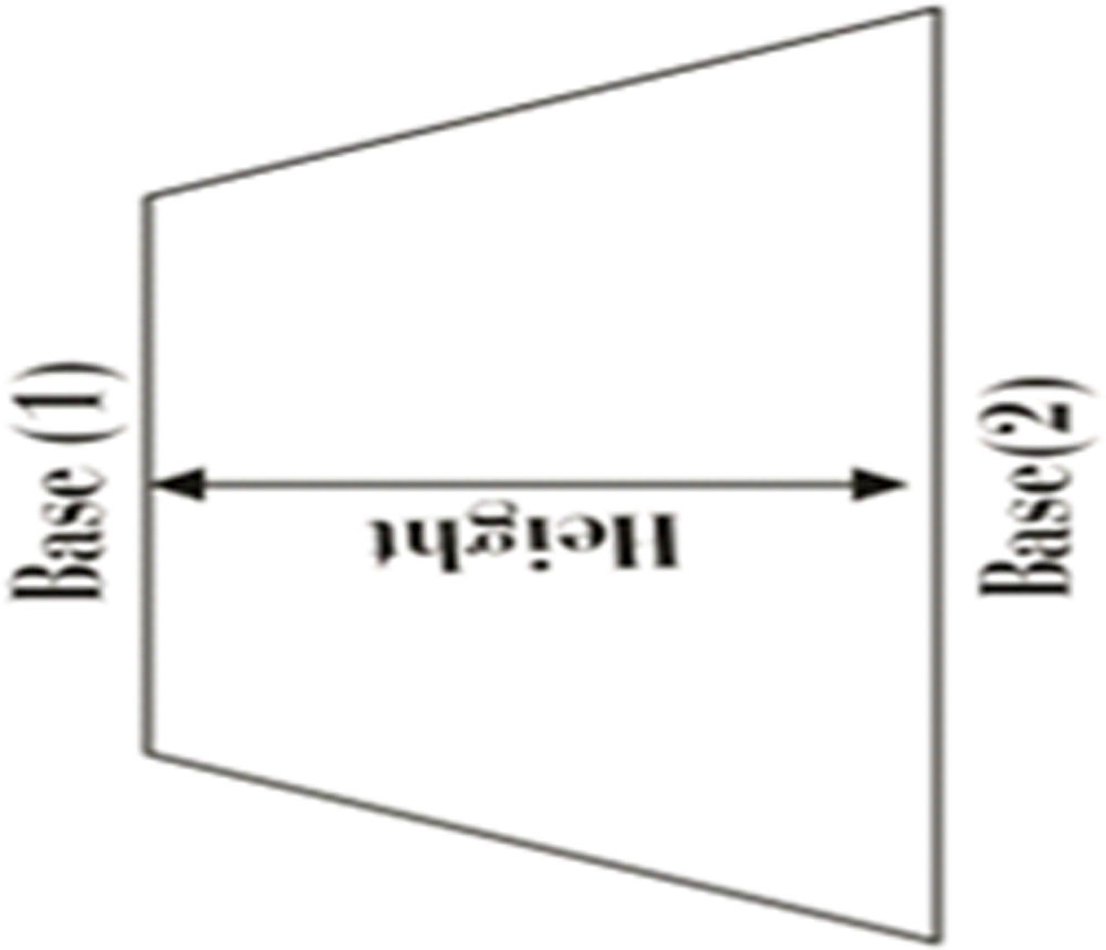
All the surgeries were performed by a single surgeon under peribulbar anaesthesia containing 4 ml of 2% lignocaine and 4 ml of 0.5% bupivacaine. With the help of a 15 number Bard Parker blade, a delineation mark is done on cornea 1 mm ahead of pterygium head and pterygium was excision. Head of pterygium was excised with gentle dissection and traction avulsion from the corneal surface, neck and body of pterygium dissected and pterygium tissue excised after delineating and separating it from overlying conjunctiva and underlying sclera.
The size of bare sclera area is measured with callipers and conjunctival autograft of size 1 mm more than bare sclera in both dimensions is harvested form superior or superotemporal quadrant. In case of free graft, limbal side of the graft is oriented to the limbal side of the graft and the defect is closed by suturing autograft with adjacent conjunctiva by nylon 10-0 sutures. Topical antibiotic eye ointment application with pad and bandage was done to the eye for 24 hours. Follow up of patients was done on 1st post operative day, 7th post operative day (1 week follow up) and 45th post operative day (1.5 month follow up). and patient followed after 7 days, 1 month, 45 days and 60 days of surgery. During each follow up,visual acuity, refraction, keratometry ,axial length and intraocular power calculation of both eyes were performed.
DATA ANALYSIS-
Statistical analysis was performed using Statistical Package for Social Sciences (SPSS) for windows. Descriptive statistics was used to describe the clinico-demographic data. Frequency and percentages were calculated for qualitative variables. Median and mean rank was used for numerical data. Paired t-test was used to compare the preoperative and postoperative keratometry, axial length and intraocular lens power. Mean ± standard deviation was used for description of age statistics. A p value < 0.05 was considered statistically significant.
RESULTS-
Seven hundred eyes with pterygium (study group) and seven hundred fellow healthy eyes (control group) were included in this study. The mean age of 489 females and 211 males was 54.87 ± 3.41 years (range 42 to 65 years). Mean pterygium length and area were 2.96±0.74 mm and 5.98±2.2 mm2. In the study group, the preoperative mean K values were lower than the control group. The AL values were similar between the 2 groups
Preoperative intraocular lens power calculation were higher than postoperative and control(p<0.001). Mean change were 0.39D for SRKII, 0.64D for SRK/T and 0.49D for Haigis. Positive and significant correlations were found between the horizontal pterygium length and area and the preoperative and postoperative intraocular lens power values. If pterygium size is larger than 2.18 mm or 6.42 mm2 and simultaneous surgery is planned , implanted IOL power should be at least 0.50 D smaller than the calculated power.
References-
- Moran DJ, Hollows FC. Pterygium and ultraviolet radiation: a positive correlation. Br J Ophthalmol 1984; 68:343-6
- Yagmur M, Özcan AA, Sari S, Ersöz TR. Visual acuity and corneal topographic changes related with pterygium surgery. J Refract Surg2005; 21:166-70
- Oldenburg JB. Conjunctivalpterygia: mechanism of corneal topographic changes. Cornea 1990; 9:200.
- Yousuf M. Role of pterygium excision in pterygium induced astigmatism. JK-Practitioner 2005; 12:91-2.
- Saleem M, Channar MS, Saleem MF. Effects of pterygium excision on corneal curvature. Pak J Med Sci2011; 27:325-8.
Leave a Comment