

Dr.Noopur Gupta, Dr.Rashmi Singh,Dr.Amreen A.Prof.(Mrs ) Radhika Tandon
ABSTRACT
PURPOSE:
Assessment of quality of vision and vision-related quality of life in patients undergoing anterior lamellar keratoplasty (ALK)
METHODS:
A prospective interventional study conducted at a tertiary eye care centre to assess the quality of vision in patients who have undergone ALK for anterior corneal opacities with a secondary objective being to assess the vision-related quality of life (using IND-VFQ-33 Questionnaire). The visual acuity, keratometric cylinder for astigmatism, contrast sensitivity, optical aberrations were measured preoperatively, and post-operatively at month 1, 3 and 6. IND-VFQ-33 questionnaire was administered preoperatively and post operatively at 6 months.
RESULTS:
15 eyes of 15 patients were included with mean age of 43.5±21.7 years. The most common indication for ALK was healed keratitis (n=5) followed by Salzmann nodular degeneration(n=3), granular corneal dystrophy (n=2), subepithelial basement membrane corneal dystrophy(n=2) and one case each of macular corneal dystrophy, gelatinous drop-like corneal dystrophy and spheroidal degeneration. 7(46.6%) patients underwent ALTK, 2 (13.3%) patients HALK, 2 (13.3%) SALK and 4 (26.6%) DALK. Out of these, 10 cases were suture-less fibrin-glue assisted ALK, donor graft in 4 cases was secured with sutures, whilein one case both sutures and glue were used.
Visual acuity improved from 1.29±0.32 preoperatively to 0.4±0.27 (month6) with a p value < 0.001. The keratometric cylinder reduced from 4.9±2.6 preoperatively to 2.18±0.67 at 6 months (p<0.001). The contrast sensitivity improved from 0.1±0.1 preoperatively to 1.13±0.2 at 6th month. The total, lower and higher order aberrations improved significantly from 4.4±2.0, 3.8±2, 1.6±1.1 to 2.9±2.04(p=0.002), 2.4±1.15(p=0.002), 0.9±0.1(0.018) at 6 months respectively. All parameters were better in the glued group. The IND VFQ improved significantly (p=0.0001) in the fibrin glue group.
CONCLUSION:
Fibrin glue aided keratoplasty is clinically better and achieves better quality of vision with a potential to improve quality of life to a greater degree.
Introduction
Corneal transplantation is the most commonly performed allograft. There has been a shift from full thickness penetrating keratoplasties to more advanced customized lamellar keratoplasties due to advances in surgical techniques and technology wherein only the diseased corneal tissue is replaced. Lamellar keratoplasty has the advantage of a low incidence of graft rejection and graft failure, faster recovery time, reduced astigmatism and superior biomechanical properties when compared to full thickness corneal graft,1 however lamellar proceduresare technically, more demanding and needs prolonged surgical time.
There have been various studies on suture-less keratoplasty which demonstrate the various advantages associated with suture-less keratoplasty but there has been no study to compare suture-less and sutured anterior lamellar keratoplasty in view of change in visual quality postoperatively and the impact on the patients’ vision-related quality of life after the surgical procedure.
We have used the standard IND-VFQ-33 questionnaire, which directly captures patients’ experiences of their vision problems upon their daily lives. It was developed in 2008 and was found to be a psychometrically sound measure of vision function, especially for populations living in low income countries.2This questionnaire has previously been used to evaluate the vision-related quality of life in patients with corneal pathology 3and uveitis.4
Materials and methods
It was a prospective comparative interventional study conducted at Dr. Rajendra Prasad Centre for Ophthalmic Sciences, AIIMS, New Delhi during the period 2017 to 2018 after obtaining ethical clearance.
Patients with corneal pathologies involving anterior to mid cornea like corneal degenerations including Salzmann nodular degeneration, spheroidal degeneration and corneal dystrophies like epithelial and subepithelial dystrophies and TGF beta 1 dystrophy and superficial corneal opacities secondary to healed keratitis, trachomatous keratopathy,and trauma were included in study. Patients with full thickness corneal pathology and those with endothelial involvement were excluded. These patients were planned for anterior lamellar therapeutic keratoplasty (ALTK)/superficial anterior lamellar keratoplasty (SALK)/ deep anterior lamellar keratoplasty (DALK) depending on the level of involvement of corneaand the pre-operative pachymetry.
Various parameters including preoperative visual acuity, corneal astigmatism, keratometry, videokeratography, contrast sensitivity, higher and lower order aberrations, visual potential, global pachymetry, anterior segment optical coherence tomography, intraoperative donor trephine size, host trephine size, donor thickness, method of dissection and use of glue/sutures was noted. Postoperative best corrected spectacle/contact lens distance acuity, graft clarity, graft host interface, contrast sensitivity, corneal aberrations using ray tracing software(itrace) and wavefront technology of Pentacam® was noted.
In addition, IND-VFQ-33 questionnaire was administered to the patients preoperatively and after 6 months of the surgery. Questionnaire items are classified into one of three scales (general functioning, psychosocial impact, and visual symptoms), 21 item section for general function, a five-item section for psychosocial impact and a seven-item section for visual symptoms. The items in the general function cover mobility, household performance, economic activity and activities of daily living.
The psychosocial scale has items concerning social, family and personal well being. The visual symptoms have items like vision, photophobia and glare. Developed as an interviewer administered questionnaire and taking an average 20–25minutes to complete, the IND-VFQ33 is suitable for use in populations of mixed literacy levels and it is short enough to keep respondent burden to a minimum level. The three subscales have four-point response scale (1=not at all, 2=a little, 3= quite a bit, 4= a lot). An extra response category (5= inability to carry out the function due to visual impairment) is included in general functioning domain. A separate composite score for the three scales is generated for analyses.
Results
Our study included 15 eyes of 15 patients with a mean age of 43.5±21.7 years. The preoperative diagnosis included various anterior to mid stromal corneal pathologies including keratitis, corneal dystrophies and degenerations. The preoperative diagnoses are shown in Table 1.
Table1: Indication for Anterior Lamellar Keratoplasty
| Diagnosis | Number of patients (n=15) | Percentage |
| Healed keratitis | 5 | 33.3% |
| Granular corneal dystrophy | 2 | 13.3% |
| Salzman nodular degeneration | 3 | 20% |
| Subepithelial corneal dystrophy | 2 | 13.3% |
| Macular corneal dystrophy | 1 | 6.6% |
| Gelatinous drop like corneal dystrophy | 1 | 6.6% |
| Spheroidal degeneration | 1 | 6.6% |
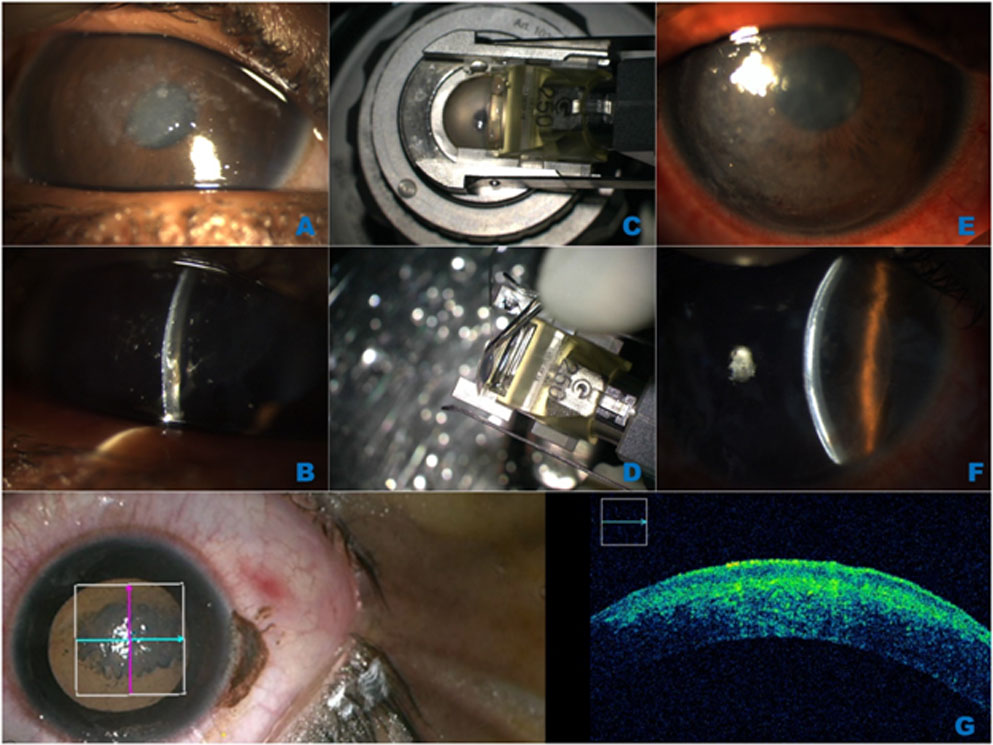
These patients underwent various anterior lamellar procedures depending on the depth of opacity. 7(46.6%) patients underwent ALTK, 2 (13.3%) patients HALK, 2 (13.3%) SALK, 4 (26.6%) DALK. Out of these, 10 cases were suture-less fibrin glue assisted, 4 cases were with sutures, and one case with both sutures and glue assisted (Figure 1 & 2).
The mean pre-operative visual acuity improved from 1.29±0.32 to 0.4±0.27 at 6 months with a significant p value of 0.002. The contrast sensitivity also showed a significant change from 0.1±0.1 to 1.13±0.2 at 6 months.The astigmatism decreased significantly from a value of 4.9±2.6 to 2.18±0.67 at 6 months. The total aberrations and lower order aberrations as measured by pentacam showed a significant decrease from 22.4±12.7 to 12.22±4.13 (p=0.012) and 19.93±13.42 to 11.4±3.99 (p=0.034) respectively. The higher order aberrations also showed a decrease though not significant from 4.93±1.78 to 4.2±1.4 (p=2.69).
We also evaluated the aberrations as measured by i-trace Ray Tracing Aberrometer. The total, higher and lower order aberrations showed a significant decrease from values of 4.4±2.0, 3.8±2, 1.6±1.1 to 2.9±2.04 (p=0.002), 2.4±1.15(p=0.002) and 0.9±0.1(p=0.018) at 6 months respectively. The measurements of various parameters at preoperative, 1stmonth,3rdmonth and 6thmonth is described in Table 2.
Table 2: Change of Visual Acuity, Contrast Sensitivity, Corneal Curvature and Optical Aberrations following Anterior Lamellar Keratoplasty
| Pre-operative Visit | Month 1 Visit | Month 3 Visit | Month 6 Visit | |
| Visual Acuity
(logMar) |
1.29±0.32 | 0.81±0.25 | 0.6±0.22 | 0.4±0.27
(p= 0.002)* |
| Contrast Sensitivity | 0.1±0.1 | 0.6±0.39 | 0.8±0.3 | 1.13±0.2 (p=0.001)* |
| Keratometric cylinder (diopters) | 4.9±2.6 | 3.07±1.5 | 2.44±1.0 | 2.18±0.67 (p=0.001)* |
| Total aberrations (Pentacam) | 22.4±12.7 | 11.44±4.1 | 12.4±3.55 | 12.22±4.13
(p=0.012) |
| Lowerorder aberrations (Pentacam) | 19.93±13.42 | 10.1±3.34 | 11.4±3.28 | 11.4±3.99
(p=0.034) |
| Higher order aberrations (Pentacam) | 4.93±1.78 | 4.39±1.11 | 4.35±1.24 | 4.2±1.4
(p=2.69) |
| Total Aberrations
(I-trace) |
4.4±2.0 | 3.8±2.0 | 4.11±2.9 | 2.9±2.04
(p=0.002)* |
| Higher order aberrations
(I-trace) |
3.8±2 | 3.35±2 | 3.6±2.5 | 2.4±1.15
(p=0.002)* |
| Lower order aberrations
(I-trace) |
1.6±1.1 | 1.3±1.1 | 1.4±0.7 | 0.9±0.1
(0.018) |
Upon comparing fibrin glue cases with the sutured cases, the visual acuity includingastigmatism, aberrations were found to be better in the fibrin glue cases.The mean pre-operative visual acuity was 1.5±0.39 and 1.17±.47 in suture-less and sutured group respectively which improved to 0.5±0.32 and 0.4±0.11 at 6 months post operatively. The mean pre-operative keratometric cylinder was 4.7±4.6 and 4.5±4.2 in suture-less and sutured group which improved to 2.09±1.03 and 4.9± 3.4 at 6 months post operatively.
The preoperative total, higher and lower aberrations as measured by pentacam in suture-less and sutured group were 20.3±12.2 and 25.2±15, 4.96±1.65 and 4.87±2.3, 18.08±12.9 and 23.62±15.49 which improved at 6 months to 11.2±4.52 and 14.2±2.5, 3.88±1.48 and 4.8±1.14, 10.4±4.45 and 13.4±2.1 respectively.
The preoperative total, higher and lower order aberrations as measured by i-trace in suture-less and sutured group was 3.5±1.42 and 6.18±1.94, 1.22±0.67 and 2.4±1.46, 3.01±1.64 and 5.5±1.64 which improved at 6 months post operative period to 1.8±1.35 and 4.9±1.52, 0.47±0.3 and 1.8±1.6, 1.6±1.2 and 4.2±1.11 respectively.All the above parameters are better in the suture-less glued group. IND-VFQ-33 scores showed a decrease in both the groups, hence signifying an improvement in quality in life with anterior lamellar keratoplasty.
DISCUSSION:
Anterior Lamellar procedures are now a choice of surgery for corneal pathologies limited to the anterior, mid to deep stroma with a healthy endothelium. 5Anterior lamellar keratoplasty are advanced procedures in which only the diseased portion of the cornea is replaced. As the amount of tissue replaced is less, the risk of graft rejection is low leading to lower chances of graft failure.6 Further, these procedures can be suture-less or with sutures. The suture-less lamellar surgeries come with the advantage of reducing suture-related complications and thus, a low chance of graft infection, rejection and failure. Suture-less procedures are also associated with a lower astigmatism and faster visual rehabilitation as compared to sutured keratoplasties.7In our study, we have evaluated not just the visual acuity post keratoplasty but also the quality of vision as measured by contrast sensitivity, clarity, and optical aberrations.
There are various tissue adhesives available for use in ophthalmology. The most commonly being used are the cyanoacrylate glue and the fibrin glue.Cyanoacrylate glue is synthetic glue which polymerizes upon coming in contact with a wet surface. It is basically used as to patch/ seal cornea in cases of small corneal perforations and avoid the risk of endophthalmitis, and reduces the need for tectonic keratoplasty. But this is associated with an eosinophilic inflammatory reaction.8,9Fibrin glue is biological glue which acts by mimicking the last step of coagulation cascade. It consists of two components, fibrinogen and thrombin component. It is better than cyanoacrylate glue as it is not associated with inflammation.10,11 In India,it is available as Tisseel Fibrin Sealant® (Baxter AG Vienna, Austria), and as Reliseal® (from Reliance life-sciences).
Tushar et al7compared the results of microkeratome-assisted suture-less anterior lamellar keratoplasty (SALK) with and without phacoemulsification. They found SALK Triple to be an effective surgery for early visual rehabilitation of patients with superficial anterior corneal opacity and concomitant cataract. They have modified the technique of glued keratoplasty as compared to the one described previously by Kaufmann et al to help prevent interface opacity in early post operative period as fibrin glue takes 2-4 weeks to clear from the interface. Instead of applying the glue to the entire stromal bed, they applied glue only to the graft host junction.
We also faced similar problems of interface haze in the early cases of glued keratoplasty. The excessive glue in the interface led not only to the haze but also led to an increased astigmatism being recorded. We then modified the technique by using the two components of glue separately. One drop of one component was applied to the host stromal bed and one drop of the second component was applied to the donor cornea. Donor cornea was then placed and centered on the host stromal bed. Excessive glue was then removed from the interface by smooth stroking movements over the cornea. This technique modification definitely helped us achieve better interface clarity and lower astigmatism.
Borderie et al6compared anterior lamellar keratoplasty and penetrating keratoplasty. They found that the risk of irreversible rejection is very low in cases of anterior lamellar keratoplasty. It also has lower graft failure rate which could be partly because of the absence of endothelial rejection. Our study also found similar results as we did not come across any episode of graft rejection in our cases.We also did IND-VFQ-33 questionnaire for both the groups and found an improvement from preoperative to post operative at 6 months in both the groups with a better outcome in suture-less group.
CONCLUSION:
There was an improvement in visual acuity, aberrations as well as astigmatism in the post operative period with the glue assisted cases showing a better outcome. Lamellar keratoplasty is a promising technique for improving visual quality as well as quality of life with improved results in glue aided cases. A larger sample size with a longer follow up are neededfor more reproducible results.
References:
- Borderie VM, Sandali O, Bullet J,Ganjoux T, Touzeau O, Laroche L. Long term results of deep anterior lamellar versus penetrating keratoplasty. Ophthalmology 2012;119(2):249-255.
- Gupta SK, Viswanath K, Thulasiraj RD, et al. The development of the Indian vision function questionnaire: field testing and psychometric evaluation. The British Journal of Ophthalmology2005;89(5):621-627.
- Vashist P, Gupta N, Tandon R, Gupta SK, Dwivedi S, Mani K. Population-based assessment of vision-related quality of life in corneal disease: results from the CORE study.Br J Ophthalmol2016;100(5):588-93.
- Venkataraman A, Rathinam S R. A pre- and post-treatment evaluation of vision-related quality of life in uveitis. Indian J Ophthalmol 2008;56:307-12.
- Vajpayee RB, Vasudendra N, Titiyal JS, Tandon R, Sharma N, Sinha R. Automated lamellar therapeutic keratoplasty (ALTK) in the treatment of anterior to mid-stromal corneal pathologies.Acta Ophthalmol Scand.2006 Dec;84(6):771-3.
- Borderie VM, Guilbert E, Touzeau O, Laroche L. Graft rejection and graft failure after anterior lamellar versus penetrating keratoplasty.Am JOphthalmol. 2011;151(6):1024-1029.e1.
- Agarwal T, Bandivadekar P, Sharma N, Sagar P, Titiyal JS. Sutureless anterior lamellar keratoplasty with phacoemulsification.2015 Jun;34(6):615-20.
- Forseth M, O’Grady K, Toriumi DM. The current status of cyanoacrylate and fibrin tissue adhesives. J Long Term Eff Med Implants 1992;2:221-33.
- Lagoutte FM, Gauthier L, Comte PR. A fibrin sealant for perforated and preperforated corneal ulcers. Br J Ophthalmol 1989;73:757-61.
- Chabbat J, Tellier M, Porte P, Steinbuch M. Properties of a new fibrin glue stable in liquid state. Thromb Res 1994;76:525-33
- Le Guéhennec L, Layrolle P, Daculsi G. A review of bioceramics and fibrin sealant. Eur Cell Mater 2004;8:1-10
Figures and Figure legends
Figure 1: Clinical photographs of the eye with healed keratitis treated with sutured hemi-automated lamellar keratoplasty.
1A; Preoperative clinical picture of the left eye showing epithelial lesions with significant stromal scarring due to healed keratitis, 1B &1C; Intraoperative picture showing intraoperative –OCT guided manual lamellar dissection, 1D; Postoperative Slit-lamp clinical picture of the left eye 5 months following hemi-automated lamellar keratoplasty.
Figure 2: Clinical photograph of a patient with Gelatinous Drop-like Corneal Dystrophy treated with suture-less anterior lamellar therapeutic keratoplasty.
2A &2B; Preoperative slit lamp bio-microscopy picture of the right eye – diffuse and Slit illumination respectively showing grape like elevated corneal lesions.
2C &2D; Intraoperative donor tissue preparation using 250 microns automated microkeratome.
2E &2F; Postoperative slit lamp bio-microscopy picture of the right eye – diffuse and Slit illumination respectively.
2G&2H; Intraoperative clinical and i-OCT guided anterior lamellar therapeutic keratoplasty
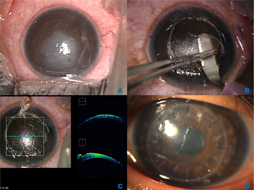
Figure 2: Clinical photograph of a patient with Gelatinous Drop-like Corneal
Dystrophy treated with suture-less anterior lamellar therapeutic keratoplasty.
2A &2B; Preoperative slit lamp bio-microscopy picture of the right eye – diffuse and Slit illumination respectively showing grape like elevated corneal lesions.
2C &2D; Intraoperative donor tissue preparation using 250 microns automated microkeratome.
2E &2F; Postoperative slit lamp bio-microscopy picture of the right eye – diffuse and Slit illumination respectively.
2G&2H; Intraoperative clinical and i-OCT guided anterior lamellar therapeutic keratoplasty
Leave a Comment